Abstract
SIRT3 modulates respiration via the deacetylation of lysine residues in electron transport chain proteins. Whether mitochondrial protein acetylation is controlled by a counter-regulatory program has remained elusive. Here we identify an essential component of this previously undefined mitochondrial acetyltransferase system. We show that GCN5L1/Bloc1s1 counters the acetylation and respiratory effects of SIRT3. GCN5L1 is mitochondrial-enriched and displays significant homology to a prokaryotic acetyltransferase. Genetic knockdown of GCN5L1 blunts mitochondrial protein acetylation, and its reconstitution in intact mitochondria restores protein acetylation. GCN5L1 interacts with and promotes acetylation of SIRT3 respiratory chain targets and reverses global SIRT3 effects on mitochondrial protein acetylation, respiration and bioenergetics. These data identify GCN5L1 as a critical, prokaryote-derived component of the mitochondrial acetyltransferase program.
Keywords: GCN5L1, SIRT3, Mitochondrial metabolism, protein acetylation
INTRODUCTION
The mitochondrial sirtuin deacetylase SIRT3 plays an important role in regulating oxidative metabolism [1–3] and redox stress [4–6]. Consequently, the regulation of SIRT3 activity is emerging as an important factor in the mitochondrial contribution towards disease susceptibility (reviewed [7]). To understand this system more fully, it is necessary to identify proteins that counteract SIRT3 activity. As such, the discovery of a mitochondrial lysine acetyltransferase has been actively pursued [3].
Given the evolutionary history of mitochondria, one avenue of research has focused on bacteria. In Salmonella enterica, the acetyltransferase Pat has been shown to counteract CobB, a sirtuin homologue, in the regulation of acetyl-CoA synthetase [8]. However, eukaryotic orthologs to Pat have not been identified in either the mitochondrial or nuclear genome [9]. An alternate scenario in eukaryotes could be that mitochondrial proteins are acetylated in the cytosol prior to mitochondrial import. However, as fasting and feeding result in a dynamic flux in these post-translational modifications [2, 10], it would be most likely that these modifications occur within mitochondria. Additionally, ATP synthase Fo subunit 8, a mitochondrial-encoded protein, is acetylated under nutrient flux conditions, further supporting the existence of an in situ mechanism [10].
In this report, we identify GCN5L1, a protein with significant homology to the nuclear acetyltransferase GCN5 (general control of amino-acid synthesis 5) [11], as a mammalian regulatory protein in the control of mitochondrial protein acetylation and respiration. We show that GCN5L1: includes prokaryote-conserved acetyltransferase substrate and acetyl-CoA binding regions; is localized within mitochondria; modulates mitochondrial electron transport chain (ETC) protein acetylation; alters mitochondrial oxygen consumption; and counters SIRT3 effects on mitochondrial protein acetylation, respiration and ATP levels.
MATERIALS AND METHODS
Phylogenetic and Structural Analysis
BLAST searches for acetyltransferases related to GCN5L1 identified several prokaryotic proteins, the closest being the xenobiotic streptogramin acetyltransferase from Burkholderia thailandensis (ZP_02389502), which we designated BtXAT. The sequences for acetyltransferases (all human, except BtXAT) described were obtained from Genbank and analyzed using the phylogenetic tool PhyML (www.phlogeny.fr.) The XAT-repeat region of GCN5L1 was identified by interrogation of the GCN5L1 protein sequence, looking for a hexapeptide repeat motif matching X-[STAV]-X-[LIV]-[GAED]-X (NCBI cd03349). Data from this region was also used to map the substrate- and acetyl-CoA-binding regions of BtXAT. Predictions of GCN5L1 protein biochemical properties were performed using a Kyte-Doolittle hydrophobicity plot.
Cell Culture and Transfection
HepG2 cells were grown in DMEM supplemented with 10% FBS. Plasmids were transfected using Fugene HD (Roche). For in vivo acetylation studies cells were starved for 4 h in HBSS and the re-fed with DMEM for 1.5 h prior to harvesting. Electroporation was used to transfect non-targeting control, GCN5L1 and SIRT3 siRNA (Dharmacon).
Contruction of plasmids
The cDNA for GCN5L1 (Open Biosystems) was cloned into p3XFLAG-CMV-14 (Sigma) to create GCN5L1-FLAG. The cDNAs for NDUFA9 and ATP5a (Open Biosystems)were cloned into pCMV3TAG-4A (Stratagene).
Polyclonal Antibody Production
A synthetic peptide corresponding to 16 amino acids of human and mouse GCN5L1, along with a conjugating cysteine at the C-terminus (MLSRLLKEHQAKQNER-C), was produced and injected into NZW rabbits (Covance). Serum was affinity purified against the immunizing peptide, and the antibody validated for recognition of human and mouse GCN5L1.
GCN5L1 Localization, Immunoblot Analysis and Co-Immunoprecipitation
Confocal microscopy was used to localize GCN5L1-FLAG and dsRed-mito (Clontech) in fixed hepatocytes, by indirect immunolabeling of FLAG using Alexa 488 (Invitrogen). Immunogold labeling and electron microscopy was used to localize endogenous GCN5L1 and the ETC protein ATP5a. Sub-mitochondrial localization was performed by osmotic pressure subfractionation and proteinase K protection assays. Antibodies used for westerns include: monoclonal acetyl-lysine (Ac-K), OPA1, GAPDH, VDAC (Cell Signaling); polyclonal Ac-K, ATP5a, NDUFA9, GDH (Abcam); FLAG (Sigma). In co-immunoprecipitation experiments between GCN5L1-FLAG and NDUFA9-Myc or ATP5a-Myc, cells were co-transfected with plasmids for 24 h. Lysates were harvested and incubated with FLAG- or Myc-conjugated beads (Sigma and Cell Signaling, respectively). Beads were washed and analyzed by western blot. Endogenous co-immunoprecipitation experiments followed a similar protocol, however lysates were incubated with the relevant antibodies overnight, and interacting proteins were captured using protein A/G beads (Santa Cruz). Western blots were quantified using image analysis software, and those shown are representative of at least three independent experiments.
Animal husbandry
The use of mice in this study was approved by the NHLBI Animal Care and Use committee, and animals were maintained according to their guidelines.
Metabolic Measurements
Oxygen consumption measurements were performed on the XF24 analyzer (Seahorse Bioscience). Control- and GCN5L1-transfected siRNA cells were transferred to 24-well plates overnight and incubated in sucrose respiration media for 1 h prior to analysis. To measure the response to the mitochondrial respiration substrates glutamate and malate, cells were permeabilized with digitonin (10 μ g per 106 cells, 5 min) and incubated in sucrose respiration media. 10 mM glutamate/5 mM malate were added after baseline measurements had been taken. ATP levels in intact cells were measured (n = 5) using the EnzyLight assay kit (Bioassay Systems).
In Vitro Acetylation Analysis and In Vitro Immunoprecipitation
An in vitro acetylation assay was adapted from a published protocol [12]. HepG2 cells were transfected with control or GCN5L1-FLAG plasmids for 24 h; or control and GCN5L1 siRNA for 72 h; at which time mitochondria were isolated. Samples were resuspended in reaction buffer (50 mM Tris-HCl pH 8.0, 50 mM NaCl, 4 mM MgCl2, 5 mM nicotinamide, pH 7.4), sonicated, then incubated for 1.5 h at 37 °C, in the absence or presence of 2.5 mM acetyl-CoA. Mitochondrial proteins were used for global acetylation analysis (reaction stopped by boiling with SDS sample buffer, followed by SDS-PAGE and immunoblot analysis with a monoclonal acetyl-lysine antibody), or immunoprecipitation from non-denatured samples. For the reconstitution of GCN5L1 in mitochondria, HepG2 cells were depleted of GCN5L1 using siRNA as described, after which intact mitochondria were isolated by sucrose gradient centrifugation. 50 μg of pure mitochondria per sample were resuspended in acetyl-CoA assay buffer (100 mM Tris-HCl pH 8.0, 50 mM NaCl, 4 mM MgCl2, 5 mM nicotinamide, pH 7.4) on ice and sonicated. Samples were incubated at either 25 °C or 95 °C for 5 mins, followed by the addition of 0.5 μg GST-GCN5L1 (Abnova) in PBS or PBS alone (control). After incubation for 1.5 h at 30 °C, the reaction was stopped by boiling in SDS sample buffer. The histone assay followed this protocol, save for the addition of 2.5 μg of recombinant human histone H3 (New England Biolabs) to the mitochondrial samples where indicated. Following the 1.5 h incubation, acetylated H3 was recovered by immunoprecipitation from non-denatured samples with a polyclonal Ac-K antibody. For in vitro immunoprecipitation, lysates from the in vitro acetylation analysis were incubated overnight with a polyclonal Ac-K antibody. Immunoprecipitated proteins were purified using protein G beads, washed and analyzed by western blot.
Statistical analysis
Where required, data were tested for normality using the Kolmogorov-Smirnov test, followed by a one-tailed Student’s t-test or a Mann-Whitney U-test using SigmaPlot 11 (Systat Software). A P value of < 0.05 was regarded as significant.
RESULTS
GCN5L1 is identified as a putative mitochondrial counter-regulator of SIRT3
To identify mitochondrial lysine acetyltransferases, we screened the human mitochondrial proteome database MitoCarta [13] for proteins harboring acetyltransferase regions, and used yeast GCN5 as ‘bait’ in BLAST searches. Fluorescent-tagged candidate proteins were expressed to evaluate mitochondrial localization, and oxygen consumption was measured following siRNA knockdown to identify candidates that counter the known respiratory effects of SIRT3 depletion [1, 14]. The strategy employed is illustrated in Figure S1A. Four candidate proteins identified included GCN5L1 (synonym BLOC1S1), NAT8b, NAT9 and NAT11. Expression of YFP-tagged constructs and dsRed-mito showed that two proteins, namely GCN5L1 and NAT9, co-localized with the mitochondrial marker (Figure S1B). As the knockdown of SIRT3 blunts mitochondrial respiration [1, 15], we reasoned that the genetic depletion of a counter-regulatory component should show the inverse phenotype. siRNA was employed to knockdown GCN5L1 and NAT9; however, only the loss of GCN5L1 resulted in increased oxygen consumption in HepG2 cells (Figure S1C). Subsequent studies focused on the role of GCN5L1 in mitochondrial protein acetylation, and as a putative counter-regulatory protein to SIRT3.
GCN5L1 is mitochondrial enriched and more highly expressed in oxidative tissues
GCN5L1 is a previously uncharacterized protein, although its sequence homology to GCN5, the nuclear acetyltransferase, has been described [11, 16]. We first confirmed its mitochondrial localization, using confocal microscopy on HepG2 cells transfected with FLAG-tagged GCN5L1 and dsRed-mito (Figure 1A); and by immuno-gold labeled electron microscopy, which showed that the endogenous protein has similar localization to the alpha subunit of ATP synthase (ATP5a) in mouse liver mitochondria (Figure 1B and S1D). Kyte-Doolittle hydropathy modeling of the GCN5L1 protein suggests that it is a non-transmembrane globular protein (Figure S1E) [17], suggesting its location within mitochondria to be in either the intermembrane space or matrix soluble fractions. Osmotic pressure subfractionation and proteinase K assays of isolated mouse mitochondria both support that GCN5L1 resides in the soluble matrix and intermembrane space fractions (Figures 1C and 1D). Its mitochondrial enrichment was further supported by the higher levels of GCN5L1 in slow- versus fast-twitch skeletal muscle (Figure 1E).
Figure 1. GCN5L1 localizes to mitochondria.
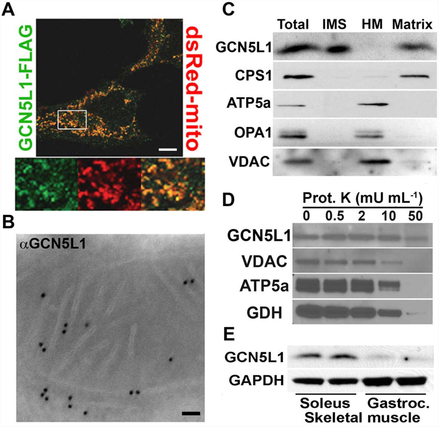
(A) Indirect immunofluorescence confocal microscopy of HepG2 cells co-expressing GCN5L1-FLAG and dsRed-mito. Scale bar = 5 μm. (B) Immuno-gold labeling of endogenous GCN5L1 in fixed mouse liver tissue. Scale bar = 5 nm. (C) Sub-mitochondrial localization of GCN5L1 by osmotic pressure analysis of isolated mouse mitochondria. Abbreviations: IMS – inter-membrane space, HM – heavy mitochondrial membranes. (D) Proteinase K protection assay to establish GCN5L1 sub-mitochondrial localization. (E) Expression of GCN5L1 in soleus (slow-twitch) and gastrocnemius (fast-twitch) skeletal muscle.
GCN5L1 possesses prokaryotic features and its knockdown disrupts mitochondrial protein acetylation
Phylogenetic mapping places GCN5L1 in a clade containing a histone acetyltransferase (HAT1), a bacterial xenobiotic acetyltransferase (BtXAT), and the nuclear lysine acetyltransferase GCN5 (Figure 2A). As such, we compared the GCN5L1 amino acid sequence with BtXAT and yeast GCN5. This comparison revealed a 53% similarity to the acetyl-CoA- and substrate-binding motifs of BtXAT. In contrast, homology to yeast GCN5 was limited to the non-catalytic N-terminal region of the protein (Figure 2B). The deduced GCN5L1 protein sequence, and especially the prokaryotic-like XAT repeat region, is highly conserved in metazoans (not shown), suggesting a functional role of this region.
Figure 2. GCN5L1 levels modulate the degree of mitochondrial protein acetylation.
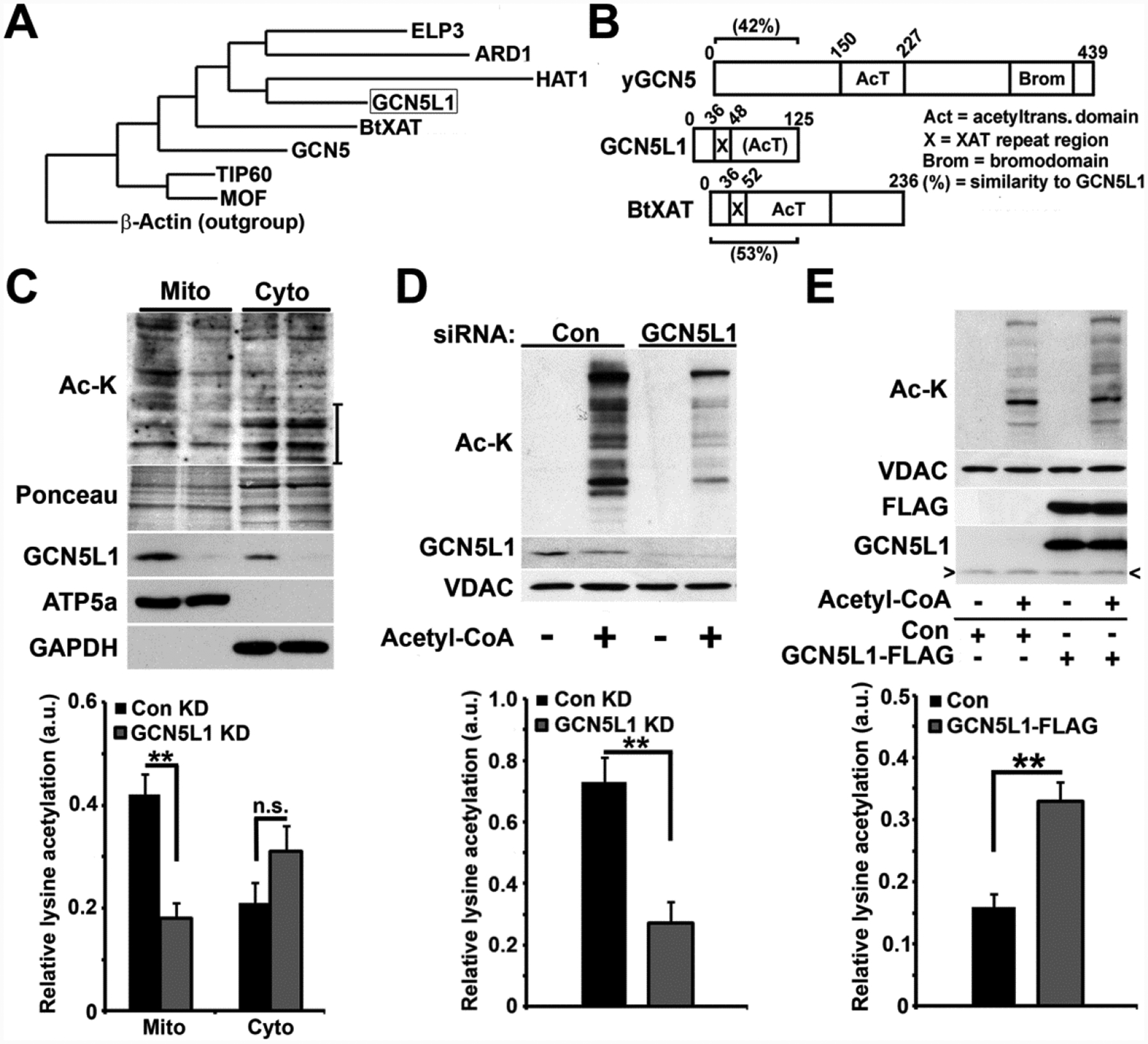
(A) Phylogenetic mapping of GCN5L1 against human and bacterial acetyltransferases identifies homology to HAT1 and BtXAT. (B) Alignment of GCN5L1 against yeast GCN5 and BtXAT acetyltransferases. (C) Lysine acetylation (Ac-K) status of endogenous mitochondrial and cytoplasmic proteins following GCN5L1 depletion; bar shows region of Ponceau stain. The accompanying histogram shows relative protein acetylation in the mitochondrial and cytosolic fractions comparing scrambled to GCN5L1 knockdown (KD) (D) Acetylation status of total mitochondrial proteins in control or GCN5L1-depleted HepG2 cells following in vitro acetylation assay with the accompanying histogram showing relative protein acetylation. (E) Acetylation status of mitochondrial proteins expressing control or GCN5L1 constructs, following an in vitro acetylation assay with the accompanying histogram showing relative protein acetylation. Arrows indicate endogenous GCN5L1. Data are expressed a mean ± s.e.m. (n≥3). n.s. – not significant, **p<0.01, compared to scrambled siRNA treated controls.
To begin exploring the functional role of GCN5L1, we determined whether siRNA knockdown of this protein would modulate protein acetylation. Although GCN5L1 is predominantly mitochondrial, cytosolic expression was also observed. GCN5L1 levels were depleted in both subcellular compartments by siRNA; however, only the mitochondrial fraction showed a significant reduction in protein acetylation in HepG2 cells, supporting its functional role in mitochondria (Figure 2C). The acetylation function of GCN5L1 was further supported by a ≈ 60% attenuation of mitochondrial protein acetylation, in an in vitro acetylation assay, using GCN5L1-depleted cells (Figure 2D). Furthermore, the overexpression of GCN5L1 in HepG2 cells increased mitochondrial protein acetylation to a similar degree in the presence of acetyl-CoA (Figure 2E).
Histone H3 is acetylated by GCN5L1-enriched mitochondrial extracts
To further characterize this effect, in vitro assays were performed in assess whether recombinant GCN5L1 can acetylate purified histones, a classic acetylation substrate. In contrast to the in vivo studies, GCN5L1 promoted only weak histone acetylation when co-incubated in vitro (data not shown). To investigate whether this discrepancy may be due to the requirement of additional mitochondrial factors in the functioning of GCN5L1, we assessed mitochondrial protein acetylation in response to the addition of recombinant GCN5L1 to mitochondrial extracts purified from GCN5L1-depleted cells. Here, the reconstitution of GCN5L1 restored protein acetylation; although this was effect was limited to native, and not denatured mitochondrial extract samples (Figure 3A). It is interesting to note in Figure 3A that the denaturing of mitochondria appears to facilitate ‘auto-acetylation,” as the boiled samples show enhanced protein acetylation in the presence and absence of GCN5L1. To further explore the requirement of additional mitochondrial cofactors in the acetylation function of GCN5L1, recombinant histone H3 was added to native GCN5L1-depleted mitochondrial extracts in parallel with the reintroduction, or not, of GCN5L1. The subsequent immunoprecipitation of acetylated lysine proteins, followed by immunoblot analysis with an antibody recognizing histone H3, shows increased H3 acetylation in the presence of GCN5L1 (Figure 3B). These data show that GCN5L1 positively regulates mitochondrial protein acetylation. However, its activity appears to require additional factors that reside in mitochondria.
Figure 3. Intact mitochondrial contents are required for GCN5L1 mediated protein acetylation.
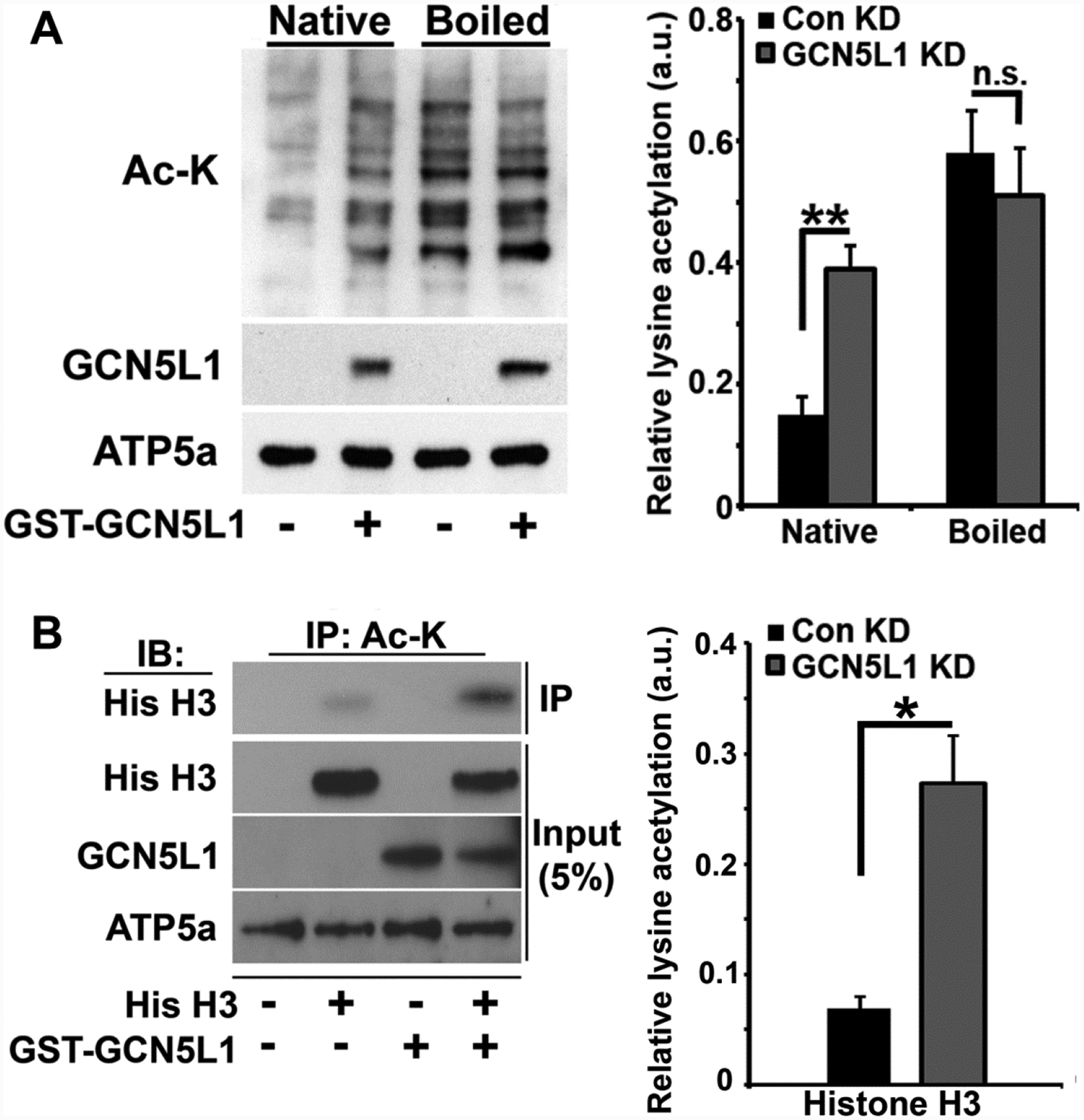
(A) Effect of the reconstitution of GCN5L1 to native and boiled mitochondrial fractions on protein acetylation (Ac-K). The accompanying histogram shows relative protein acetylation in the native or boiled (denatured) mitochondrial fractions with or without the restoration of GCN5L1. (B) The extent of recombinant histone H3 acetylation in mitochondrial extracts in the presence or absence of GCN5L1. The relative acetylation of histone H3 in the presence or absence of GCN5L1 is shown in the accompanying histogram. Data are expressed a mean ± s.e.m. (n≥3). n.s. – not significant, *p<0.05 and **p<0.01, compared to scrambled siRNA treated controls.
GCN5L1 Promotes Electron Transport Chain Protein Acetylation
As the predominant mitochondrial protein lysine deacetylase, SIRT3, activates ETC proteins [1, 18], we investigated whether known SIRT3 ETC targets interact with, and are modulated by, GCN5L1. Co-expression of GCN5L1 with Myc-tagged complex I protein NDUFA9, or complex V protein ATP5a, followed by reciprocal immunoprecipitation, shows that there is a physical interaction between both NDUFA9 and ATP5a with GCN5L1 (Figures S2A and S2B). To confirm these interactions occur between the endogenous proteins in vivo, untransfected HepG2 cell extracts were used for immunoprecipitation studies. Here the antibody directed against GCN5L1 was employed for immunoprecipitation, and antibodies directed against NDUFA9 and ATP5a were used in immunoblot analysis (Figure 4A). These data confirm the protein-protein interaction between GCN5L1 and the ETC proteins. To functionally characterize whether these ETC proteins are targets of GCN5L1 mediated acetylation, we then undertook in vitro acetylation procedures using total mitochondrial protein in cells displaying either knockdown or overexpression of GCN5L1. Using mitochondria from control and GCN5L1-depleted cells, followed by immunoprecipitation with an acetylated lysine antibody and immunoblot analysis, showed that acetylation of endogenous NDUFA9 and ATP5a is diminished following GCN5L1 knockdown (Figure 4B). In parallel, immunoprecipitation with an antibody against acetylated lysine demonstrates that endogenous NDUFA9 and ATP5a exhibit increased acetylation in cells overexpressing GCN5L1 relative to control samples (Figure 4C and 4D).
Figure 4. GCN5L1 modulates the acetylation of mitochondrial electron transport chain proteins.
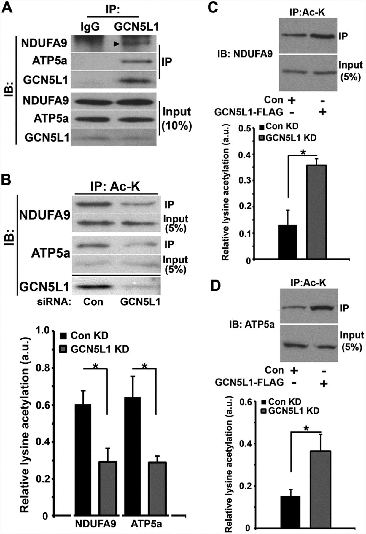
(A) In vivo interaction of GCN5L1 with the ETC proteins NDUFA9 and ATP5a. The arrow shows the specific band for NDUFA9 relative to the non-specific band seen in the IgG immunoprecipitation control. (B) In vitro immunoprecipitation acetylation assay of endogenous NDUFA9 and ATP5a with accompanying histogram showing the relative levels of the respective protein acetylation (Ac-K) levels in control or GCN5L1-depleted HepG2 cells. (C) In vitro immunoprecipitation acetylation assay using an acetylated-lysine antibody to assay endogenous NDUFA9 acetylation with the accompanying histogram showing relative acetylation in HepG2 cells expressing control or GCN5L1 plasmids . (D) In vitro immunoprecipitation acetylation assay using an acetylated-lysine antibody to assay endogenous ATP5a acetylation with the accompanying histogram showing relative acetylation in HepG2 cells expressing control or GCN5L1 plasmids. n ≥ 3 for all experiments. *p<0.05, compared to respective controls. Results are mean ± s.e.m.
Mitochondrial Respiration is Attenuated Following GCN5L1 Knockdown
As GCN5L1 targets ETC proteins, we then evaluated the effect of GCN5L1 knockdown on mitochondrial respiration. Following knockdown of GCN5L1 compared to scrambled control siRNA in HepG2 cells (Figure 5A), oxygen consumption was assessed using the Seahorse apparatus. Basal cellular oxygen consumption was increased by GCN5L1 knockdown, as was the maximal oxygen consumption following mitochondrial uncoupling (Figure 5B). The depletion of GCN5L1 in HepG2 cells resulted in a 36% increase in oxygen consumption (Figure 5C). To confirm that this oxygen was consumed in the mitochondria, the HepG2 cells were permeabilized with digitonin and oxygen consumption was determined in the presence of glutamate and malate as specific mitochondrial respiratory substrates. Here, oxygen consumption was increased by 32% in GCN5L1 knockdown cells (Figure 5D). In parallel with these changes in oxygen consumption, the depletion of GCN5L1 results in significantly higher cellular ATP levels (Figure 5E).
Figure 5. GCN5L1 knockdown increases mitochondrial respiration.
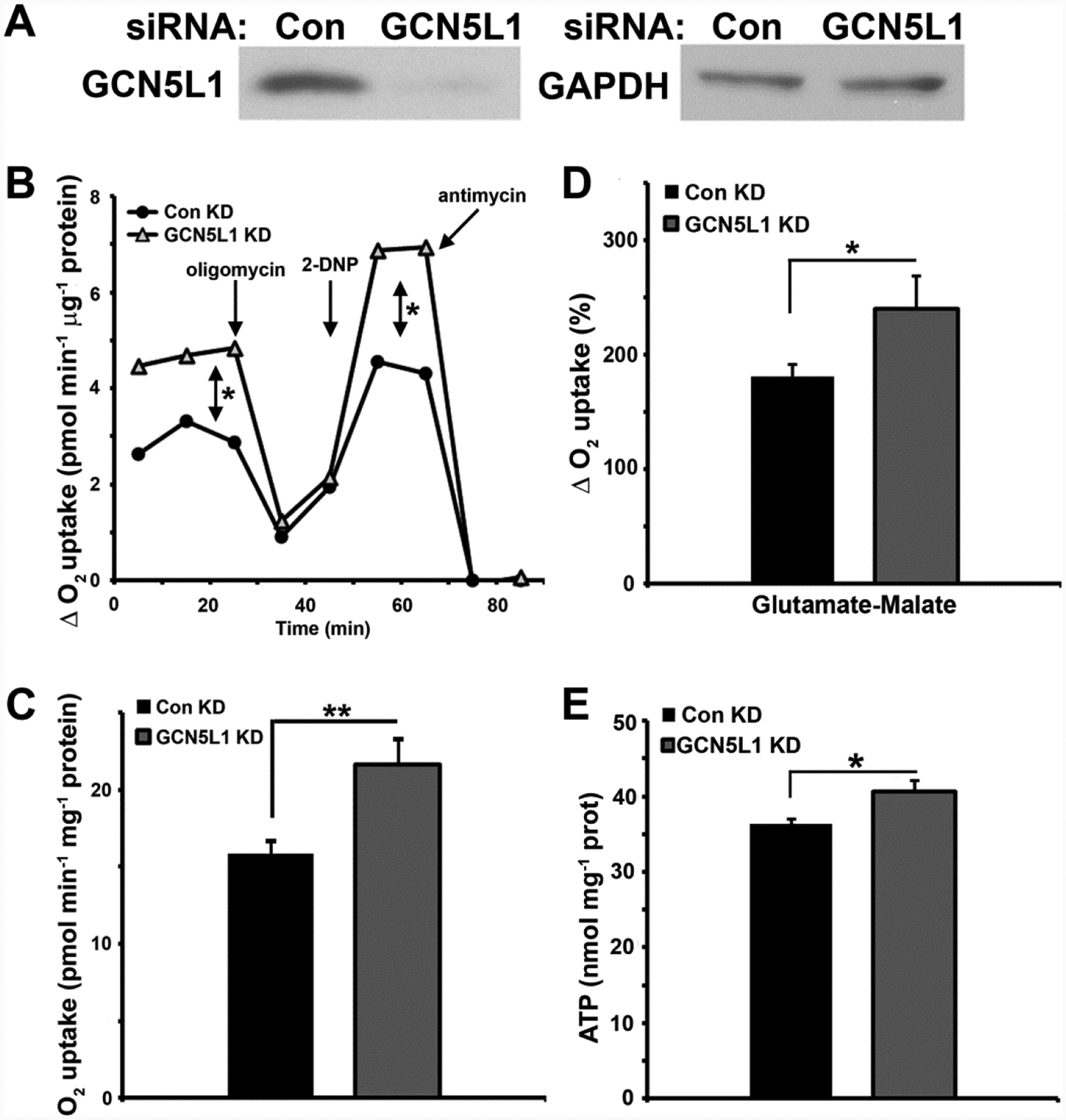
(A) Steady-state GCN5L1 levels in HepG2 cells following control or GCN5L1 knockdown (KD). (B) Representative tracing of basal oxygen consumption and maximal oxygen consumption induced by the uncoupler dinitrophenol (2-DNP) comparing control and GCN5L1 KD HepG2 cells. (C) The absolute differences in basal oxygen consumption comparing control and GCN5L1 KD HepG2 cells, taken as a mean of the first three data points. (D) Relative differences in oxygen consumption in control and GCN5L1 KD cells following digitonin administration and the use of glutamate and malate as mitochondrial respiration substrates. (E) Differences in cellular ATP levels in control and GCN5L1 KD HepG2 cells. n ≥ 3 for all experiments. *p<0.05 and **p<0.01, compared to respective controls. Results are mean ± s.e.m.
GCN5L1 Functionally Opposes SIRT3 Effects
The above results show that the acetylation and bioenergetic phenotype of GCN5L1 depletion is in direct contrast to that observed following the loss of SIRT3. As these findings suggest that GCN5L1 may counteract SIRT3 function, we explored the response to the combined genetic manipulation of these proteins. We employed an in vivo model where SIRT3 was knocked down in HepG2 cells, with or without the concurrent knockdown of GCN5L1. Knockdown of SIRT3 alone increased mitochondrial protein acetylation, which was significantly reversed by the concurrent knockdown of GCN5L1 (Figure 6A). Accordingly, the knockdown of SIRT3 diminished mitochondrial oxygen consumption and cellular ATP levels [1, 14], and the concurrent knockdown of GCN5L1 reversed these metabolic phenotypes (Fig. 6B–C). The ability of GCN5L1 knockdown to reverse SIRT3 acetylation and respiratory phenotypes was confirmed in SIRT3−/− MEF cells (data not shown).
Figure 6. GCN5L1 counteracts the effects of SIRT3.
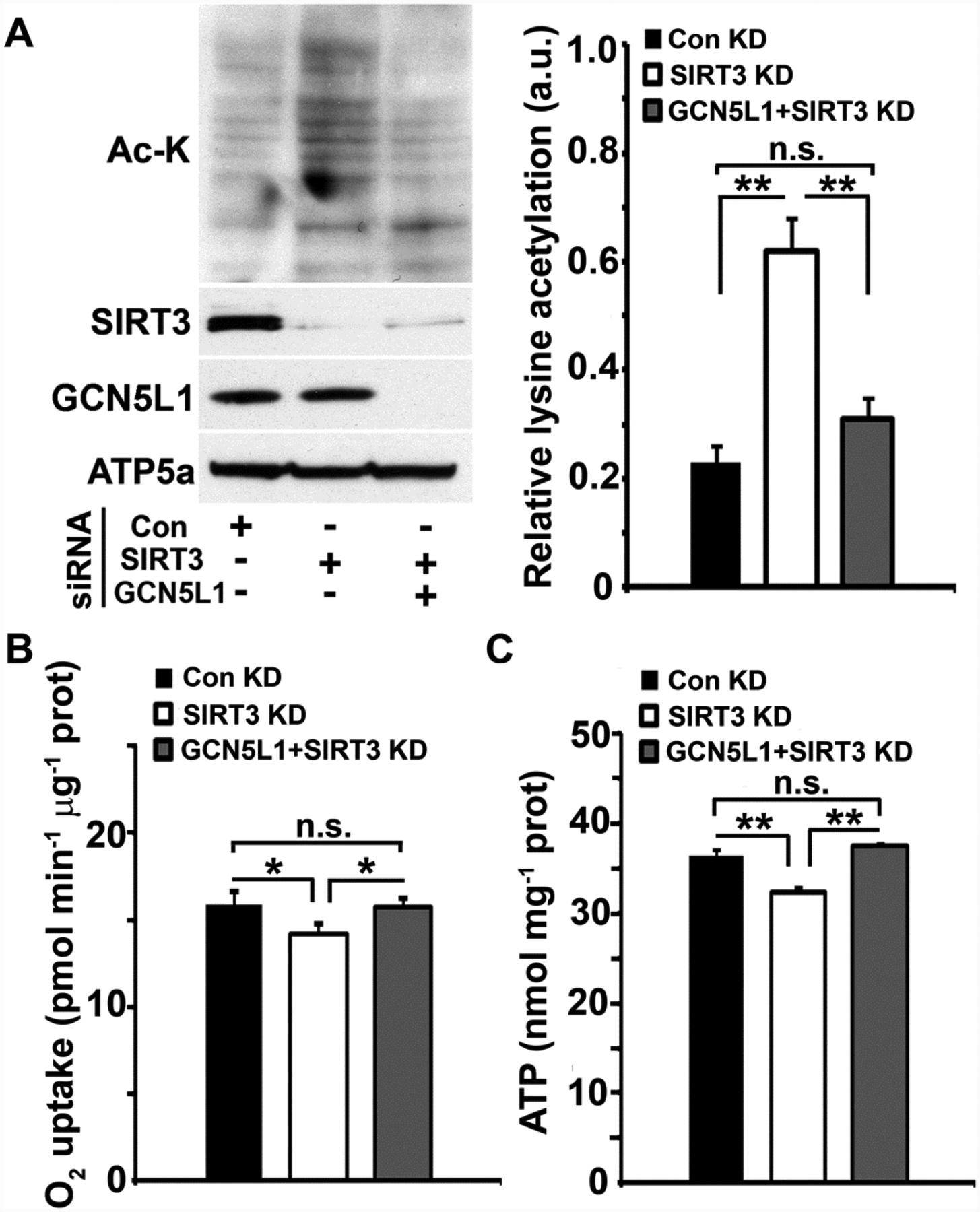
(A) Total mitochondrial protein acetylation (Ac-K) in HepG2 cells transfected with control, SIRT3 or SIRT3+GCN5L1 siRNA with an accompanying histogram showing the relative differences in mitochondrial protein acetylation under the three different conditions. Metabolic measurements of (B) mean total oxygen consumption (n = 5) and (C) mean total cellular ATP (n=5) of HepG2 cells transfected with control, SIRT3 or SIRT3+GCN5L1 siRNA. Western analyses of mitochondrial acetylation were performed at least five times, and representative blots are shown. n.s. – not significant, *p<0.05 and **p<0.01, compared to the defined controls. Results are mean ± s.e.m.
DISCUSSION
Identification of the mitochondrial acetyltransferase machinery has proven elusive. This is in contrast to the nuclear compartment, where the acetyltransferases GCN5, p300 and TIP60 have been found to function as counter-regulatory enzymes to SIRT1 [19]. The identification GCN5L1 in this report reveals a novel mediator of mitochondrial protein acetylation to counter sirtuin deacetylase function.
The maintenance of sequence homology, from prokaryotes to nuclear-encoded eukaryotic mitochondrial proteins, is consistent with the symbiotic hypothesis of mitochondrial evolution [20]. This phylogenetic fidelity is also evident, for example, in eukaryotic mitochondrial kinases [21], and in the iron/sulphur assembly regulatory protein frataxin [22]. As with these examples, the conservation of the BtXAT protein acetyl-CoA- and substrate-binding regions in GCN5L1 supports the strong evolutionary pressure for retention of prokaryotic features in mitochondrial regulatory proteins.
The characterization of GCN5L1 in this study shows that its expression, in isolation, is insufficient to significantly augment protein acetylation. However, the loss of acetylation following GCN5L1 depletion, and the re-establishment of this activity after its reconstitution, both demonstrate the requirement for GCN5L1 in mitochondrial protein acetylation. The findings presented here suggest that GCN5L1 is one critical component of the mitochondrial acetyltransferase machinery. This concept is compatible with the function of yeast GCN5, where integration of this protein into a multi-subunit complex is required for maximal acetyltransferase activity [23]. Together, these data have compelled us to begin to investigate whether additional mitochondrial proteins form multimeric complexes with GCN5L1 in order to orchestrate acetyltransferase activity.
The early characterization of the bioenergetic role of the mitochondrial deacetylase SIRT3 was performed in genetic knockout or knockdown conditions in cell and murine models. The initial studies showed that the absence of SIRT3 resulted in a marked reduction in mitochondrial oxygen consumption and a decrease in cellular ATP levels [1, 14]. Subsequent studies in an in vivo context showed that the bioenergetic effects of SIRT3 were most pronounced under fasted conditions, and that the activity of numerous enzymes in multiple mitochondrial metabolic pathways are modified by SIRT3-mediated protein deacetylation. In this manuscript we employed a similar approach, and identified that the absence of GCN5L1 inversely perturbs mitochondrial oxygen consumption and cellular ATP levels. However, more comprehensive biochemical analysis of the effects of mitochondrial protein acetylation will need to be performed as additional substrates of GCN5L1 are delineated, particularly following the identification of the putative functional partners of GCN5L1.
In summary, this study shows that GCN5L1 functions as an essential component of the mitochondrial lysine acetyltransferase machinery, has phylogenetic linkage to prokaryotic acetyltransferases, and modulates mitochondrial respiration via acetylation of ETC proteins. GCN5L1 also counters the mitochondrial deacetylase function of SIRT3. Further investigation of GCN5L1, its putative interacting proteins and substrates, and the role of acetyl-CoA, should enhance our understanding of how acetylation modulates mitochondrial function.
Supplementary Material
ACKNOWLEDGEMENTS
This research was supported by the Division of Intramural Research of the NHLBI, National Institutes of Health. We thank Mary McKee (Partners Healthcare, Boston MA) for her help with electron microscopy studies, Michael Lazarou (NINDS, Bethesda MD) for advice on Proteinase K assays and Toren Finkel (NHLBI) and Richard Youle (NINDS) for their critical review of this manuscript.
Abbreviations used:
- Ac-K
acetyl-lysine
- ATP5a
ATP synthase subunit 5a
- ETC
electron transport chain
- GCN5L1
general control of amino acid synthesis 5 (GCN5) like 1
- HAT
histone acetyltransferase
- NAT
N-terminal acetyltransferase
- NDUFA9
NADH dehydrogenase subunit A9
- SIRT3
sirtuin family member 3
- BtXAT
bacterial xenobiotic acetyltransferase
Footnotes
Conflict of interest statement: No conflicts of interests to report.
REFERENCES
- 1.Ahn BH, Kim HS, Song S, Lee IH, Liu J, Vassilopoulos A, Deng CX and Finkel T (2008) A role for the mitochondrial deacetylase Sirt3 in regulating energy homeostasis. Proc.Natl.Acad.Sci.U.S.A 105, 14447–14452 [DOI] [PMC free article] [PubMed] [Google Scholar]
- 2.Hirschey MD, Shimazu T, Goetzman E, Jing E, Schwer B, Lombard DB, Grueter CA, Harris C, Biddinger S, Ilkayeva OR, Stevens RD, Li Y, Saha AK, Ruderman NB, Bain JR, Newgard CB, Farese RV Jr., Alt FW, Kahn CR and Verdin E (2010) SIRT3 regulates mitochondrial fatty-acid oxidation by reversible enzyme deacetylation. Nature. 464, 121–125 [DOI] [PMC free article] [PubMed] [Google Scholar]
- 3.Hallows WC, Yu W, Smith BC, Devires MK, Ellinger JJ, Someya S, Shortreed MR, Prolla T, Markley JL, Smith LM, Zhao S, Guan KL and Denu JM (2011) Sirt3 Promotes the Urea Cycle and Fatty Acid Oxidation during Dietary Restriction. Mol Cell. 41, 139–149 [DOI] [PMC free article] [PubMed] [Google Scholar]
- 4.Tao R, Coleman MC, Pennington JD, Ozden O, Park SH, Jiang H, Kim HS, Flynn CR, Hill S, Hayes McDonald W, Olivier AK, Spitz DR and Gius D (2010) Sirt3-mediated deacetylation of evolutionarily conserved lysine 122 regulates MnSOD activity in response to stress. Mol Cell. 40, 893–904 [DOI] [PMC free article] [PubMed] [Google Scholar]
- 5.Someya S, Yu W, Hallows WC, Xu J, Vann JM, Leeuwenburgh C, Tanokura M, Denu JM and Prolla TA (2010) Sirt3 mediates reduction of oxidative damage and prevention of age-related hearing loss under caloric restriction. Cell. 143, 802–812 [DOI] [PMC free article] [PubMed] [Google Scholar]
- 6.Lu Z, Bourdi M, Li JH, Aponte AM, Chen Y, Lombard DB, Gucek M, Pohl LR and Sack MN (2011) SIRT3-dependent deacetylation exacerbates acetaminophen hepatotoxicity. EMBO Rep. 12, 840–846 [DOI] [PMC free article] [PubMed] [Google Scholar]
- 7.Webster BR, Lu Z, Sack MN and Scott I (2011) The role of sirtuins in modulating redox stressors. Free Radic Biol Med [DOI] [PMC free article] [PubMed] [Google Scholar]
- 8.Starai VJ and Escalante-Semerena JC (2004) Identification of the protein acetyltransferase (Pat) enzyme that acetylates acetyl-CoA synthetase in Salmonella enterica. J Mol Biol. 340, 1005–1012 [DOI] [PubMed] [Google Scholar]
- 9.Huang JY, Hirschey MD, Shimazu T, Ho L and Verdin E (2010) Mitochondrial sirtuins. Biochim Biophys Acta. 1804, 1645–1651 [DOI] [PubMed] [Google Scholar]
- 10.Kim SC, Sprung R, Chen Y, Xu Y, Ball H, Pei J, Cheng T, Kho Y, Xiao H, Xiao L, Grishin NV, White M, Yang XJ and Zhao Y (2006) Substrate and functional diversity of lysine acetylation revealed by a proteomics survey. Mol.Cell 23, 607–618 [DOI] [PubMed] [Google Scholar]
- 11.Inoue M, Isomura M, Ikegawa S, Fujiwara T, Shin S, Moriya H and Nakamura Y (1996) Isolation and characterization of a human cDNA clone (GCN5L1) homologous to GCN5, a yeast transcription activator. Cytogenet.Cell Genet 73, 134–136 [DOI] [PubMed] [Google Scholar]
- 12.Hirschey MD, Shimazu T, Huang JY and Verdin E (2009) Acetylation of mitochondrial proteins. Methods Enzymol. 457, 137–147 [DOI] [PubMed] [Google Scholar]
- 13.Pagliarini DJ, Calvo SE, Chang B, Sheth SA, Vafai SB, Ong SE, Walford GA, Sugiana C, Boneh A, Chen WK, Hill DE, Vidal M, Evans JG, Thorburn DR, Carr SA and Mootha VK (2008) A mitochondrial protein compendium elucidates complex I disease biology. Cell. 134, 112–123 [DOI] [PMC free article] [PubMed] [Google Scholar]
- 14.Bao J, Lu Z, Joseph JJ, Carabenciov D, Dimond CC, Pang L, Samsel L, McCoy JP Jr., Leclerc J, Nguyen P, Gius D and Sack MN (2010) Characterization of the murine SIRT3 mitochondrial localization sequence and comparison of mitochondrial enrichment and deacetylase activity of long and short SIRT3 isoforms. J.Cell Biochem 110, 238–247 [DOI] [PMC free article] [PubMed] [Google Scholar]
- 15.Bao J, Scott I, Lu Z, Pang L, Dimond CC, Gius D and Sack MN (2010) SIRT3 is regulated by nutrient excess and modulates hepatic susceptibility to lipotoxicity. Free Radic Biol Med. 49, 1230–1237 [DOI] [PMC free article] [PubMed] [Google Scholar]
- 16.Driessen CA, Winkens HJ, Kuhlmann LD, Janssen BP, Van Vugt AH, Deutman AF and Janssen JJ (1997) Cloning and structural analysis of the murine GCN5L1 gene. Gene. 203, 27–31 [DOI] [PubMed] [Google Scholar]
- 17.Kyte J and Doolittle RF (1982) A simple method for displaying the hydropathic character of a protein. J.Mol.Biol 157, 105–132 [DOI] [PubMed] [Google Scholar]
- 18.Law IK, Liu L, Xu A, Lam KS, Vanhoutte PM, Che CM, Leung PT and Wang Y (2009) Identification and characterization of proteins interacting with SIRT1 and SIRT3: implications in the anti-aging and metabolic effects of sirtuins. Proteomics. 9, 2444–2456 [DOI] [PubMed] [Google Scholar]
- 19.Lu Z, Scott I, Webster BR and Sack MN (2009) The emerging characterization of lysine residue deacetylation on the modulation of mitochondrial function and cardiovascular biology. Circ.Res 105, 830–841 [DOI] [PMC free article] [PubMed] [Google Scholar]
- 20.Margulis L (1970) Recombination of non-chromosomal genes in Chlamydomonas: assortment of mitochondria and chloroplasts? J.Theor.Biol 26, 337–342 [DOI] [PubMed] [Google Scholar]
- 21.Popov KM, Kedishvili NY, Zhao Y, Shimomura Y, Crabb DW and Harris RA (1993) Primary structure of pyruvate dehydrogenase kinase establishes a new family of eukaryotic protein kinases. J.Biol.Chem 268, 26602–26606 [PubMed] [Google Scholar]
- 22.Bedekovics T, Gajdos GB, Kispal G and Isaya G (2007) Partial conservation of functions between eukaryotic frataxin and the Escherichia coli frataxin homolog CyaY. FEMS Yeast Res. 7, 1276–1284 [DOI] [PubMed] [Google Scholar]
- 23.Grant PA, Eberharter A, John S, Cook RG, Turner BM and Workman JL (1999) Expanded lysine acetylation specificity of Gcn5 in native complexes. J Biol Chem. 274, 5895–5900 [DOI] [PubMed] [Google Scholar]
Associated Data
This section collects any data citations, data availability statements, or supplementary materials included in this article.


