

Abstract
Objectives:
This study aimed to propose an improved scan method to shorten irradiation time and reduce radiation exposure.
Methods:
The maxilla of a human head CT phantom and a Catphan phantom were used for qualitative and quantitative assessment, respectively. The phantoms were scanned by a 160-row multidetector CT scanner using volumetric and helical scanning. In volumetric scanning, the tube current varied from 120 to 60 to 30 to 20 mA with a tube voltage of 120 kV. Images were reconstructed with a bone kernel using iterative reconstruction (IR) and filtered back projection. As a reference protocol, helical scanning was performed using our clinical setting with 120 kV. Two dental radiologists independently graded the quality of dental images using a 4-point scale (4, superior to reference; 1, unacceptable). For the quantitative assessment, we assessed the system performance from each scan.
Results:
There was no significant difference between the image quality of volumetric scanning using the 60 mA protocol reconstructed with IR and that of the reference (3.08 and 3.00, p = 0.3388). The system performance values at 1.0 cycles/mm of volumetric scanning and 60 mA protocol reconstructed with IR and reference were 0.0038 and 0.0041, respectively. The effective dose of volumetric scanning using the 60 mA protocol was 51.8 µSv, which is a 64.2% reduction to that of the reference.
Conclusions:
We proposed an improved scan method resulting in a 64.2% reduction of radiation dose with one-fourth of irradiation time by combining volumetric scanning and IR technique in multidetector CT.
Keywords: dental imaging, dentomaxillofacial CT, radiation dose, image quality
Introduction
Multidetector CT (MDCT) and cone beam CT (CBCT) are considered crucial for the diagnostic imaging of the dentomaxillofacial region.1–4 MDCT provides superior contrast of soft tissues, while CBCT has superior spatial resolution to MDCT. Thus, CBCT has been utilized for pre-operative assessment in oral/maxillofacial surgery.2 However, CBCT use is limited to patients who can remain stationary for a long time (approximately 10–70 s) while in the sitting position for image acquisition.2,5 Therefore, MDCT, with its short irradiation time, could be an alternative for patients with involuntary movements and children. However, the radiation dose received with MDCT is generally higher than that of CBCT.6 During dentomaxillofacial scanning, radiosensitive organs such as the thyroid gland and salivary grands are around the oral cavity; therefore, the irradiated volume was chosen carefully and the radiation dose should be minimised while maintaining the diagnostic performance.7,8
In recent years, MDCT has improved in terms of acquisition and reconstruction techniques to reduce the radiation dose and/or improve the image quality. For the acquisition technique, a non-helical volumetric scanning has been installed in wide-coverage MDCT. Volumetric scanning can acquire an entire dentomaxillofacial image within 1 s and a single rotation MDCT scan.1 Therefore, this scan technique leads to further reduction of the effects caused by patient motion due to shorter irradiation time compared to conventional helical scanning. Moreover, compared to helical scanning, volumetric scanning is more useful for reducing the radiation dose since there is no irradiation burden caused by overrange that occurs in helical scanning.9 Iterative reconstruction (IR) technique could be utilised in MDCT for reconstruction technique. IR technique drastically reduces image noise and improves the image quality.10–13 Since spatial resolution is dependent on the object contrast and image noise when using IR algorithm, task-based assessment is recommended for the evaluation of spatial resolution, such as task-based modulation transfer function (MTFtask).14
To date, the usefulness of volumetric scanning1,9 and the IR technique10–13 has been reported separately. However, there have been no studies demonstrating the utility of combining volumetric scanning and the IR technique in the dentomaxillofacial region. The technical novelty of this study is the demonstration of the potential usefulness of high-speed and low-dose MDCT imaging combining volumetric scanning and IR technique in the dentomaxillofacial region. This study aimed to propose an improved scanning method that shortens irradiation time, leading to reduced radiation dose while maintaining diagnostic image quality in MDCT.
Methods and materials
Phantoms
The maxilla of a human head CT phantom (PB-1; Kyotokagaku, Kyoto, Japan; Figure 1a)15 was used for observer evaluation.
Figure 1.
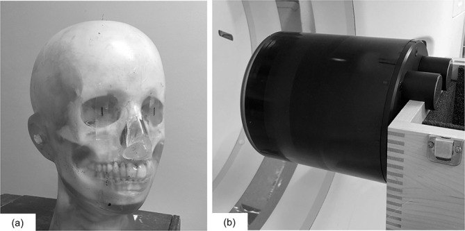
Picture of the PB-1 head CT phantom (a) and Catphan phantom (b).
A Catphan phantom 600 (Phantom Laboratory, Greenwich)16 was used for physical characteristics analysis (Figure 1b). This 200-mm-diameter phantom consists of two modules: the sensitometry module (CTP 404) and the image uniformity module (CTP 486).
Image acquisition and reconstruction conditions
The phantoms were scanned by a 160-row MDCT scanner (Aquilion Precision; Canon Medical Systems, Otawara, Japan) using volumetric and helical scanning. In volumetric scanning, all scans were performed with the following conditions: tube voltage 120 kV, tube rotation time 0.5 s/rot., and 0.5 mm collimation × 80 rows. The tube current varied from 120 to 60 to 30 to 20 mA, and hereafter, these protocols were referred to as Volume-120, Volume-60, Volume-30, and Volume-20, respectively. Images were reconstructed using 0.5 mm thickness with an 80 mm field of view. In all acquisitions, IR (adaptive iterative dose reduction three-dimensional enhanced mild; Canon Medical Systems, Otawara, Japan) and filtered back projection (FBP) technique were performed with the FC 81 (bone) kernel. This IR technique reduces the image noise and enhances the edge sharpness for high-contrast objects.17 As a reference protocol, helical scanning was performed using a tube current of 100 mA corresponding to our clinical setting. Detailed acquisition and reconstruction parameters are summarised in Table 1.
Table 1.
Acquisition and reconstruction parameters
| Parameter | Volume protocol | Reference protocol |
|---|---|---|
| Scanning method | Volumetric scanning | Helical scanning |
| Tube voltage (kV) | 120 | 120 |
| Tube current (mA) | 120, 60, 30, 20 | 100 |
| Rotation time (s/rot.) | 0.5 | 0.5 |
| Pitch factor | - | 0.825 |
| Effective tube current-time product (mAs) | 60, 30, 20, 15 | 60 |
| Focus size (mm ×mm) | 0.4 × 0.5 | 0.4 × 0.5 |
| Beam collimation | 0.5 mm ×80 rows | 0.5 mm ×40 rows |
| Calibrated-FOV (mm) | 320 | 320 |
| Irradiation time (s) | 0.5 | 2.0 |
| Reconstruction method | IR, FBP | FBP |
| Matrix size | 512 × 512 | 512 × 512 |
| Slice thickness/intervals (mm) | 0.5/0.5 | 0.5/0.5 |
| Reconstruction function | FC 81 | FC 81 |
| Displayed-FOV (mm) | 80 | 80 |
FOV field of view, FBP filtered back projection, IR iterative reconstruction.
The irradiation time in helical scanning was defined as the time displayed on the MDCT scanner.
Qualitative assessment
For the qualitative assessment, the maxilla of PB-1 phantom was scanned parallel to the occlusal plane. Two dental radiologists with 10 and 8 years of experience independently graded the 54 maxilla images (each six slice position of four tube current settings and two reconstruction methods and reference) by comparing with the image acquired by reference using a 4-point scale (4, superior to reference; 3, comparable to reference; 2, acceptable; and 1, unacceptable). The images were randomised, and the observers were blinded to the acquisition and reconstruction conditions. Images were evaluated using a liquid-crystal display (RadiForce GX550, EIZO, Nanao, Ishikawa, Japan) with a preset window level of 500 HU and a window width of 3500 HU. Reading time was not limited.
Quantitative assessment
For the quantitative assessment, we compared the spatial resolution, image noise, frequency characteristics of the image noise, and system performance (SP) in all of the acquisition and reconstruction conditions using an appropriate software (CT measure version 098f, Japanese Society of CT Technology, Hiroshima, Japan).18,19
Modulation transfer function
For in-plane spatial resolution, MTFtask curves for all reconstruction algorithms were calculated from the phantom experiments using the CTP 404 module. The MTFtask curves were calculated using an inserted disk-shaped object (Teflon, CT number at 120 kV approximately 940 HU) surrounded by a square region of interest (ROI) according to the circular edge method (Figure 2a).14 The “Task” considered in this study was bone, and a Teflon insert was used to reproduce the bone contrast. Data from the 400 slices were used to produce clear MTFtask curve in each acquisition and reconstruction condition.
Figure 2.

(a) The square region of interest placed in contact with Teflon (940 HU at 120 kV) for measurement of MTFtask curve, and (b) square region of interest (256 × 256 pixels) positioned in the center of the image for measurement of image noise and NPS curve. HU, Hounsfield unit; MTF,modulation transfer function; NPS, noise power spectrum.
Image noise
For the image noise, the standard deviation (SD) of the CT number was calculated. We placed a square ROI (256 × 256 pixels) in the centre of the CTP 486 image (Figure 2b). The mean image noise was acquired from 50 CT slices in each acquisition and reconstruction condition.
Noise power spectrum
To evaluate the frequency characteristics of the image noise, the noise power spectrum (NPS) was calculated using the CTP 486 module. To acquire the NPS curve, square ROI (256 × 256 pixels) was placed in the centre of each image (Figure 2b). Subsequently, the NPS curve was obtained by the radial frequency method.20 The NPS curves were acquired from 400 CT slices to improve the accuracy of NPS data.
System performance
For a comprehensive evaluation of image quality, we measured the SP as a function of spatial frequency.21 SP as a function of spatial frequency, u, was expressed as follows:
Radiation dose calculation
The values of volume CT dose index (CTDIvol) and dose–length product (DLP) were recorded from the corresponding dose report. We estimated the effective dose by multiplying the DLP by the effective dose coefficients of 0.0031 and 0.0042 (mSvmGy-1cm-1) estimating the head and neck region of adult and paediatric 10-year-old patients, respectively.22
Statistical analysis
Intra- and interobserver agreement were calculated using the κ statistic. A time interval of 3 months was set for evaluating the intraobserver agreement. Statistical analysis was performed to compare the image quality evaluated by observers between each volumetric scanning protocol reconstructed with IR/FBP and reference using the Wilcoxon signed-rank test. A p-value < 0.05 was considered statistically significant. We assessed the association between visual scores and SP values at 1.0 cycles/mm. Statistical analysis was performed using JMP Pro 13.0.0 (SAS Institute, Cary, NC).
Ethical consideration
This study was an experimental phantom study and did not require an approval by an institutional review board.
Results
Qualitative evaluation
Table 2 shows the results of the observer evaluation test, and Figure 3 shows maxilla images. Observers rated higher scores for images acquired with a higher tube current. Images reconstructed with IR were evaluated as superior than those reconstructed with FBP in the corresponding tube current setting. There was no significant difference between the image quality of Volume-60 reconstructed with IR and that of the reference (3.08 and 3.00, p = 0.3388). Image quality of Volume-120 reconstructed with IR was significantly better than that of the reference (3.42 and 3.00, p = 0.0172). Although the image quality of Volume-20 reconstructed with IR was inferior than that of the reference (2.33 and 3.00, p = 0.0046), image quality of this protocol was within acceptable limits with the lowest tube current. The two observers in our study showed good intraobserver (κ = 0.696 and κ = 0.631) and interobserver agreements (κ = 0.616).
Table 2.
Image quality in each acquisition and reconstruction condition evaluated by two observers
| Protocol | Reconstruction method | |||
|---|---|---|---|---|
| IR | p-value | FBP | p-value | |
| Volume-120 | 3.42 ± 0.49 | 0.0172 | 3.17 ± 0.37 | 0.1661 |
| Volume-60 | 3.08 ± 0.28 | 0.3388 | 2.83 ± 0.37 | 0.1661 |
| Volume-30 | 2.67 ± 0.62 | 0.1039 | 2.00 ± 0.00 | 0.0005 |
| Volume-20 | 2.33 ± 0.62 | 0.0046 | 1.75 ± 0.43 | <0.0001 |
| Reference | - | - | 3.00 ± 0.00 | - |
FBP, filtered back projection; IR, iterative reconstruction.
Each scan in Volume-120, Volume-60, Volume-30, and Volume-20 was conducted by volumetric scanning with tube current of 120, 60, 30, and 20 mA, respectively.
Data are given as mean ± standard deviation.
Statistical analysis was conducted between each volumetric scanning protocol and reference.
Figure 3.

Phantom images: Volume-120 reconstructed with (a) IR, (b) FBP; Volume-60 reconstructed with (c) IR, (d) FBP; Volume-30 reconstructed with (e) IR, (f) FBP; Volume-20 reconstructed with (g) IR, (h) FBP; and (i) reference. Each scan in Volume-120, Volume-60, Volume-30, and Volume-20 was conducted by volumetric scanning with tube current of 120, 60, 30, and 20 mA, respectively. FBP, filtered backprojection; IR, iterative reconstruction.
Quantitative evaluation
Modulation transfer function
The comparisons of the MTFtask curves between volumetric scanning protocols reconstructed with IR and reference and between volumetric scanning protocols reconstructed with FBP and reference are shown in Figure 4a and b, respectively. The shapes of MTFtask curves reconstructed with FBP were almost the same in all protocols (Figure 4b), while the shape of MTFtask curves reconstructed with IR indicated differences depending on the tube currents (Figure 4a). The 10% MTF values of Volume-120, Volume-60, Volume-30, and Volume-20 reconstructed with IR/FBP and reference were 1.25/1.11, 1.18/1.11, 1.12/1.10, 1.04/1.07, and 1.11, respectively.
Figure 4.

Comparisons of MTFtask curves between (a) volumetric scanning protocols reconstructed with IR and reference and (b) volumetric scanning protocols reconstructed with FBP and reference. Each scan in Volume-120, Volume-60, Volume-30, and Volume-20 was conducted by volumetric scanning with tube current of 120, 60, 30, and 20 mA, respectively. FBP, filtered backprojection; IR, iterative reconstruction; MTF, modulation transfer function.
Image noise
The mean and SD of image noise acquired by Volume-120, Volume-60, Volume-30, and Volume-20 reconstructed with IR/FBP and reference were 183.5 ± 2.0/147.7 ± 2.1, 210.6 ± 2.0/211.9 ± 3.0, 199.1 ± 3.0/306.8 ± 4.5, 178.7 ± 6.0/385.0 ± 5.3, and 152.3 ± 1.6, respectively. The image noise of the images reconstructed with FBP increased as the tube current decreased. The image noise of Volume-120 reconstructed with FBP was comparable to that of the reference, while the image noise of Volume-120 reconstructed with IR was higher than that of the reference.
Noise power spectrum
Figure 5a shows the comparisons of the NPS curves between volumetric scanning protocols reconstructed with IR and reference, and Figure 5b shows the comparisons of the NPS curves between volumetric scanning protocols reconstructed with FBP and reference. The NPS curves reconstructed with IR showed that the peaks of NPS curves shifted to low spatial frequency, specifically in Volume-20 and Volume-30 (Figure 5a). The NPS curves reconstructed with FBP showed that the NPS values were inversely proportional to the tube current, but had a similar shape (Figure 5b).
Figure 5.

Comparisons of NPS curves between (a) volumetric scanning protocols reconstructed with IR and reference and (b) volumetric scanning protocols reconstructed with FBP and reference. Each scan in Volume-120, Volume-60, Volume-30, and Volume-20 was conducted by volumetric scanning with tube current of 120, 60, 30, and 20 mA, respectively. FBP, filtered backprojection; NPS, noise power spectrum.
System performance
The SP curves between volumetric scanning protocols reconstructed with IR and reference and between volumetric scanning protocols reconstructed with FBP and reference are shown in Figure 6a and b, respectively. The SP values were proportional to tube currents. The SP value of Volume-120 reconstructed with FBP was almost similar to that of the reference (Figure 6b), while the SP value of Volume-120 reconstructed with IR was slightly higher than that of the reference (Figure 6a). Table 3 shows the SP values at 1.0 cycles/mm in each acquisition and reconstruction condition. The SP values at 1.0 cycles/mm of Volume-120 reconstructed with IR, Volume-60 reconstructed with IR, Volume-20 reconstructed with IR, and reference were 0.0051, 0.0038, 0.0021 and 0.0041, respectively. In each protocol, SP value at 1.0 cycles/mm for the image reconstructed with IR was higher than that of the image reconstructed with FBP. Figure 7 shows the association between visual scores and SP values at 1.0 cycles/mm, and a strong association between these values (R2 = 0.9230) was observed.
Figure 6.

Comparisons of SP curves between (a) volumetric scanning protocols reconstructed with IR and reference and (b) volumetric scanning protocols reconstructed with FBP and reference. Each scan in Volume-120, Volume-60, Volume-30, and Volume-20 was conducted by volumetric scanning with tube current of 120, 60, 30, and 20 mA, respectively. IR, iterativereconstruction; FBP, filtered back projection; SP, systemperformance.
Table 3.
SP values at 1.0 cycles/mm in each acquisition and reconstruction condition
| Protocol | Reconstruction method | |
|---|---|---|
| IR | FBP | |
| Volume-120 | 0.0051 | 0.0040 |
| Volume-60 | 0.0038 | 0.0028 |
| Volume-30 | 0.0028 | 0.0019 |
| Volume-20 | 0.0021 | 0.0013 |
| Reference | - | 0.0041 |
FBP, filtered back projection; IR, iterative reconstruction; SP, system performance.
Each scan in Volume-120, Volume-60, Volume-30, and Volume-20 was conducted by volumetric scanning with tube current of 120, 60, 30, and 20 mA, respectively.
Data are given as mean ± standard deviation.
Figure 7.

Relationship between visual scores and SP values at 1.0 cycles/mm. Pearson’s correlation coefficient was 0.9230. SP, system performance.
Radiation dose assessment
Table 4 shows the CTDIvol, DLP, effective dose, and reduction ratio from reference in each protocol. The effective doses of Volume-120, Volume-60, and Volume-20 in adults were 103.5, 51.8, and 17.4 µSv, which indicated 28.5%, 64.2%, and 88.0% reductions of the reference, respectively.
Table 4.
Comparison of radiation dose in each acquisition protocol
| Protocol | CTDIvol [mGy] | DLP [mGycm] | Effective dose for adult patient [µSv] | Effective dose for paediatric patient [µSv] | Reduction ratio from reference [%] |
|---|---|---|---|---|---|
| Volume-120 | 8.3 | 33.4 | 103.5 | 140.3 | 28.5 |
| Volume-60 | 4.2 | 16.7 | 51.8 | 70.1 | 64.2 |
| Volume-30 | 2.1 | 8.3 | 25.7 | 34.9 | 82.3 |
| Volume-20 | 1.4 | 5.6 | 17.4 | 23.5 | 88.0 |
| Reference | 8.4 | 46.7 | 144.8 | 196.1 | - |
CTDIvol, volume computed tomography dose index; DLP, dose–length product.
Each scan in Volume-120, Volume-60, Volume-30, and Volume-20 was conducted by volumetric scanning with tube current of 120, 60, 30, and 20 mA, respectively.
Discussions
We proposed an improved scanning method that requires a short irradiation time and reduces the radiation dose while maintaining diagnostic image quality in MDCT. The present study revealed that combining Volume-60 protocol and IR technique achieved an effective dose reduction of 64.2% and required a quarter of the time compared to the reference protocol. Volumetric scanning could reduce the radiation dose caused by overranging, and IR could achieve further reduction of the radiation dose. Furthermore, our method reduced the irradiation time that correlated with potential motion artefacts.
According to the visual evaluation, there was no significant difference between Volume-60 reconstructed with IR and the reference protocol, which indicated that 64.2% of the radiation dose could be reduced using Voluma-60 protocol with IR technique. This was because IR improved the overall image quality by enhancing spatial resolution of the image of Volume-60. Compared to the image of Volume-60 reconstructed with FBP, image noise of Volume-60 reconstructed with IR was almost similar, while the 10% MTF value of Volume-60 reconstructed with IR was higher. Thus, the SP value of 1.0 cycles/mm of Volume-60 reconstructed with IR improved at a similar level to that of the reference.
The volumetric scanning reduced 28.5% of the effective dose. This was because there was no overranging associated with helical scanning.9,23,24 In helical scanning, the smaller helical pitch is useful to reduce the additional radiation dose caused by overranging. However, smaller helical pitch results in a longer irradiation time. Therefore, we considered that the use of volumetric scanning is the best option to reduce radiation dose and irradiation time.
There was a strong association between visual scores and SP values at 1.0 cycles/mm, indicating that our quantitative assessment had a reliable result. We considered that the use of SP value was appropriate for the quantitative assessment of image quality due to the strong association with visual score. To propose an improved scan method, a comprehensive evaluation index that considers both the image quality and effective dose is needed. However, no comprehensive evaluation index that can be used for IR images has been proposed so far. Therefore, further studies are needed to develop a comprehensive evaluation index to propose an improved scan method considering both the image quality and effective dose for IR images.
MTFtask increased above 1.0 at low spatial frequencies. This was because the edge sharpness kernel (FC81 in our study) enhanced the spatial resolution. A similar result was found in a previous report.25
Among all protocols proposed in this study, the effective dose of Volume-20 reconstructed with IR was 17.4 µSv, and the image quality of this protocol was acceptable. Ludlow et al26 reported that the average effective dose of CBCT for maxillary views was 53 (range, from 5 to 140) µSv. Thus, the effective dose by Volume-20 (17.4 µSv) was lower or comparable to that of CBCT. Additionally, our method has a shorter irradiation time compared to CBCT. Although image quality of Volume-20 reconstructed with IR was inferior to that of the reference, the image quality of this protocol was acceptable. Therefore, this protocol could be utilised as an alternative for children where radiation exposure and patient motion during acquisition can pose serious issues.
This study has several limitations. First, in the qualitative assessment, only the general impressions of the dental images were evaluated using a phantom. In dentomaxillofacial radiology, specific clinical applications should be considered. Furthermore, the method used here may not be feasible for some clinical applications requiring a sharpness that only CBCT can achieve. Therefore, further studies are needed to clarify which clinical applications would be compatible with our proposed method, as well as to confirm how much could the tube current be lowered in clinical examinations. Second, we could not evaluate the difference of motion artefact between volumetric scanning and helical scanning since we evaluated the image quality using a phantom only. Therefore, we have to evaluate the difference of motion artefacts in the clinical images. Third, we could not reveal the balance of the effect of IR technique for the spatial resolution and image noise. The detailed algorithm of IR technique was not disclosed to the users, and the effect of IR technique on the spatial resolution and image noise has complex variations depending on acquisition and reconstruction conditions. However, we revealed that, in our experimental environment, the IR technique improved the comprehensive image quality. Fourth, scan range of volumetric scanning will be limited by detector width.
Conclusion
We propose an improved scan method that achieves a 64.2% reduction of the radiation dose, requiring only a quarter of the normal irradiation time, and with preserving image quality by combining volumetric scanning and IR technique in MDCT.
Footnotes
Funding: This research did not receive any specific grant from funding agencies in the public, commercial, or not-for-profit sectors.
REFERENCES
- 1.Lin W-C, Wang H-H, Hsu W-L, Cao B-H, Chen D-J, Tsai C-J. Investigation of an optimized scanning protocol for the dentomaxillofacial region using 320-slice multidetector computed tomography. Dentomaxillofac Radiol 2017; 46: 20160395. doi: 10.1259/dmfr.20160395 [DOI] [PMC free article] [PubMed] [Google Scholar]
- 2.Veldhoen S, Schöllchen M, Hanken H, Precht C, Henes FO, Schön G, et al. Performance of cone-beam computed tomography and multidetector computed tomography in diagnostic imaging of the midface: a comparative study on phantom and cadaver head scans. Eur Radiol 2017; 27: 790–800. doi: 10.1007/s00330-016-4387-2 [DOI] [PubMed] [Google Scholar]
- 3.Lascala CA, Panella J, Marques MM. Analysis of the accuracy of linear measurements obtained by cone beam computed tomography (CBCT-NewTom. Dentomaxillofac Radiol 2004; 33: 291–4. doi: 10.1259/dmfr/25500850 [DOI] [PubMed] [Google Scholar]
- 4.Demiriz L, Hazar Bodrumlu E, İçen M, Durmuşlar MC. Evaluation of the accuracy of cone beam computed tomography on measuring impacted supernumerary teeth. Scanning 2016; 38: 579–84. doi: 10.1002/sca.21303 [DOI] [PubMed] [Google Scholar]
- 5.Abdelkarim A. Myths and facts of cone beam computed tomography in orthodontics. J World Fed Orthod 2012; 1: e3–8. doi: 10.1016/j.ejwf.2012.04.002 [DOI] [Google Scholar]
- 6.Miracle AC, Mukherji SK. Conebeam CT of the head and neck, part 1: physical principles. AJNR Am J Neuroradiol 2009; 30(1088-95): 1088–95. doi: 10.3174/ajnr.A1653 [DOI] [PMC free article] [PubMed] [Google Scholar]
- 7.Brenner D, Elliston C, Hall E, Berdon W. Estimated risks of radiation-induced fatal cancer from pediatric CT. AJR Am J Roentgenol 2001; 176: 289–96. doi: 10.2214/ajr.176.2.1760289 [DOI] [PubMed] [Google Scholar]
- 8.Pearce MS, Salotti JA, Little MP, McHugh K, Lee C, Kim KP, et al. Radiation exposure from CT scans in childhood and subsequent risk of leukaemia and brain tumours: a retrospective cohort study. Lancet 2012; 380: 499–505. doi: 10.1016/S0140-6736(12)60815-0 [DOI] [PMC free article] [PubMed] [Google Scholar]
- 9.Kroft LJM, Roelofs JJH, Geleijns J. Scan time and patient dose for thoracic imaging in neonates and small children using axial volumetric 320-detector row CT compared to helical 64-, 32-, and 16- detector row CT acquisitions. Pediatr Radiol 2010; 40: 294–300. doi: 10.1007/s00247-009-1436-x [DOI] [PMC free article] [PubMed] [Google Scholar]
- 10.Kalra MK, Woisetschläger M, Dahlström N, Singh S, Lindblom M, Choy G, et al. Radiation dose reduction with Sinogram Affirmed iterative reconstruction technique for abdominal computed tomography. J Comput Assist Tomogr 2012; 36: 339–46. doi: 10.1097/RCT.0b013e31825586c0 [DOI] [PubMed] [Google Scholar]
- 11.Joemai RMS, Veldkamp WJH, Kroft LJM, Hernandez-Giron I, Geleijns J. Adaptive iterative dose reduction 3D versus filtered back projection in CT: evaluation of image quality. AJR Am J Roentgenol 2013; 201: 1291–7. doi: 10.2214/AJR.12.9780 [DOI] [PubMed] [Google Scholar]
- 12.Nagatani Y, Takahashi M, Murata K, Ikeda M, Yamashiro T, Miyara T, et al. Lung nodule detection performance in five observers on computed tomography (CT) with adaptive iterative dose reduction using three-dimensional processing (AIDR 3D) in a Japanese multicenter study: comparison between ultra-low-dose CT and low-dose CT by receiver-operating characteristic analysis. Eur J Radiol 2015; 84: 1401–12. doi: 10.1016/j.ejrad.2015.03.012 [DOI] [PubMed] [Google Scholar]
- 13.Klink T, Obmann V, Heverhagen J, Stork A, Adam G, Begemann P. Reducing CT radiation dose with iterative reconstruction algorithms: the influence of scan and reconstruction parameters on image quality and CTDIvol. Eur J Radiol 2014; 83: 1645–54. doi: 10.1016/j.ejrad.2014.05.033 [DOI] [PubMed] [Google Scholar]
- 14.Richard S, Husarik DB, Yadava G, Murphy SN, Samei E. Towards task-based assessment of CT performance: system and object MTf across different reconstruction algorithms. Med Phys 2012; 39: 4115–22. doi: 10.1118/1.4725171 [DOI] [PubMed] [Google Scholar]
- 15.Watanabe H, Honda E, Tetsumura A, Kurabayashi T. A comparative study for spatial resolution and subjective image characteristics of a multi-slice CT and a cone-beam CT for dental use. Eur J Radiol 2011; 77: 397–402. doi: 10.1016/j.ejrad.2009.09.023 [DOI] [PubMed] [Google Scholar]
- 16.Davis AT, Palmer AL, Pani S, Nisbet A. Assessment of the variation in CT scanner performance (image quality and Hounsfield units) with scan parameters, for image optimisation in radiotherapy treatment planning. Phys Med 2018; 45(59-64): 59–64. doi: 10.1016/j.ejmp.2017.11.036 [DOI] [PubMed] [Google Scholar]
- 17.Minamishima K, Sugisawa K, Yamada Y, Jinzaki M. Quantitative and qualitative evaluation of hybrid iterative reconstruction, with and without noise power spectrum models: a phantom study. J Appl Clin Med Phys 2018; 19: 318–25. doi: 10.1002/acm2.12304 [DOI] [PMC free article] [PubMed] [Google Scholar]
- 18.Ichikawa K. CTmeasure, Japanese Society of CT Technology, Kasumi, Minami-ku, Hiroshima, JPN. 2012–2014. Available from: http://www.jsct-tech.org.
- 19.Funama Y, Taguchi K, Utsunomiya D, Oda S, Katahira K, Tokuyasu S, et al. Image quality assessment of an iterative reconstruction algorithm applied to abdominal CT imaging. Phys Med 2014; 30: 527–34. doi: 10.1016/j.ejmp.2014.02.005 [DOI] [PMC free article] [PubMed] [Google Scholar]
- 20.Ghetti C, Ortenzia O, Serreli G. Ct iterative reconstruction in image space: a phantom study. Phys Med 2012; 28: 161–5. doi: 10.1016/j.ejmp.2011.03.003 [DOI] [PubMed] [Google Scholar]
- 21.Miura Y, Ichikawa K, Fujimura I, Hara T, Hoshino T, Niwa S, et al. Comparative evaluation of image quality among different detector configurations using area detector computed tomography. Radiol Phys Technol 2018; 11: 54–60. doi: 10.1007/s12194-017-0437-y [DOI] [PubMed] [Google Scholar]
- 22.ICRP publication 102: managing patient dose in MDCT. Ann ICRP 2007;. [DOI] [PubMed] [Google Scholar]
- 23.Sorantin E, Riccabona M, Stücklschweiger G, Guss H, Fotter R. Experience with volumetric (320 rows) pediatric CT. Eur J Radiol 2013; 82: 1091–7. doi: 10.1016/j.ejrad.2011.12.001 [DOI] [PubMed] [Google Scholar]
- 24.Nievelstein RAJ, van Dam IM, van der Molen AJ. Multidetector CT in children: current concepts and dose reduction strategies. Pediatr Radiol 2010; 40: 1324–44. doi: 10.1007/s00247-010-1714-7 [DOI] [PMC free article] [PubMed] [Google Scholar]
- 25.Ohkubo M, Wada S, Ida S, Kunii M, Kayugawa A, Matsumoto T, et al. Determination of point spread function in computed tomography accompanied with verification. Med Phys 2009; 36: 2089–97. doi: 10.1118/1.3123762 [DOI] [PubMed] [Google Scholar]
- 26.Ludlow JB, Timothy R, Walker C, Hunter R, Benavides E, Samuelson DB, et al. Effective dose of dental CBCT-a meta analysis of published data and additional data for nine CBCT units. Dentomaxillofac Radiol 2015; 44: 20140197. doi: 10.1259/dmfr.20140197 [DOI] [PMC free article] [PubMed] [Google Scholar]