

A 56-year-old male patient with chronic bronchitis reported worsening of his usual dyspnea for one week prior to hospital admission, evolving to fever, productive cough, and non-massive hemoptysis, as well as a progressive need for oxygen supplementation, in the last 24 h.
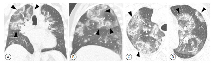
Multidetector CT scans of the chest showed diffuse, extensive ground-glass opacities, together with foci of consolidation, many of them representing reversed halo signs, in both lungs. Screening for multiple respiratory pathogens was positive for SARS-CoV-2, indicating that the patient had COVID-19.
The reversed halo sign, defined as an area with ground-glass attenuation surrounded by partial or complete rings of consolidation, is a radiological finding present in patients with pneumonia caused by the new coronavirus. The reversed halo sign has been reported in other forms of viral pneumonia. 1 , 2 When identified, the reversed halo sign typically occurs longer after symptom onset, suggesting that this CT finding correlates with the underlying pathophysiology of the disease process as it organizes. 2 Such findings indicate that organizing pneumonia is one of the mechanisms of lung injury. 3
Figure 1. Images in coronal (A), sagittal (B), and axial (C and D) reconstructions from multidetector CT, showing multiple diffuse ground glass areas in both lungs, surrounded by partial or complete rings of consolidation, known as the reversed halo sign (arrows), in a 56-year-old male patient with COVID-19 pneumonia.
REFERENCES
- 1.Marchiori E, Zanetti G, Escuissato DL, Souza AS, Jr, Meirelles GSP, Fagundes J. Reversed halo sign high-resolution CT scan findings in 79 patients. Chest. 2012;141(5):1260–1266. doi: 10.1378/chest.11-1050. [DOI] [PubMed] [Google Scholar]
- 2.Bernheim A, Mei X, Huang M, Yang Y, Fayad ZA, Zhang N, et al. Chest CT Findings in Coronavirus Disease-19 (COVID-19): Relationship to Duration of Infection [published online ahead of print, 2020 Feb 20] Radiology. 2020:200463–200463. doi: 10.1148/radiol.2020200463. [DOI] [PMC free article] [PubMed] [Google Scholar]
- 3.Wu Y, Xie YL, Wang X. Longitudinal CT Findings in COVID-19 Pneumonia Case Presenting Organizing Pneumonia Pattern. Radiol Cardiothorac Imaging. 2020;2(1):e200031. doi: 10.1148/ryct.2020200031. [DOI] [PMC free article] [PubMed] [Google Scholar]