Abstract
Dairy cattle health, wellbeing and productivity are deeply affected by stress. Its influence on metabolism and immune response is well known, but the underlying epigenetic mechanisms require further investigation. In this study, we compared DNA methylation and gene expression signatures between two dairy cattle populations falling in the high- and low-variant tails of the distribution of milk cortisol concentration (MC), a neuroendocrine marker of stress in dairy cows. Reduced Representation Bisulfite Sequencing was used to obtain a methylation map from blood samples of these animals. The high and low groups exhibited similar amounts of methylated CpGs, while we found differences among non-CpG sites. Significant methylation changes were detected in 248 genes. We also identified significant fold differences in the expression of 324 genes. KEGG and Gene Ontology (GO) analysis showed that genes of both groups act together in several pathways, such as nervous system activity, immune regulatory functions and glucocorticoid metabolism. These preliminary results suggest that, in livestock, cortisol secretion could act as a trigger for epigenetic regulation and that peripheral changes in methylation can provide an insight into central nervous system functions.
Keywords: cortisol secretion, DNA methylation, bisulfite sequencing, gene expression, dairy cattle health
1. Introduction
Dairy animals experience a large variety of stressors that can modify normal behavior and growth, leading to a decrease in productive performances. Normal physiological events such as calving, onset of lactation, lactation, weaning and group rearrangement can cause metabolic and environmental conditions which lead to stress, an impairment of animal wellbeing and a consequent decrease in the quality of animal products. Under these stressful conditions, the hypothalamic–pituitary–adrenal (HPA) axis, the autonomic nervous system, and the immune system are called into action to re-establish homeostasis [1]. Stress modifies the secretion of various hormones, as glucocorticoids, which play an important role in immunity [2,3,4]. A common feature of stress situations is an increase in cortisol secretion that produces a mobilization of energy reserves [5]. For example Bertulat et al. [6] showed higher concentration of glucocorticoid metabolites in the feces of drying off cows with higher milk yield while Horst and Jorgensen [7] reported an increase in plasma cortisol in cows with milk fever, associated with immune suppression and an increased risk of clinical mastitis and high somatic cell count in milk. To date, since blood sampling is often a source of stress, milk can be viewed as a viable and non-invasive way to measure cortisol levels in lactating cows, as it can be measured without the manipulation of animals.
Even though many methods of wellbeing evaluation are reported in the literature, cortisol is still considered one of the gold standards to describe animal response to stress [8]. However, until now, no systematic studies have investigated the epigenetic mechanisms related to cortisol secretion during stress conditions in lactating cows.
Epigenetics are defined as heritable changes in gene activity and expression and consequently in phenotype, which occur without alteration in the underlying DNA sequence [9]. At least four molecular systems, including DNA methylation [10], histone post-translational modifications [11], non-coding RNAs [12] and chromatin remodeling [13], control gene expression. The epigenome refers to the entirety of the epigenetic features possessed by an organism’s genome. DNA methylation was the first-recognized and is the most-studied epigenetic regulatory mechanism that plays a key role in the control of gene expression. In mammalian cells, DNA methylation mostly occurs at the cytosine of a CpG dinucleotide. CpG islands are genomic regions enriched in CpG dinucleotides. CpG islands cluster most at the 5’ promoter regions of many housekeeping genes and are generally hypomethylated to permit transcription, with the exception of genes involved in imprinting, X chromosome inactivation and cancer process [14,15,16]. The hyper-methylation of DNA combined with histone H3-lysine 9 [17] or histone H4-lysine 20 methylation [18] can result in inaccessible chromatin by recruiting chromatin remodeling proteins and generally causes the inhibition of downstream gene expression by physically blocking the binding of transcription factors [19]. Epigenetic modifications can be inherited from one cell generation to the next (mitotic inheritance) and between generations of a species (meiotic inheritance) and can be natural and essential for many developmental functions, but can also be influenced by several factors including age, environment, lifestyle or a disease state [20]. In mammalian cells, the changes in gene expression in response to external factors, such as viral infection, stress, drug delivery, temperature changes, dietary components, may have long-lasting effects on development, metabolism and health, sometimes even across generations. Epigenetics represent a link between the environment and gene expression [21].
In this study, we investigated the potential regulatory roles of epigenome signatures in the blood of dairy cows falling in the extreme category of the distribution of cortisol concentration in milk. We focused on DNA methylation in blood cells since blood is quickly accessible and highly informative of animal response to environmental challenges. Moreover, it was previously demonstrated that methylation levels at CpG sites in leukocytes could serve as an accurate predictor of CpG site variability in other tissues [22]. In parallel, we used transcriptome sequencing data to integrate the relationship between DNA methylation and transcriptional regulation on a genome-wide scale to identify novel genes involved in glucocorticoids secretion.
2. Material and Methods
2.1. Ethics Statement
The farms involved in the present study adhere to a high standard of veterinary care based on best practice manual, under the supervision of the official veterinary service. All experimental procedures and the care of animals complied to the Italian legislation on animal care (DL n.116, 27/1/1992) in force at the time the study was carried out and adhered to the bioethical rules of the University of Udine. The approval for conducting this study was also granted by the veterinarian responsible of animal welfare of the Department of Agricultural and Environmental Science of the University of Udine (Prot. N. 2/2015, OPBA—Organismo Preposto al Benessere degli Animali).
2.2. Animals Treatment and Experimental Design
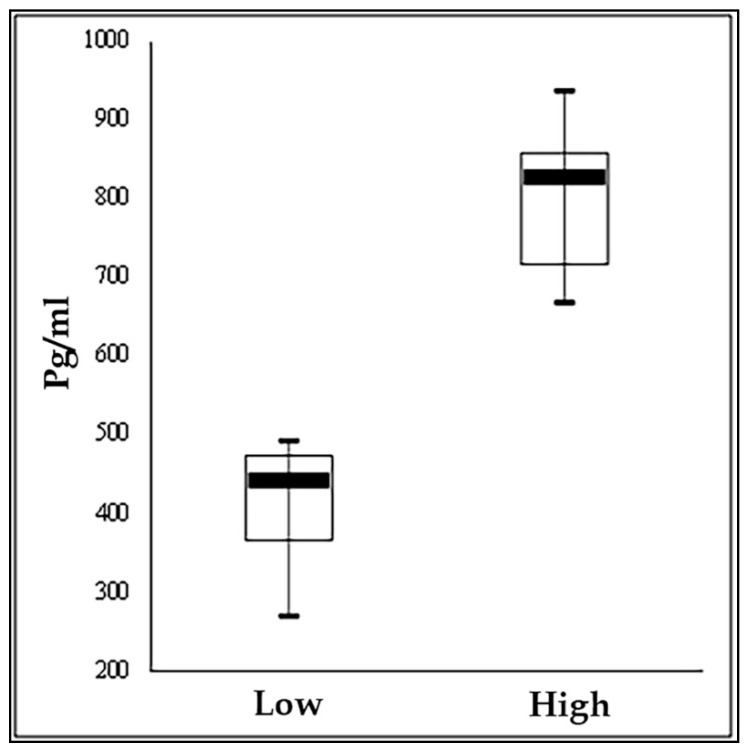
The study was carried out ona commercial farm of the Italian Simmental breed located in the North East of Italy (Latitude: 45°76′491 N; Longitude: 13°10′466 E) in the month of May. From the herd, 126 lactating cows with days in milk (DIM) between 70 and 250, clinically healthy and with a parity from 2 to 6 (mean 3.0 ± 1.1) were initially selected for the study. One hundred ml of milk was collected from each cow at morning milking and an aliquot of 50 mL was transferred into a tube containing preservative and used for protein, fat, lactose analyses and for somatic cell count (SCC) determinations with FT-NIR (FOSS instrument, Hillerød, Denmark). The other aliquot of milk was transferred to a tube without preservative, frozen within 2 hand stored at −20 °C for cortisol analyses [23]. Milk cortisol was analyzed in skimmed milk, after centrifugation (1500 g, 4 °C, 15 min). Skim milk extracts were assayed by a solid-phase microtiter RIA [24], using a Microscint 20 instrument (Perkin-Elmer Life Sciences, Monza, Italy) and counted on the β-counter (Top-Count, Perkin-Elmer Life Sciences, Monza, Italy). All samples were assayed in duplicate. The sensitivity of the assay was defined as the dose of hormone at 90% binding (B/B0) and was 3.125 pg/well. The intra-assay and inter-assay coefficients of variation in high and low cortisol pooled skim milk samples were 5.9% and 9.1% and 13.5% and 15.1%, respectively. On the same day, after milking and before feeding, blood was sampled form the tail vein in a tube with K3-EDTA (Venoject, Terumo Europe N.V., Leuven, Belgium) and in one PAXgene Blood RNA System tube (Preanalytix, Hombrechtikon, Switzerland) for analysis. The K3-EDTA tubes were centrifuged within 1 h at 755× g for 10 min at 20 °C and the blood cells stored at −20 °C for DNA analysis. The PAX gene tubes were handled and stored following the manufacturer’s instructions for RNA extraction. The body condition score (BCS) of each cow was also recorded using a scale from 1 (thin) to 5 (fat) with 0.25-point intervals [25] (Table 1). Cows were ranked on the basis of cortisol in their milk and the 10 cows with the highest and the 10 cows with the lowest concentrations were selected for transcriptome and epigenome analysis (Figure 1).
Table 1.
Body condition score (BCS), parity, days in milking (DIM) and milk yield and quality in the 10 cows with lowest and 10 cows with highest cortisol concentration in milk.
| Item | Low Cortisol | High Cortisol | ||||
|---|---|---|---|---|---|---|
| Mean | Sd | Mean | Sd | p Value | ||
| BCS | Score | 3.23 | 0.38 | 3.10 | 0.47 | 0.262 |
| Parity | n° | 1.70 | 0.95 | 2.10 | 0.88 | 0.170 |
| DIM | Days | 156.2 | 62.8 | 143.3 | 69.3 | 0.334 |
| Milk | Kg/d | 26.20 | 6.54 | 30.38 | 6.23 | 0.080 |
| Fat | % | 3.68 | 1.03 | 3.95 | 0.70 | 0.254 |
| Protein | % | 3.61 | 0.25 | 3.59 | 0.39 | 0.441 |
| Casein | % | 2.84 | 0.20 | 2.81 | 0.32 | 0.415 |
| Urea | mmol/L | 21.39 | 5.12 | 21.01 | 6.13 | 0.442 |
| SCC | Number | 133.50 | 89.09 | 153.40 | 110.02 | 0.331 |
| Cortisol | pg/mL | 399.9 | 79.7 | 814.8 | 88.6 | 0.000 |
Before ranking, cows with somatic cell counts (SCC) higher than 200,000 cells/mL were excluded.
Figure 1.
Box plot of milk cortisol distribution of 10 cows with the higher and the 10 cows with the lower values.
2.3. DNA Isolation, Library Preparation and RRBs Sequencing
Blood samples from these 20 animals were collected. Peripheral blood mononuclear cells (PBMC) were isolated from heparinized blood samples with Ficoll-Paque. CD4+ lymphocyte cells were isolated with the CD4 anti-bovine monoclonal antibody (VMRD, Pullman, WA, USA) and positively selected with GAM microspheres using the MACS MiltenyiBiotec system (BergischGladbach, Germany). DNA was extracted from CD4+ cells following the QIAamp DNA Blood Midi Kit (Qiagen) procedures. gDNA concentration was measured with Quant-it Picogreen dsDNA assay and 1 μg input was used in the MSP1 digest. Following overnight incubation at 37 °C, digestion reactions were terminated by adding 0.5 M EDTA and purified on a GeneJET PCR purification column. Libraries were prepared using the NEB Next Ultra DNA library preparation kit for Illumina and methylated adapters. Subsequently, ligated products corresponding to DNA fragments 150–400-bp long were isolated and purified using 2.5% agarose gel electrophoresis. The recovered DNA was bisulfite converted using the EZ DNA Methylation Gold kit (Zymo Research). DNA with a known methylation level was used as a spike control, and all conversion rates were >99%, ranging from 99.01 to 99.39% (Table S1 (Supplementary Material)). Fourteen cycles of PCR were performed, and the products were purified using AMPureXP beads. Reduced Representation Bisulfite Sequencing (RRBS) libraries were used for cluster generation and subsequent sequencing on Illumina HiSeq2500 PE 2 × 50 bp (NXT-Dx Ghent, Belgium, http://www.nxt-dx.com/).
2.4. Data Analysis
Preliminary quality control of the raw reads was carried out with FASTQC v0.11.9 (http://www.bioinformatics.babraham.ac.uk/projects/fastqc/). The FASTQ sequence reads were generated using the Illumina Casava pipeline 1.8.2. The quality and adapter trimming of Illumina raw sequences was performed with Trim Galore v0.6.1 (http://www.bioinformatics.babraham.ac.uk/projects/trim_galore/) using a two-step approach, which allowed us to remove two additional bases containing a cytosine, which were artificially introduced in the end-repair step during the library preparation. Bismark software (version 0.22.1) [26] was used to align each bisulfite-treated read to the bovine reference genome (ARS-UCD1.2) with option-N 1 (maximum number of mismatches allowed). The reference genome was first transformed into a bisulfite-converted version (C-to-T and G-to-A conversions) and then indexed using bowtie2 software [27]. Sequence reads were also transformed into fully bisulfite-converted versions (C-to-T and G-to-A conversions) before they were aligned to similarly converted versions of the genome in a directional manner. Sequence reads that produced the best unique alignment from the two alignment processes (original top and bottom strand) were then compared to the normal genomic sequence, and the methylation state of all cytosine positions in the reads was inferred using the Bismark_methylation_extractor function. Read duplicates were marked and removed using Picard Tools v2.2 (http://broadinstitute.github.io/picard).
2.5. Identification of DMRs and DMGs
Differentially methylated regions (DMRs) were identified within the statistical environment R using the library methylKit [28], which applies a sliding-window approach. The window was set to 1000 bp and the step size to 500 bp. In order to ensure the good quality of the data and great confidence in the methylation percentage, we first filtered out bases with less than 10 reads or more than the 99.9th percentile of coverage distribution. Coverage values were normalized by default and bases were merged in order to retain the ones that were covered in all samples. A logistic regressionwas then implemented to calculate p values, that were adjusted to q values using the Sliding Linear Model (SLIM) method [29]. We excluded covariates and overdispersion correction from the model since they showed not to provide significant changes to the results. DMRs regions were defined those that had q value of less than 0.05 and a read coverage of greater than ten in all samples. When a region where a DMR and a specific gene function element overlapped, the corresponding gene was selected as the DMR-related gene, namely a differentially methylated gene (DMG). Gene features within differential methylated genes (DMGs) were annotated by matching information available in the General Feature Format (GFF) file downloaded from the Ensembl database, release 99 (ftp://ftp.ensembl.org/pub/release-99/gff3/bos_taurus/Bos_taurus.ARS-UCD1.2.99.gff3.gz). The promoter region was defined as an area 2-kb upstream of the transcription start sites.
2.6. Association Analysis
Correlations between DNA methylation levels and gene density, chromosome length and GC percentage were measured using Pearson’s product–moment correlation coefficient. Correlations between DNA methylation and gene expression were measured using Spearman’s rank correlation coefficient because the relationship of DNA methylation with gene expression data was not necessarily expected to be linear. All these analyses were performed with custom R scripts.
2.7. GO and KEGG Enrichment Analysis of DMR-Related Genes
Gene Ontology (GO) and KEGG pathway enrichment analysis of DMGs were performed by clusterProfiler, an ontology-based R package able to automate the process of biological term classification and enrichment analysis of gene clusters and to provide a visualization module for displaying analysis results [30]. GO terms and KEGG with p values of less than 0.01 and q values of less than 0.2 were considered significantly enriched by DMR-related genes. The DMGs involved in the GO pathways related to development and metabolism were analyzed with the R package enrichplot [31] to highlight links between genes and GO terms in a gene concept network.
2.8. Sample Preparation for RNA—Seq Analysis
Blood samples collected in the PAX gene Blood RNA Tubes (PreAnalytiX GmbH, Hombrechtikon, Switzerland) were frozen 4 h after the collection and stored at −80 °C until the RNA isolation. Prior to RNA isolation, blood samples were thawed at +4 °C for at least 12 h. RNA was isolated according to the PAX gene Blood RNA Kit (PreAnalytiX GmbH, Hombrechtikon, Switzerland) protocol. The quantity and quality of RNA were analyzed with Agilent 2100 Bioanalyzer (Agilent Technologies, Santa Clara, CA, USA) before sequencing and the RNA integrity number (RIN) score was ≥7 for all the samples. Since the RNA-Seq is highly reproducible within a large dynamic range of detection and provides an accurate estimation of RNA concentration in peripheral whole blood [32]; globin depletion was avoided, as it could significantly reduce the amount and quality of isolated RNA. Sequencing was performed with the Illumina pipeline.
2.9. Quantification of Gene Expression Levels and Differential Expression Analysis
After quality control and the filtering of raw data, clean reads were aligned to the bovine reference genome (ARS-UCD1.2) using STAR v2.7.3 [33], a splice-aware aligner. featureCounts (Subread package v1.6.5) [34] was used to count the read numbers mapped to each gene. Differential gene expression between the high and low groups was performed using the DESeq2 R package (1.26.0) [35]. The p values were adjusted using the Benjamini–Hochberg method [36]. A corrected p value of 0.05 was set as the threshold for significantly different expression, while genes with extremely low expression (fold changes (FC) values between +1 and −1) were filtered out.
3. Results
3.1. Global Mapping of DNA Methylation
In the present study, blood samples from 20 Italian Simmental dairy cows falling in the high and low tails of the distribution of milk cortisol concentration (Figure 1) were used to investigate genome-wide DNA methylation. A total of 278 and 262 million raw reads were generated from the bisulfite sequencing of the high and low groups, respectively. After data filtering, 139 and 132 million clean reads were generated; of these, approximately 48 and 46 million mapped to the reference genome with a unique best fit. The mapping efficiency of high- and low-variant samples ranged from 25.6% to 41.5% and from 30.9% to 42.8% of the cattle genome, respectively (Table 2 and Table S1 (Supplementary Material)).
Table 2.
Data generated by genome-wide bisulfite sequencing. high- and low-variant tails of the distribution of milk cortisolconcentration.
| Samples | Raw Reads | Clean Reads | Mapped Paired End Reads | Average Mapping Rate (%) |
|---|---|---|---|---|
| High | 278,881,124 | 139,639,195 | 36,779,144 | 35.15 |
| Low | 262,917,162 | 132,458,581 | 35,048,383 | 35.71 |
3.2. DNA Methylation Patterns
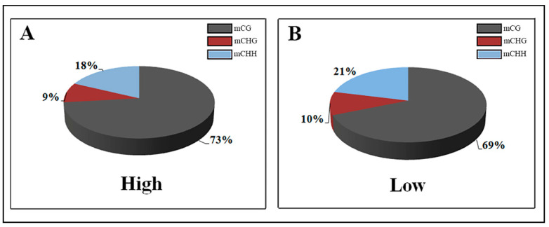
In the genome of each group, over 50% of the CpG sites were methylated, which is the primary DNA sequence context of cytosine methylation. Specifically, we observed, on average, genome-wide levels of 50.5% CG, 6.8% CHG, and 7.8% CHH (where H is A, C, or T) methylation in the high-variant group and 53.6% CG,7.1% CHG, and 8.1% CHH methylation in the low-variant group. (Table 3). Compared with the low group, among the methylated cytosines the relative proportion of CG in the high group was greater (73.24% vs. 68.84%) and consequently that the proportion of methylated CHH and CHG was lower (17.94% vs. 21.16% and 8.83% vs. 10% respectively; Figure 2).
Table 3.
Genome-wide methylation levels of the two high- and low-cortisol groups for CpG and non-CpG sites.
| Samples | mCpG(%) | mCHG(%) | mCHH (%) |
|---|---|---|---|
| High | 50.48 | 6.82 | 7.82 |
| Low | 53.58 | 7.11 | 8.14 |
Figure 2.
Comparison of DNA methylation patterns in the two high-and low-cortisol groups.
We noted that a high cortisol concentration in milk resulted in a decrease in overall methylation at all sites in comparison to low cortisol concentrations, but in an increase in the methylation rate of CpG sites compared to non-CpG sites, especially in the CHH context.
3.3. DNA Methylation Levels of Gene Features
To disentangle the differences identified in global DNA methylation profiles across high- and low-cortisol dairy cows, we compared the average DNA methylation levels of different gene features along the genome (Figure 3).
Figure 3.
DNA methylation levels of different functional regions between the two high- and low-cortisol groups. (A) CG regions. (B) CHG regions. (C) CHH regions. H = A, C, or T.
A major proportion of CG methylated sites were present in the regions of 3’UTR followed by intron regions, while the average methylation levels of promoters,5’UTR and exon regions were the lowest. Conversely, all five regions exhibited a more balanced distribution of methylation at CHG and CHH sites.
3.4. Differential Methylated Regions (DMR) and Genes (DMG)
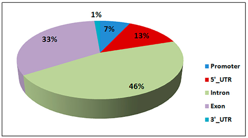
A total of 897 DMRs were identified between the two groups (q value ≤ 0.05), falling into 248 DMGs (Table S2 (Supplementary Material)). Of these DMGs, 146 were up-methylated, and 102 genes were down-methylated in the high group. Moreover, we also detected a negative correlation of methylation levels with gene density (Pearson’s r = −0.712, p value < 0.001) and with chromosome length (Pearson’s r = −0.754, p value < 0.001) and a positive correlation with the GC percentage (Pearson’s r = 0.958, p value < 0.001). The differentially methylated regions were mainly located in introns, whose proportion covers nearly half of the total percentage of methylation, followed by the exons and the promoter regions (Figure 4).
Figure 4.
Distribution of differentially methylated regions (DMRs).
3.5. Functional Enrichment Analysis of the DMGs
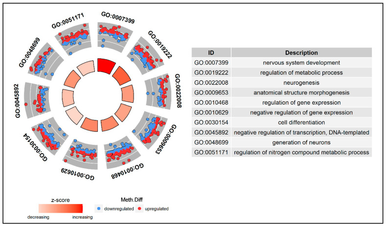
To investigate the potential biological functions of the DMGs, a GO enrichment analysis and a KEGG pathway analysis were performed. All DMGs were annotated in three GO categories: biological process; cellular component; and molecular function. Some of these DMGs were enriched in the following biological process terms: cellular process (129; 52%); single-organism process (123; 49.6%); and metabolic process (103; 41.5%). In addition, most of the top 10 significantly enriched pathways in GO analysis were strictly related to nervous system activity and metabolic responses to stress (Figure 5 and Table S3 (Supplementary Material)).
Figure 5.
Gene Ontology (GO) circle plot for differentially methylated genes (DMGs). The inner ring is a bar plot where the height of the bar indicates the significance of the term (q value), and color corresponds to the z-score. The outer wheel shows a scatter plot of methylation difference for each gene under the Gene Ontology(GO) terms. Red dots indicate hyper-methylated genes and blue dots show hypo-methylated genes.
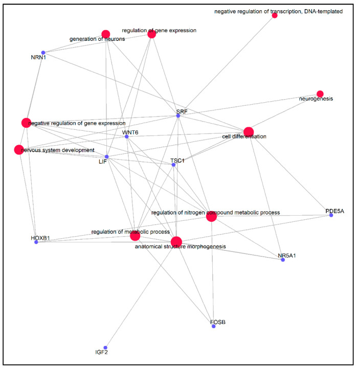
Several DMGs, such as IGF2, LIF, NR5A1, SRF, FOSB, PDE5A, TSC1, NRN1, WNT6 and HOXB1, were involved in biological processes significant for stress response, inflammatory reactions and the immune system. Furthermore, the gene–gene interaction network analysis showed that these DMGs were highly correlated with each other (Figure 6).
Figure 6.
Gene-pathway association network indicating genes affiliated to significantly GO enriched pathways. Red dots indicate GO pathways and blue dots indicate genes.
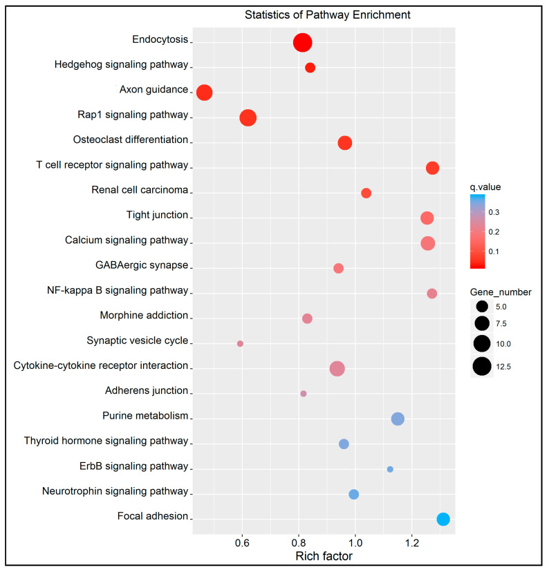
The KEGG enrichment analysis identified 62 pathways (top 20 in Figure 7 and Table S4 (Supplementary Material)).
Figure 7.
Scatter plot of KEGG pathway enrichment statistics. Top 20 statistics of pathways, enrichment in the KEGG cattle database. The y-axis represents the name of pathway and the x-axis represents the rich factor, the proportion of differentially methylated genes to all the genes that are annotated in a specific pathway term. Dot size represents the number of genes and the color indicates the q value.
Of these pathways, some were associated with immune system and glucocorticoid secretion, such as the T cell receptor signaling pathway (q value = 0.051), the hedgehog signaling pathway (q value = 0.032), axon guidance (q value = 0.041), the calcium signaling pathway (q value = 0.157) and the GABAergic synapse pathway (q value = 0.188). Eleven differentially methylated genes participated in these five pathways.
3.6. Differentially Expressed Genes (DEGs)
A total of 324 DEGs were also identified in the present study between the two groups (q value ≤ 0.05). Among these, 149 genes were over-expressed and 175 were under-expressed in the high group (Table S5 (Supplementary Materials)). We mapped these genes to the GO and KEGG pathway. Again, they were annotated in three GO categories: biological process; cellular component; and molecular function. Some of these DEGs were enriched in the following biological process terms: cellular process (104; 36.1%); single-organism process (95; 33%); and metabolic process (75; 26%). For all the DEGs, only the GO term “binding”, with 58 genes in the categories of molecular function, was significantly enriched (q value < 0.05), implying that a broad range of genes experienced transcriptional regulation during the activation process of cortisol secretion. A KEGG pathway analysis was also performed to investigate pathways in which DEGs might be involved. In the top 20 list (Table S6 (Supplementary Materials)), pathways implicated in inflammatory reactions (Inflammatory bowel disease (IBD), q value= 0.014), and immune defense functions against viral disease (Influenza A, q value = 0.011, Hepatitis B, q value = 0.109, Hepatitis C, q value = 0.103) are found. This confirms several studies that recently indicated a strong correlation between stressors and the progression and outcome of liver inflammatory diseases, such as chronic viral hepatitis [37]. To examine the relationship between DNA methylation and genome-wide gene expression, an association analysis was also performed between these DMGs and differentially expressed genes (DEGs) obtained from transcriptome data from the same animals (p value < 0.001). We found six overlapping genes, four were up-methylated, TRIM26, PAX2, SYNGR1 and SNCB, and two down-methylated, UPP1 and HTRA1 (Figure S1 (Supplementary Material) and Table 4).
Table 4.
Six DMGs that overlapped with differentially expressed genes (DEGs) (q value < 0.05 in both analyses).
| Gene ID | Gene Name | DMRs | Methylation Stat (High vs. Low) | UP/DOWN Regulation (High vs. Low) |
|---|---|---|---|---|
| ENSBTAG00000035744 | TRIM26 | exon | Hyper | Down |
| ENSBTAG00000021566 | PAX2 | exon, intron | Hyper | Up |
| ENSBTAG00000005765 | SYNGR1 | intron | Hyper | Up |
| ENSBTAG00000009803 | SNCB | utr3, exon, promoter | Hyper | Down |
| ENSBTAG00000008428 | UPP1 | intron | Hypo | Down |
| ENSBTAG00000008389 | HTRA1 | exon | Hypo | Up |
4. Discussion
There is a growing debate in the livestock industry about stress and its detrimental effects on animal welfare and psychological and physiological responses of animals to traditional agriculture procedures and new production technologies [38]. The measurement of corticosteroid hormones is commonly used as an indicator of the animal’s response to stress. Blood sampling is a stressful factor, so, in recent years, several efforts have beenmade in many species to find alternative and non-invasive ways to measure cortisol level [39,40,41]. Moreover, the cortisol concentration in the blood of cattle can vary due to circadian rhythmicity and several extrinsic factors, such as cold, heat, humidity and wind [42]. On the contrary, milk sampling can be achieved directly in a milking parlor without animal handling and overcomes some of the problems associated with other sampling sites, such as blood, urine and feces. Furthermore, several studies showed that free cortisol concentrations in milk aredirectly related to free cortisol in the blood in cows, especially after adrenal stimulation [43,44]. These works suggest that milk cortisol concentration can be considered a valid proxy of blood cortisol.The use of milk cortisol as a biomarker of environmental stimuli in dairy cows was also reported by Tsukada et al. (2008) [45], Waki et al. (1987) [46], Verkerk et al. (1998) [47] and Poscic et al. (2018) [20]. For this reason, milk can be considered a preferential site of sampling in dairy cows to point out the short-term stimulation of the HPA axis [48].
Blood is a peripheral tissue and is not necessarily related to changes in DNA methylation in the central nervous system. Despite this, we focused on DNA methylation in blood cells since several studies recently proved that peripheral tissues methylation can be informative for the neurobiological mechanisms underlying high cortisol levels. First, cortisol is released into the periphery by the pituitary and is known to affect multiple tissue types [49]. Second, HPA axis genes are highly expressed in peripheral blood mononuclear cells [50]. Third, it was previously demonstrated that methylation levels at CpG sites in leukocytes could serve as an accurate predictor of CpG site variability in other tissues, especially the brain [22,51]. Peripheral changes in methylation may therefore at least partially be considered as proxies of epigenetic processes in the brain and provide highly informative evidence of animal responses to environmental challenges.
To the best of our knowledge, this work is the first attempt to investigate the underlying epigenetic mechanism related to the expression of genes triggered by glucocorticoid secretion and compare the genome-wide methylation profiles between two groups of dairy cattle with high and low levels of milk cortisol. Usually, stressful factors lead to the adrenal secretion of glucocorticoids initiated by the hypothalamic neuropeptide corticotropin-releasing hormone (CRH), which stimulates pituitary release of the adrenocorticotropic hormone (ACTH). The activation of ACTH receptors in the adrenal cortex stimulate glucocorticoid synthesis and secretion; glucocorticoids then act on a wide range of target tissues [52]. Most of the diseases induced by an impaired stress response arise from primary defects in the adrenal glands that are usually associated with significantly decreased neutrophils and natural killers (NKs) [53].
It is well known that DNA methylation, especially in the promoter regions, can change gene expression via different modes [54]. Our results indicated that only a small proportion of the DMRs were located in the 5′UTR, 3′UTR, and promoter regions of the cattle genome, while nearly half of the total percentage of methylation falls within intron regions (Figure 4).
4.1. Key Differentially Methylated Genes Associated with Cellular Defense and Stress Response
In the present study, we identified several DMGs associated with stress response as assessed by cortisol level. Some of these, including PDE5A, IGF2, NR5A1, FOSB, NRN1 and WNT6 appear to be of particular interest because of their functions with cattle immune system. Furthermore, these DMGs, which are already known to be involved in adaptive responses to numerous stressors from human studies, could participate to the same molecular mechanisms in cattle [55,56]. Genes that act as regulators in immune mechanisms are strong candidates for differential methylation as DNA is isolated from white blood cells. Heat stress is certainly one of the most problematic kinds of stress able to decrease the welfare and productive performance of dairy and beef cattle [57]. PDE5A, which encodes for cGMP-binding, a cGMP-specific phosphodiesterase, besides having one of the top hypo-methylated intronic regions, as shown in our experiment, has also been identified as hypo-methylated in the liver and mammary gland tissues of bull calves and heifers exposed to heat stress during pregnancy, again with the hypo-methylated window mainly located in an intronic region, as reported by Skibiel et al. (2018) [58]. This gene is also known to be part of different pathways, biological functions, and molecular processes such as cell signaling, protein binding, phosphorylation, cell activation and cGMP binding [58]. Insulin-Like Growth Factor 2 (IGF2) belongs to the IGF signaling pathway, a highly conserved evolutionarily network that regulates cell proliferation, differentiation, survival and longevity [59,60,61]. Interestingly, there is evidence that this gene increases the expression of interleukin (IL)-10 from specific B cells and plays a crucial role in inhibiting intestinal allergic inflammation, as demonstrated by Geng XR et al. (2014) [62] in an experiment with a mouse model. It has also previously been proven that IGF2 is one of the most complex and well-characterized imprinted genes, both in mice and humans [63]. A paternally expressed quantitative trait loci (eQTL) affecting muscle growth, fat deposition and the size of the heart in pigs maps to IGF2 and is caused by a nucleotide substitution in intron 3 of this gene [64]. Normally, IGF2 is highly expressed during embryo development and then its expression drops at weaning and becomes undetectable, but Barroca et al. (2017) [65] also recently observed a significant gene activity in mice during adulthood for maintaining tissue homeostasis. They also noted that reducing or slowing down the IGF2 level would represent a means for stem cells to survive when faced with cellular stressors, resulting in increased cellular health. Vangeel et al. (2015) [66] demonstrated in humans that maternal emotional stress during pregnancy, as defined by cortisol measurements, is associated with fetal DNA methylation of IGF2.
Nuclear receptor subfamily 5 group A member 1 (NR5A1) encodes for steroidogenic transcription factor 1 (SF-1), a key regulator of adrenal function and reproductive development. It was suggested that a normal dosage of SF-1is required for mounting an adequate stress response in mice since a reduced expression of this transcription factor would lead to adrenal failure [67]. Another interesting transcription factor that has been found to be differentially methylated in our gene list is FOSB. This gene encodes for a protein implicated as a regulator of cell proliferation, differentiation, and transformation. Previous studies carried out in human shave shed light on its role as regulator in stress response, since it has been observed that, during repeated stress events, ΔFosB, an alternative splice product of the FOSB gene, accumulates in several brain areas and start to induce a reduction in the deleterious effects of chronic stress, such as depression-like behaviors and despair [68,69]. Interestingly, this transcription factor activity seems to be strictly linked to CREB, another stimulus-induced transcription factor, which acts together with FOSB in histone acetylation and deacetylation, mediating long-lasting forms of synaptic plasticity [70]. Furthermore, the CREB gene has been previously identified as a differentially methylated gene in pigs during a heat stress study by Hao et al. (2016) [71]. This evidence suggests that FOSB and CREB could act together in an epigenetic mechanism involved in stress response.
We found two other interesting genes which exhibited hypomethylation following an increase in cortisol level. They are also involved in all the top GO significant pathways linked to nervous system development and neurogenesis. The first is NRN1, which encodes a member of the neuritin family, an extracellular glycophosphatidylinositol-linked protein that promotes neuronal survival, differentiation, function, and repair, even if the exact mechanism of this neuroprotective effect remains unclear [72]. Recent studies demonstrated that the atrophy of neuronal processes contributes to the negative effects of stress, but also that this process is reversible. Hyeon Son et al. (2012) [73] suggested a connection between neuritin and the prevention of stress effects by protecting the brain from the atrophy of dendrites and spines. The authors used a viral vector to increment neuritin expression in the hippocampus of some rats, and those with an increase in neuritin mRNA levels did not show adecrease in sucrose preference resulting from chronic stress and also did not display the reduction in dendritic spine density that appears with chronic stress either. Besides being crucial for the development, survival and function of neurons, this gene also promotes the maintenance of regulatory T cells (Tregs), a subpopulation of T cells that modulate the immune system, maintain tolerance to self-antigens, and prevent autoimmune disease [74]. Barbi et al. (2016) [75] proved that a knockout of the neuritin gene in Tregs could lead to the onset of inflammatory/autoimmune diseases. The second gene that belongs to GO nervous system pathways is WNT6. It belongs to gene family of structurally related genes that encode secreted signaling proteins. These proteins have been implicated in oncogenesis and in several developmental processes, including the regulation of cell fate and patterning during embryogenesis [76]. Most functional studies indicate that the Wnt family, including WNT6, exerts pro-inflammatory functions on different cellular targets, including various types of immune and non-immune cells [77]. This is consistent with recent studies about glucocorticoids, since they can influence a broad range of both innate and acquired immune responses, while a variety of regulatory proteins may also mediate the anti-inflammatory effects of glucocorticoids [78]. Apart from IGF2, all of the above mentioned genes exhibited hypo-methylation within intronic regions. We also found that homeobox genes were significantly overrepresented in the differentially methylated gene list. This is not surprising, since the methylated activation or deactivation of these transcription regulators starts a cascade reaction that targets different pathways, thus allowing the fast response of the organism to the stressful situations. Furthermore, homeobox genes are reported to be differentially expressed in leukocytes [79]. The homeobox genes are clearly better studied in embryonic development where they orchestrate the body plan [80]. Recent evidence, however, points out the essential regulation role of the homeobox genes in adulthood, both in normal and pathological physiological processes. We will now provide a brief description of each of the differentially methylated homeobox genes, focusing on their biological role in adult vertebrate, where known. Ceramide synthase 4 (CERS4) synthesizes ceramides containing C18-22 fatty acids. Thanks to this participation in the sphingo lipid metabolism, it likely plays a role in the control of body weight and food intake [81]. The function of the homeodomain in this protein is unknown but its deletion in CerS5 does not affect activity [82]. The cut-like homeobox 1 protein (cux1) is a transcription factor that regulates a large number of genes and microRNAs involved in multiple cellular processes [83]. CUX1 encodes two main isoforms with p200, which binds to DNA with extremely fast kinetics (rapid “on” and “off” rates), while, usually, the classical transcription factor binds stably to DNA [84]. The p100 CUX1 isoform shows normal slow DNA binding kinetics and it functions as a transcriptional repressor or activator [85]. Distal-less homeobox 3 gene (DLX3) is a transcriptional activator that regulates keratinocyte proliferation and differentiation, with the assistance of the tumor suppressors p53 [86] and p63 [87]. A better knowledge of the biological mechanism of keratinocyte growth and differentiation could produce important returns for bovine production [88,89]. The transcriptional role of homeobox B1 (HOXB1) during embryonic development involves the anterior bodily structures. A relatively high level of expression has been registered in the brain stem of the adult brain, also in territories where HOX genes are detected during development. For example, the developmental expression of HOXB1 is linked to a subpopulation of noradrenergic neurons, a collection of neurons located in the central nervous system. Once activated, they are able to decrease anxiety-like behavior and induce an active coping strategy in response to acute stressors [90]. The iroquois homeobox 6 gene (IRX6) is involved not only in lactation [91], but also in neuronal development [92]. The LIM homeobox 5 gene (LHX5) is, again, involved in brain development, both of the mammillary body [93] and the forebrain [94]. Its methylation level has been found altered in a mouse model of neuro-developmental disorders [95].
4.2. Key Differentially Methylated Genes Associated with KEGG Pathways
The regulation of the stress response system is a complex biological process involving not only different functional areas of the brain like the amygdala, hypothalamus and prefrontal cortex, but also different tissues like the adipose tissue, bones, liver, muscles and pancreas. Therefore, examining regulatory networks is the preferred method of analysis. In the present study, the DMGs were enriched in several KEGG pathways, including the calcium signaling pathway, T cell receptor signaling pathway, axon guidance and GABAergic synapse.
Glucocorticoids can influence a broad range of both innate and acquired immune responses and it is well known that they not only have anti-inflammatory effects, but also induce pro-inflammatory responses [96]. Among the significant pathways, the T cell receptor signaling pathway has been studied in cattle for its role in stress defense or stress-related diseases. For example, a variation in T-lymphocyte levels following heat stress in two Bos taurus and Bos indicus crossbreeds was previously reported [97]. Interestingly, we found, within this pathway, two DMGs, PAK4 and LCK, related to the nervous system both in normal and pathological physiological processes [53,56]. PAK4 regulates a wide range of cellular functions, it is essential for embryonic brain development and has a neuroprotective function. Lately, the transcription factor CREB has emerged as a novel effector of PAK4 [98]. As mentioned before, this gene seems to also be involved with FOSB in a mechanism that modulates synaptic plasticity. This finding has broad implications for the role of PAK4 in health and disease and suggests the presence of a complex interaction between several genes that could play a role in stress response. Several studies demonstrated that GABAergic synapse and axon guidance pathway activity undergo significant changes in response to acute stress, which remodel the excitability of neurons and the activation of the HPA axis [99,100,101]. In addition, increases in dexamethasone corticosteroids are also associated with a decline in the Ca2+concentration within the endoplasmic reticulum lumen, likely an effect of increased aldosterone secretion, which contributes to the imbalance of total cellular Ca2+ [102]. All this confirms previous studies showing that changes in methylation obtained from peripheral blood mononuclear cells were significantly enriched for central nervous system pathways [103]. Additional studies of the translational and posttranslational effects together with the expression and function of the proteins encoded by the genes identified here will be required to provide a global view of the methylation mechanisms undergoing variations in cortisol secretion.
5. Conclusions
In summary, this is the first study to compare comprehensive DNA methylation profiles as well as transcriptome data of dairy cattle populations in relation to cortisol secretion. Further studies are also needed to explore the function of non-CpG methylation, which might help to improve our knowledge about the biological significance of the non-CpG methylation changes that occur during stress processes.
We identified DMRs and genes associated with these regions. Pathway and network analyses of these differentially methylated genes revealed a number of candidate genes that might affect different cortisol levels or at least could be activated as a consequence of glucocorticoid synthesis and secretion. The results of this study might therefore provide additional insight into the epigenetic genome mechanisms related to stress response and will likely contribute to the improvement of animal welfare.
Supplementary Materials
The following are available online at https://www.mdpi.com/2073-4425/11/8/850/s1. The raw reads of Reduced Representation Bisulfite Sequencing (RRBS) and RNA-Seq data from this study have been deposited in NCBI Sequence Read Archive with accession numbers GSE146877 and GSE146971, respectively. Table S1: Sequencing summary, Table S2: Differentially methylated genes (DMGs) between high group andlow group. (q value ≤ 0.05), Table S3: Top 10 significantly enriched Gene Ontology (GO) pathways from DMG analysis, Table S4: Top 20 significantly enriched KEGG pathways from DMG analysis, Table S5: Differentially expressed genes (DEGs) between high group and low group. (q value ≤ 0.05), Table S6: Top20 significantly enriched KEGG pathways from DEG analysis, Figure S1: Overlap of DMGs and DEGs in the two high and low-cortisol groups.
Author Contributions
Conceived and designed the experiments: B.S., A.V., P.A.M. Performed the experiments: S.B., B.S., S.S. Provided help for blood and milk sample collections and milk analysis: S.S., B.S. Carried out RNA extraction and preparation: B.S., S.S., S.B. Carried out DNA extraction and preparation: S.B. Analyzed the data: M.D.C., G.C. Prepared the manuscript: M.D.C., G.C., B.S., P.A.M. All authors reviewed and approved the final manuscript.
Funding
This research was supported by the “Departments of Excellence-2018” Program (Dipartimenti di Eccellenza) of the Italian Ministry of Education, University and Research, the DIBAFDepartment of the University of Tuscia, the Project “Landscape 4.0—food, wellbeing and environment” by the CEF Highlander project, co-financed by the Connecting European Facility Programme of the European Union, grant agreement no. INEA/CEF/ICT/A2018/1815462 and by the PRIN—GEN2PHEN project.
Conflicts of Interest
The authors declare no conflict of interest.
References
- 1.Kirovski D., Snoj T., Nedic S., Nedic D., Cebulj-Kadunc N., Kobal S. Non-invasive methods for detecting chronic stress in dairy cows; Proceedings of the First Dairy Care Conference; Copenhagen, Denmark. 22–23 August 2014; p. 46. [Google Scholar]
- 2.Pelt A.C. Glucocorticoids: Effects, Action Mechanisms, and Therapeutic Uses. Nova Science; Hauppauge, NY, USA: 2011. [Google Scholar]
- 3.Rhen T., Cidlowski J.A. Antiinflammatory Action of Glucocorticoids—New Mechanisms for Old Drugs. N. Engl. J. Med. 2005;353:1711–1723. doi: 10.1056/NEJMra050541. [DOI] [PubMed] [Google Scholar]
- 4.Pazirandeh A., Xue Y., Prestegaard T., Jondal M., Okret S. Effects of altered glucocorticoid sensitivity in the T-cell lineage on thymocyte and T-cell homeostasis. FASEB J. 2002;16:727–729. doi: 10.1096/fj.01-0891fje. [DOI] [PubMed] [Google Scholar]
- 5.Qaid M.M., Abdelrahman M.M. Role of insulin and other related hormones in energy metabolism—A review. Cogent Food Agric. 2016;2:1267691. doi: 10.1080/23311932.2016.1267691. [DOI] [Google Scholar]
- 6.Bertulat S., Fischer-Tenhagen C., Suthar V., Möstl E., Isaka N., Heuwieser W. Measurement of fecal glucocorticoid metabolites and evaluation of udder characteristics to estimate stress after sudden dry-off in dairy cows with different milk yields. J. Dairy Sci. 2013;96:3774–3787. doi: 10.3168/jds.2012-6425. [DOI] [PubMed] [Google Scholar]
- 7.Horst R., Jorgensen N. Elevated Plasma Cortisol during Induced and Spontaneous Hypocalcemia in Ruminants. J. Dairy Sci. 1982;65:2332–2337. doi: 10.3168/jds.S0022-0302(82)82505-8. [DOI] [PubMed] [Google Scholar]
- 8.Sgorlon S., Fanzago M., Guiatti D., Gabai G., Stradaioli G., Stefanon B. Factors affecting milk cortisol in mid lactating dairy cows. BMC Vet. Res. 2015;11:259. doi: 10.1186/s12917-015-0572-9. [DOI] [PMC free article] [PubMed] [Google Scholar]
- 9.Bird A.P. Perceptions of epigenetics. Nature. 2007;447:396–398. doi: 10.1038/nature05913. [DOI] [PubMed] [Google Scholar]
- 10.Goldberg A.D., Allis C.D., Bernstein E. Epigenetics: A Landscape Takes Shape. Cell. 2007;128:635–638. doi: 10.1016/j.cell.2007.02.006. [DOI] [PubMed] [Google Scholar]
- 11.Strahl B.D., Allis C.D. The language of covalent histone modifications. Nature. 2000;403:41–45. doi: 10.1038/47412. [DOI] [PubMed] [Google Scholar]
- 12.Mitchell G., Amit I., Garber M., French C., Lin M.F., Feldser D., Huarte M., Zuk O., Carey B.W., Cassady J.P., et al. Chromatin signature reveals over a thousand highly conserved large non-coding RNAs in mammals. Nature. 2009;458:223–227. doi: 10.1038/nature07672. [DOI] [PMC free article] [PubMed] [Google Scholar]
- 13.Cosma M.P., Tanaka T., Nasmyth K. Ordered Recruitment of Transcription and Chromatin Remodeling Factors to a Cell Cycle- and Developmentally Regulated Promoter. Cell. 1999;97:299–311. doi: 10.1016/S0092-8674(00)80740-0. [DOI] [PubMed] [Google Scholar]
- 14.Feinberg A.P., Tycko B. The history of cancer epigenetics. Nat. Rev. Cancer. 2004;4:143–153. doi: 10.1038/nrc1279. [DOI] [PubMed] [Google Scholar]
- 15.Esteller M. Epigenetics in Cancer. N. Engl. J. Med. 2008;358:1148–1159. doi: 10.1056/NEJMra072067. [DOI] [PubMed] [Google Scholar]
- 16.Laird P.W. The power and the promise of DNA methylation markers. Nat. Rev. Cancer. 2003;3:253–266. doi: 10.1038/nrc1045. [DOI] [PubMed] [Google Scholar]
- 17.Cowell I.G., Aucott R., Mahadevaiah S.K., Huskisson N., Bongiorni S., Prantera G., Fanti L., Pimpinelli S., Wu R., Gilbert D.M., et al. Heterochromatin, HP1 and methylation at lysine 9 of histone H3 in animals. Chromosoma. 2002;111:22–36. doi: 10.1007/s00412-002-0182-8. [DOI] [PubMed] [Google Scholar]
- 18.Kourmouli N., Jeppesen P., Mahadevhaiah S., Burgoyne P., Wu R., Gilbert D.M., Bongiorni S., Prantera G., Fanti L., Pimpinelli S., et al. Heterochromatin and tri-methylated lysine 20 of histone H4 in animals. J. Cell Sci. 2004;117:2491–2501. doi: 10.1242/jcs.01238. [DOI] [PubMed] [Google Scholar]
- 19.Suzuki M.M., Bird A.P. DNA methylation landscapes: Provocative insights from epigenomics. Nat. Rev. Genet. 2008;9:465–476. doi: 10.1038/nrg2341. [DOI] [PubMed] [Google Scholar]
- 20.Siddeek B., Mauduit C., Simeoni U., Benahmed M. Sperm epigenome as a marker of environmental exposure and lifestyle, at the origin of diseases inheritance. Mutat. Res. 2018;778:38–44. doi: 10.1016/j.mrrev.2018.09.001. [DOI] [PubMed] [Google Scholar]
- 21.Feil R., Fraga M.F. Epigenetics and the environment: Emerging patterns and implications. Nat. Rev. Genet. 2012;13:97–109. doi: 10.1038/nrg3142. [DOI] [PubMed] [Google Scholar]
- 22.Ma B., Wilker E.H., Willis-Owen S.A.G., Byun H.-M., Wong K.C.C., Motta V., Baccarelli A., Schwartz J., Cookson W.O.C.M., Khabbaz K., et al. Predicting DNA methylation level across human tissues. Nucleic Acids Res. 2014;42:3515–3528. doi: 10.1093/nar/gkt1380. [DOI] [PMC free article] [PubMed] [Google Scholar]
- 23.Pošćić N., Gabai G., Stefanon B., Da Dalt L., Sgorlon S. Milk cortisol response to group relocation in lactating cows. J. Dairy Res. 2017;84:36–38. doi: 10.1017/S0022029916000790. [DOI] [PubMed] [Google Scholar]
- 24.Gabai G., Mollo A., Marinelli L., Badan M., Bono G. Endocrine and Ovarian Responses to Prolonged Adrenal Stimulation at the Time of Induced Corpus Luteum Regression. Reprod. Domest. Anim. 2006;41:485–493. doi: 10.1111/j.1439-0531.2006.00697.x. [DOI] [PubMed] [Google Scholar]
- 25.Edmonson A., Lean I., Weaver L., Farver T., Webster G. A Body Condition Scoring Chart for Holstein Dairy Cows. J. Dairy Sci. 1989;72:68–78. doi: 10.3168/jds.S0022-0302(89)79081-0. [DOI] [Google Scholar]
- 26.Krueger F., Andrews S.R. Bismark: A flexible aligner and methylation caller for Bisulfite-Seq applications. Bioinformatics. 2011;27:1571–1572. doi: 10.1093/bioinformatics/btr167. [DOI] [PMC free article] [PubMed] [Google Scholar]
- 27.Langmead B., Salzberg S. Fast gapped-read alignment with Bowtie 2. Nat. Methods. 2012;9:357–359. doi: 10.1038/nmeth.1923. [DOI] [PMC free article] [PubMed] [Google Scholar]
- 28.Akalin A., Kormaksson M., Li S., Garrett-Bakelman F.E., Figueroa M.E., Melnick A., Mason C. methylKit: A comprehensive R package for the analysis of genome-wide DNA methylation profiles. Genome Biol. 2012;13:R87. doi: 10.1186/gb-2012-13-10-r87. [DOI] [PMC free article] [PubMed] [Google Scholar]
- 29.Wang H.-Q., Tuominen L., Tsai C.-J. SLIM: A sliding linear model for estimating the proportion of true null hypotheses in datasets with dependence structures. Bioinformatics. 2010;27:225–231. doi: 10.1093/bioinformatics/btq650. [DOI] [PubMed] [Google Scholar]
- 30.Yu G., Wang L.-G., Han Y., He Q.-Y. clusterProfiler: An R Package for Comparing Biological Themes Among Gene Clusters. OMICS J. Integr. Biol. 2012;16:284–287. doi: 10.1089/omi.2011.0118. [DOI] [PMC free article] [PubMed] [Google Scholar]
- 31.Yu G. Enrichplot: Visualization of Functional Enrichment Result, R Package Version 1.1.2. [(accessed on 24 July 2020)];2018 Available online: https://github.com/GuangchuangYu/enrichplot.
- 32.Shin H., Shannon C.P., Fishbane N., Ruan J., Zhou M., Balshaw R., Wilson-McManus J.E., Ng R.T., McManus B.M., Tebbutt S.J. Variation in RNA-SeqTranscriptome Profiles of Peripheral Whole Blood from Healthy Individuals with and without Globin Depletion. PLoS ONE. 2014;9:e91041. doi: 10.1371/journal.pone.0091041. [DOI] [PMC free article] [PubMed] [Google Scholar]
- 33.Dobin A., Davis C.A., Schlesinger F., Drenkow J., Zaleski C., Jha S., Batut P., Chaisson M., Gingeras T.R. STAR: Ultrafast universal RNA-seq aligner. Bioinformatics. 2012;29:15–21. doi: 10.1093/bioinformatics/bts635. [DOI] [PMC free article] [PubMed] [Google Scholar]
- 34.Liao Y., Smyth G.K., Shi W. featureCounts: An efficient general purpose program for assigning sequence reads to genomic features. Bioinformatics. 2013;30:923–930. doi: 10.1093/bioinformatics/btt656. [DOI] [PubMed] [Google Scholar]
- 35.Love M.I., Huber W., Anders S. Moderated estimation of fold change and dispersion for RNA-seq data with DESeq2. Genome Biol. 2014;15:31. doi: 10.1186/s13059-014-0550-8. [DOI] [PMC free article] [PubMed] [Google Scholar]
- 36.Benjamini Y., Hochberg Y. Controlling the False Discovery Rate: A Practical and Powerful Approach to Multiple Testing. J. R. Stat. Soc. Ser. B (Methodol.) 1995;57:289–300. doi: 10.1111/j.2517-6161.1995.tb02031.x. [DOI] [Google Scholar]
- 37.Nagano J., Nagase S., Sudo N., Kubo C. Psychosocial Stress, Personality, and the Severity of Chronic Hepatitis, C. Psychosomatics. 2004;45:100–106. doi: 10.1176/appi.psy.45.2.100. [DOI] [PubMed] [Google Scholar]
- 38.Lyles J.L., Calvo-Lorenzo M.S. BILL E. KUNKLE INTERDISCIPLINARY BEEF SYMPOSIUM: Practical developments in managing animal welfare in beef cattle: What does the future hold? J. Anim. Sci. 2014;92:5334–5344. doi: 10.2527/jas.2014-8149. [DOI] [PubMed] [Google Scholar]
- 39.Heimbürge S., Kanitz E., Otten W. The use of hair cortisol for the assessment of stress in animals. Gen. Comp. Endocrinol. 2018;270:10–17. doi: 10.1016/j.ygcen.2018.09.016. [DOI] [PubMed] [Google Scholar]
- 40.Palme R., Robia C.H., Messmann S., Hofer J., Möstl E. Measurement of faecal cortisol metabolites in ruminants: A non-invasive parameter of adrenocortical function. Wien.Tierarztl. Monatsschr. 1999;86:237–241. [Google Scholar]
- 41.Schwinn A.-C., Knight C.H., Bruckmaier R., Gross J.J. Suitability of saliva cortisol as a biomarker for hypothalamic–pituitary–adrenal axis activation assessment, effects of feeding actions, and immunostimulatory challenges in dairy cows1. J. Anim. Sci. 2016;94:2357–2365. doi: 10.2527/jas.2015-0260. [DOI] [PubMed] [Google Scholar]
- 42.Lefcourt A., Bitman J., Kahl S., Wood D. Circadian and Ultradian Rhythms of Peripheral Cortisol Concentrations in Lactating Dairy Cows. J. Dairy Sci. 1993;76:2607–2612. doi: 10.3168/jds.S0022-0302(93)77595-5. [DOI] [PubMed] [Google Scholar]
- 43.Shutt D., Fell L. Comparison of Total and Free Cortisol in Bovine Serum and Milk or Colostrum. J. Dairy Sci. 1985;68:1832–1834. doi: 10.3168/jds.S0022-0302(85)81035-3. [DOI] [PubMed] [Google Scholar]
- 44.Thinh N., Yoshida C., Long S., Yusuf M., Nakao T. Adrenocortical Response in Cows after Intramuscular Injection of Long-Acting Adrenocorticotropic Hormone (Tetracosactide Acetate Zinc Suspension) Reprod. Domest. Anim. 2011;46:296–300. doi: 10.1111/j.1439-0531.2010.01666.x. [DOI] [PubMed] [Google Scholar]
- 45.Fukasawa M., Tsukada H., Kosako T., Yamada A. Effect of lactation stage, season and parity on milk cortisol concentration in Holstein cows. Livest. Sci. 2008;113:280–284. doi: 10.1016/j.livsci.2007.05.020. [DOI] [Google Scholar]
- 46.Waki T., Nakao T., Moriyoshi M., Kawata K. A practical test of adrenocortical function in dairy cows: Cortisol levels in defatted milk and its response to ACTH. J. Coll. Dairy. 1987;12:231–243. [Google Scholar]
- 47.Verkerk G.A., Phipps A.M., Carragher J.F., Matthews L.R., Stelwagen K. Characterization of milk cortisol concentrations as a measure of short-term stress responses in lactating dairy cows. Anim. Welf. 1998;7:77–86. [Google Scholar]
- 48.Colitti M., Sgorlon S., Licastro D., Stefanon B. Differential expression of miRNAs in milk exosomes of cows subjected to group relocation. Res. Vet. Sci. 2019;122:148–155. doi: 10.1016/j.rvsc.2018.11.024. [DOI] [PubMed] [Google Scholar]
- 49.Stachowicz M., Lebiedzińska A. The effect of diet components on the level of cortisol. Eur. Food Res. Technol. 2016;242:2001–2009. doi: 10.1007/s00217-016-2772-3. [DOI] [Google Scholar]
- 50.Gladkevich A., Kauffman H.F., Korf J. Lymphocytes as a neural probe: Potential for studying psychiatric disorders. Prog. Neuropsychopharmacol. Biol. Psychiatry. 2004;28:559–576. doi: 10.1016/j.pnpbp.2004.01.009. [DOI] [PubMed] [Google Scholar]
- 51.Braun P.R., Han S., Hing B., Nagahama Y., Gaul L.N., Heinzman J.T., Grossbach A.J., Close L., Dlouhy B.J., Howard M.A., et al. Genome-wide DNA methylation comparison between live human brain and peripheral tissues within individuals. Transl. Psychiatry. 2019;9:1–10. doi: 10.1038/s41398-019-0376-y. [DOI] [PMC free article] [PubMed] [Google Scholar]
- 52.Ramamoorthy S., Cidlowski J.A. Corticosteroids. Rheum. Dis. Clin. N. Am. 2016;42:15–31. doi: 10.1016/j.rdc.2015.08.002. [DOI] [PMC free article] [PubMed] [Google Scholar]
- 53.Bancos I., Hazeldine J., Chortis V., Hampson P., Taylor A.E., Lord J.M., Arlt W. Primary adrenal insufficiency is associated with impaired natural killer cell function: A potential link to increased mortality. Eur. J. Endocrinol. 2017;176:471–480. doi: 10.1530/EJE-16-0969. [DOI] [PMC free article] [PubMed] [Google Scholar]
- 54.Lorincz M.C., Dickerson D.R., Schmitt M., Groudine M. Intragenic DNA methylation alters chromatin structure and elongation efficiency in mammalian cells. Nat. Struct. Mol. Biol. 2004;11:1068–1075. doi: 10.1038/nsmb840. [DOI] [PubMed] [Google Scholar]
- 55.Won S.-Y., Park M.-H., You S.-T., Choi S.-W., Kim H.K., McLean C., Bae S.-C., Kim S.R., Jin B.-K., Lee K.H., et al. Nigral dopaminergic PAK4 prevents neurodegeneration in rat models of Parkinsons disease. Sci. Transl. Med. 2016;8:367ra170. doi: 10.1126/scitranslmed.aaf1629. [DOI] [PubMed] [Google Scholar]
- 56.Trovato A., Panelli S., Strozzi F., Cambulli C., Barbieri I., Martinelli N., Lombardi G., Capoferri R., Williams J.L. Expression of genes involved in the T cell signalling pathway in circulating immune cells of cattle 24 months following oral challenge with Bovine Amyloidotic Spongiform Encephalopathy (BASE) BMC Vet. Res. 2015;11:1–9. doi: 10.1186/s12917-015-0412-y. [DOI] [PMC free article] [PubMed] [Google Scholar]
- 57.Summer A., Lora I., Formaggioni P., Gottardo F. Impact of heat stress on milk and meat production. Anim. Front. 2018;9:39–46. doi: 10.1093/af/vfy026. [DOI] [PMC free article] [PubMed] [Google Scholar]
- 58.Skibiel A.L., Peñagaricano F., Amorín R., Ahmed B.M., Dahl G.E., LaPorta J. In Utero Heat Stress Alters the Offspring Epigenome. Sci. Rep. 2018;8:14609. doi: 10.1038/s41598-018-32975-1. [DOI] [PMC free article] [PubMed] [Google Scholar]
- 59.Gems D. Insulin/IGF signalling and ageing: Seeing the bigger picture. Curr. Opin. Genet. Dev. 2001;11:287–292. doi: 10.1016/S0959-437X(00)00192-1. [DOI] [PubMed] [Google Scholar]
- 60.Kenyon C. The genetics of ageing. Nature. 2010;464:504–512. doi: 10.1038/nature08980. [DOI] [PubMed] [Google Scholar]
- 61.Leroith D., Yakar S. Mechanisms of Disease: Metabolic effects of growth hormone and insulin-like growth factor 1. Nat. Clin. Pr. Endocrinol. Metab. 2007;3:302–310. doi: 10.1038/ncpendmet0427. [DOI] [PubMed] [Google Scholar]
- 62.Geng X.-R., Yang G., Li M., Song J.-P., Liu Z.-Q., Qiu S., Liu Z., Yang P.-C. Insulin-like Growth Factor-2 Enhances Functions of Antigen (Ag)-specific Regulatory B Cells. J. Biol. Chem. 2014;289:17941–17950. doi: 10.1074/jbc.M113.515262. [DOI] [PMC free article] [PubMed] [Google Scholar]
- 63.Chao W., D’Amore P.A. IGF2: Epigenetic regulation and role in development and disease. Cytokine Growth Factor Rev. 2008;19:111–120. doi: 10.1016/j.cytogfr.2008.01.005. [DOI] [PMC free article] [PubMed] [Google Scholar]
- 64.Van Laere A.-S., Nguyen M., Braunschweig M., Nezer C., Collette C., Moreau L., Archibald A.L., Haley C.S., Buys N., Tally M., et al. A regulatory mutation in IGF2 causes a major QTL effect on muscle growth in the pig. Nature. 2003;425:832–836. doi: 10.1038/nature02064. [DOI] [PubMed] [Google Scholar]
- 65.Barroca V., Lewandowski D., Jaracz-Ros A., Hardouin S.-N. Paternal Insulin-like Growth Factor 2 (Igf2) Regulates Stem Cell Activity During Adulthood. EBioMedicine. 2016;15:150–162. doi: 10.1016/j.ebiom.2016.11.035. [DOI] [PMC free article] [PubMed] [Google Scholar]
- 66.Vangeel E., Izzi B., Hompes T., Vansteelandt K., Lambrechts D., Freson K., Claes S. DNA methylation in imprinted genesIGF2andGNASXLis associated with prenatal maternal stress. Genes Brain Behav. 2015;14:573–582. doi: 10.1111/gbb.12249. [DOI] [PubMed] [Google Scholar]
- 67.Bland M.L., Jamieson C.A.M., Akana S.F., Bornstein S.R., Eisenhofer G., Dallman M.F., Ingraham H.A. Haploinsufficiency of steroidogenic factor-1 in mice disrupts adrenal development leading to an impaired stress response. Proc. Natl. Acad. Sci. USA. 2000;97:14488–14493. doi: 10.1073/pnas.97.26.14488. [DOI] [PMC free article] [PubMed] [Google Scholar]
- 68.Berton O., Covington H.E., III, Ebner K., Tsankova N.M., Carle T.L., Ulery P., Bhonsle A., Barrot M., Krishnan V., Singewald G.M., et al. Induction of ΔFosB in the periaqueductal gray by stress promotes active coping responses. Neuron. 2007;55:289–300. doi: 10.1016/j.neuron.2007.06.033. [DOI] [PubMed] [Google Scholar]
- 69.Vialou V., Robison A.J., LaPlant Q.C., Iii H.E.C., Dietz D.M., Ohnishi Y.N., Mouzon E., Rush A.J., Watts E.L., Wallace D.L., et al. ΔFosB in brain reward circuits mediates resilience to stress and antidepressant responses. Nat. Neurosci. 2010;13:745–752. doi: 10.1038/nn.2551. [DOI] [PMC free article] [PubMed] [Google Scholar]
- 70.Levine A.A., Guan Z., Barco A., Xu S., Kandel E.R., Schwartz J.H. CREB-binding protein controls response to cocaine by acetylating histones at the fosB promoter in the mouse striatum. Proc. Natl. Acad. Sci. USA. 2005;102:19186–19191. doi: 10.1073/pnas.0509735102. [DOI] [PMC free article] [PubMed] [Google Scholar]
- 71.Hao Y., Cui Y., Gu X. Genome-wide DNA methylation profiles changes associated with constant heat stress in pigs as measured by bisulfite sequencing. Sci. Rep. 2016;6:27507. doi: 10.1038/srep27507. [DOI] [PMC free article] [PubMed] [Google Scholar]
- 72.Sun X., Dai L., Zhang H., He X., Hou F., He W., Tang S., Zhao D. Neuritin Attenuates Neuronal Apoptosis Mediated by Endoplasmic Reticulum Stress In Vitro. Neurochem. Res. 2018;43:1383–1391. doi: 10.1007/s11064-018-2553-4. [DOI] [PMC free article] [PubMed] [Google Scholar]
- 73.Son H.-T., Banasr M., Choi M., Chae S.Y., Licznerski P., Lee B., Voleti B., Li N., Lepack A., Fournier N.M., et al. Neuritin produces antidepressant actions and blocks the neuronal and behavioral deficits caused by chronic stress. Proc. Natl. Acad. Sci. USA. 2012;109:11378–11383. doi: 10.1073/pnas.1201191109. [DOI] [PMC free article] [PubMed] [Google Scholar]
- 74.Bettelli E., Carrier Y., Gao W., Korn T., Strom T.B., Oukka M., Weiner H.L., Kuchroo V.K. Reciprocal developmental pathways for the generation of pathogenic effector TH17 and regulatory T cells. Nature. 2006;441:235–238. doi: 10.1038/nature04753. [DOI] [PubMed] [Google Scholar]
- 75.Barbi J., Vignali P., Yu H., Pan F., Pardoll D. The Neurotrophic Factor Neuritin Maintains and Promotes the Function of Regulatory T cells in Autoimmunity and Cancer. J. Immunol. 2016;196(Suppl. 1):58.12. [Google Scholar]
- 76.Gonçalves C.S., De Castro J.V., Pojo M., Martins E.P., Queirós S., Chautard E., Taipa R., Pires M.M., Pinto A.A., Pardal F., et al. WNT6 is a novel oncogenic prognostic biomarker in human glioblastoma. Theranostics. 2018;8:4805–4823. doi: 10.7150/thno.25025. [DOI] [PMC free article] [PubMed] [Google Scholar]
- 77.Brandenburg J., Reiling N. The Wnt Blows: On the Functional Role of Wnt Signaling in Mycobacterium tuberculosis Infection and Beyond. Front. Immunol. 2016;7 doi: 10.3389/fimmu.2016.00635. [DOI] [PMC free article] [PubMed] [Google Scholar]
- 78.Ayroldi E., Cannarile L., Migliorati G., Nocentini G., Delfino D.V., Riccardi C. Mechanisms of the anti-inflammatory effects of glucocorticoids: Genomic andnongenomic interference with MAPK signaling pathways. FASEB J. 2012;26:4805–4820. doi: 10.1096/fj.12-216382. [DOI] [PubMed] [Google Scholar]
- 79.Morgan R., Whiting K. Differential expression of HOX genes upon activation of leukocyte sub-populations. Int. J. Hematol. 2008;87:246–249. doi: 10.1007/s12185-008-0057-8. [DOI] [PMC free article] [PubMed] [Google Scholar]
- 80.Garcia-Fernàndez J. The genesis and evolution of homeobox gene clusters. Nat. Rev. Genet. 2005;6:881–892. doi: 10.1038/nrg1723. [DOI] [PubMed] [Google Scholar]
- 81.Bonzon-Kulichenko E., Schwudke D., Gallardo N., Moltó E., Fernández-Agulló T., Shevchenko A., Andrés A. Central Leptin Regulates Total Ceramide Content and Sterol Regulatory Element Binding Protein-1C Proteolytic Maturation in Rat White Adipose Tissue. Endocrinology. 2009;150:169–178. doi: 10.1210/en.2008-0505. [DOI] [PubMed] [Google Scholar]
- 82.Mesika A., Ben-Dor S., Laviad E.L., Futerman A.H. A New Functional Motif in Hox Domain-containing Ceramide Synthases. J. Biol. Chem. 2007;282:27366–27373. doi: 10.1074/jbc.M703487200. [DOI] [PubMed] [Google Scholar]
- 83.Hulea L., Nepveu A. CUX1 transcription factors: From biochemical activities and cell-based assays to mouse models and human diseases. Gene. 2012;497:18–26. doi: 10.1016/j.gene.2012.01.039. [DOI] [PubMed] [Google Scholar]
- 84.Sansregret L., Nepveu A. The multiple roles of CUX1: Insights from mouse models and cell-based assays. Gene. 2008;412:84–94. doi: 10.1016/j.gene.2008.01.017. [DOI] [PubMed] [Google Scholar]
- 85.Vadnais C., A Awan A., Harada R., Clermont P.-L., Leduy L., Bérubé G., Nepveu A. Long-range transcriptional regulation by the p110 CUX1 homeodomain protein on the ENCODE array. BMC Genom. 2013;14:258. doi: 10.1186/1471-2164-14-258. [DOI] [PMC free article] [PubMed] [Google Scholar]
- 86.Palazzo E., Kellett M., Cataisson C., Gormley A., Bible P.W., Pietroni V., Radoja N., Hwang J., Blumenberg M., Yuspa S.H., et al. The homeoprotein DLX3 and tumor suppressor p53 co-regulate cell cycle progression and squamous tumor growth. Oncogene. 2015;35:3114–3124. doi: 10.1038/onc.2015.380. [DOI] [PMC free article] [PubMed] [Google Scholar]
- 87.Radoja N., Guerrini L., Iacono N.L., Merlo G.R., Costanzo A., Weinberg W.C., La Mantia G., Morasso M.I., Calabrò V. Homeobox gene Dlx3 is regulated by p63 during ectoderm development: Relevance in the pathogenesis of ectodermal dysplasias. Development. 2007;134:13–18. doi: 10.1242/dev.02703. [DOI] [PubMed] [Google Scholar]
- 88.Mills J., Zarlenga D., Dyer R. Bovine coronary region keratinocyte colony formation is supported by epidermal-dermal interactions. J. Dairy Sci. 2009;92:1913–1923. doi: 10.3168/jds.2008-1422. [DOI] [PubMed] [Google Scholar]
- 89.Refaai W., Ducatelle R., Geldhof P., Mihi B., El-Shair M., Opsomer G. Digital dermatitis in cattle is associated with an excessive innate immune response triggered by the keratinocytes. BMC Vet. Res. 2013;9:193. doi: 10.1186/1746-6148-9-193. [DOI] [PMC free article] [PubMed] [Google Scholar]
- 90.Chen Y.-W., Das M., Oyarzabal E.A., Cheng Q., Plummer N.W., Smith K.G., Jones G.K., Malawsky D., Yakel J.L., Shih Y.-Y.I., et al. Genetic identification of a population of noradrenergic neurons implicated in attenuation of stress-related responses. Mol. Psychiatry. 2018;24:710–725. doi: 10.1038/s41380-018-0245-8. [DOI] [PMC free article] [PubMed] [Google Scholar]
- 91.Lewis M.T., Ross S., Strickland P.A., Snyder C.J., Daniel C.W. Regulated expression patterns of IRX-2, an Iroquois-class homeobox gene, in the human breast. Cell Tissue Res. 1999;296:549–554. doi: 10.1007/s004410051316. [DOI] [PubMed] [Google Scholar]
- 92.Star E.N., Zhu M., Shi Z., Liu H., Pashmforoush M., Sauve Y., Bruneau B.G., Chow R.L. Regulation of retinal interneuron subtype identity by the Iroquois homeobox gene Irx6. Development. 2012;139:4644–4655. doi: 10.1242/dev.081729. [DOI] [PMC free article] [PubMed] [Google Scholar]
- 93.Miquelajauregui A., Sandoval-Schaefer T., Martínez-Armenta M., Perez-Martinez L., Cárabez A., Zhao Y., Heide M., Alvarez-Bolado G., Varela-Echavarria A. LIM homeobox protein 5 (Lhx5) is essential for mamillary body development. Front. Neuroanat. 2015;9 doi: 10.3389/fnana.2015.00136. [DOI] [PMC free article] [PubMed] [Google Scholar]
- 94.Cepeda-Nieto A.C., Pfaff S.L., Varela-Echavarria A. Homeodomain transcription factors in the development of subsets of hindbrain reticulospinal neurons. Mol. Cell. Neurosci. 2005;28:30–41. doi: 10.1016/j.mcn.2004.06.016. [DOI] [PubMed] [Google Scholar]
- 95.Richetto J., Massart R., Weber-Stadlbauer U., Szyf M., Riva M.A., Meyer U. Genome-wide DNA Methylation Changes in a Mouse Model of Infection-Mediated Neurodevelopmental Disorders. Biol. Psychiatry. 2017;81:265–276. doi: 10.1016/j.biopsych.2016.08.010. [DOI] [PubMed] [Google Scholar]
- 96.Chen Y., Arsenault R., Napper S., Griebel P.J. Models and Methods to Investigate Acute Stress Responses in Cattle. Animals. 2015;5:1268–1295. doi: 10.3390/ani5040411. [DOI] [PMC free article] [PubMed] [Google Scholar]
- 97.Morrow-Tesch J., Woollen N., Hahn L. Response of gamma delta T-lymphocytes to heat stress in Bostaurus and Bosindicus crossbred cattle. J. Therm. Biol. 1996;21:101–108. doi: 10.1016/0306-4565(95)00030-5. [DOI] [Google Scholar]
- 98.Won S.-Y., Park J.-J., Shin E.-Y., Kim E.-G. PAK4 signaling in health and disease: Defining the PAK4–CREB axis. Exp. Mol. Med. 2019;51:1–9. doi: 10.1038/s12276-018-0204-0. [DOI] [PMC free article] [PubMed] [Google Scholar]
- 99.Hewitt S.A., Wamsteeker J.I., Kurz E.U., Bains J.S. Altered chloride homeostasis removes synaptic inhibitory constraint of the stress axis. Nat. Neurosci. 2009;12:438–443. doi: 10.1038/nn.2274. [DOI] [PubMed] [Google Scholar]
- 100.Lerch J.K., Alexander J.K., Madalena K.M., Motti D., Quach T., Dhamija A., Zha A., Gensel J.C., Webster-Marketon J., Lemmon V.P., et al. Stress Increases Peripheral Axon Growth and Regeneration through Glucocorticoid Receptor-Dependent Transcriptional Programs. Eneuro. 2017;4 doi: 10.1523/ENEURO.0246-17.2017. [DOI] [PMC free article] [PubMed] [Google Scholar]
- 101.Jie F., Yin G., Yang W., Yang M., Gao S., Lv J., Li B. Stress in Regulation of GABA Amygdala System and Relevance to Neuropsychiatric Diseases. Front. Mol. Neurosci. 2018;12 doi: 10.3389/fnins.2018.00562. [DOI] [PMC free article] [PubMed] [Google Scholar]
- 102.Davis M.C., McColl K.S., Zhong F., Wang Z., Malone M.H., Distelhorst C.W. Dexamethasone-induced Inositol 1,4,5-Trisphosphate Receptor Elevation in Murine Lymphoma Cells Is Not Required for Dexamethasone-mediated Calcium Elevation and Apoptosis. J. Biol. Chem. 2008;283:10357–10365. doi: 10.1074/jbc.M800269200. [DOI] [PMC free article] [PubMed] [Google Scholar]
- 103.Mehta D., Klengel T., Conneely K.N., Smith A.K., Altmann A., Pace T.W., Rex-Haffner M., Loeschner A., Gonik M., Mercer K.B., et al. Childhood maltreatment is associated with distinct genomic and epigenetic profiles in posttraumatic stress disorder. Proc. Natl. Acad. Sci. USA. 2013;110:8302–8307. doi: 10.1073/pnas.1217750110. [DOI] [PMC free article] [PubMed] [Google Scholar]
Associated Data
This section collects any data citations, data availability statements, or supplementary materials included in this article.


