Abstract
Current information from conventional microbiological methods on the microbial diversity of table olives is insufficient. Next-generation sequencing (NGS) technologies allow comprehensive analysis of their microbial community, providing microbial identity of table olive varieties and their designation of origin. The purpose of this study was to evaluate the bacterial and yeast diversity of fermented olives of two main Greek varieties collected from different regions—green olives, cv. Halkidiki, from Kavala and Halkidiki and black olives, cv. Konservolia, from Magnesia and Fthiotida—via conventional microbiological methods and NGS. Total viable counts (TVC), lactic acid bacteria (LAB), yeast and molds, and Enterobacteriaceae were enumerated. Microbial genomic DNA was directly extracted from the olives’ surface and subjected to NGS for the identification of bacteria and yeast communities. Lactobacillaceae was the most abundant family in all samples. In relation to yeast diversity, Phaffomycetaceae was the most abundant yeast family in Konservolia olives from the Magnesia region, while Pichiaceae dominated the yeast microbiota in Konservolia olives from Fthiotida and in Halkidiki olives from both regions. Further analysis of the data employing multivariate analysis allowed for the first time the discrimination of cv. Konservolia and cv. Halkidiki table olives according to their geographical origin.
Keywords: table olives, Halkidiki olives, Konservolia olives, NGS, Greek-style fermentation, Spanish-style fermentation, microbiological analysis, metagenomic analysis
1. Introduction
Table olives are an important fermented food in Mediterranean countries with great nutritional and economic significance. Their content in bioactive compounds, vitamins, dietary fibers, unsaturated fatty acids, minerals, and antioxidants with demonstrated positive effects on human health meets the consumers’ needs toward natural or minimal processed foods that, beyond basic nutrition, offer additional health benefits [1]. Raised awareness of the health benefits of olives may be partially the driving force for the increased global table olive consumption that has doubled over the past three decades and is expected to increase by 2.1 percent in 2020, as predicted by the International Olive Council (IOC) [2].
In Greece, the table olive industry has evolved in recent years into a dynamic sector of the national economy. With an annual production of 215,000 tons, 85% of which is exported, Greece is the second largest producer of olives in Europe after Spain. The most economically important varieties grown in Greece for table olive processing are Halkidiki and Konservolia, and their final products are sold under the names “green olives Halkidiki variety” and “Greek black olives”, respectively [3].
The Halkidiki variety is primarily grown in the prefecture of Halkidiki, but also in other regions (e.g., Central and Eastern Macedonia). Green olives of the Halkidiki variety have a characteristically large fruit, cylindrical–conical shape, a bright green or greenish-yellow color (it does not turn completely black when it reaches maturity), and outstanding organoleptic characteristics that render the end product a major export product [3]. After harvesting, these olives undergo the Spanish-style processing method by treating the fruit with a diluted NaOH solution (2–3%) to reduce bitterness and also to increase the permeability of olive pericarp. A water wash follows to remove the excess alkali, and olives are then placed in brine (NaCl 8–12%) where the fermentation, driven by lactic acid bacteria, takes place and lasts 3–7 months [4,5].
The Konservolia variety is primarily grown in Central Greece in the prefectures of Fthiotida, Fokida, Magnesia, Aitoloakarnania, Arta, and Evia. Konservolia olives are round to oval in shape, large in size with a high ratio of flesh to pit, and can be transformed into a range of different types of table olives, though the most common type is natural black olives in brine [3]. For this type of preparation, known as Greek-style, olives are immersed directly in a brine solution of about 6–10% NaCl (w/v) where they undergo natural fermentation for 8–12 months, mainly promoted by yeasts and LAB. The debittering is achieved through the enzymatic activities (mainly β-glucosidase and esterase) of indigenous microorganisms [6,7].
Table olive fermentation processes are a complex microbial ecosystem in which the closely related roles of the LAB and yeast populations are of fundamental importance to obtain high-quality products [8,9]. Currently, two main approaches are adopted to investigate the microbial ecology of table olive fermentations. The culture-dependent techniques that rely on the prior cultivation of the microorganisms are usually applied for the characterization of microbiota present in a specific food ecosystem, however the complete profile of the microbial diversity is underestimated, since they fail to detect populations that are not culturable or in stressed/injured states [10,11]. Recently, culture-independent methods have arisen to overcome the limitations of the classical culture-based approach. The study of microbial diversity is achieved using NGS technologies after direct nucleic acid extraction from the food matrix [12]. Regarding table olive fermentations, culture-independent techniques have been extensively applied in the investigation of the microbial ecology of green olives, belonging mainly to Italian and Spanish varieties, fermented naturally or using the Spanish method [13,14,15,16,17,18]. Furthermore, these studies are usually performed with brines, not taking into consideration the study of the microbial population adhered to olive surface, which is finally the food intake by consumers [19]. However, information about the microbial diversity of natural black and Spanish-style green olive fermentations for Greek table olive varieties is scarce. Recently, Kazou et al. [20] performed 16S and internal transcribed spacer (ITS) metataxonomic analysis to unravel the microbiota of natural black cv. Kalamata fermented olives on both olives and brines.
In the last few years, a large body of scientific research supports the claim that “environment selects”, implying that different contemporary environments maintain distinctive microbial distributions [21]. The idea that free-living microbial taxa exhibit biogeographic patterns was confirmed recently by Lucena-Padros and Ruiz Barba [22], who examined the biogeographic distribution of microorganisms associated to Spanish-style green olive fermentations in the province of Seville. On the other hand, recent studies dispute the idea that “everything is everywhere”, implying that microorganisms have enormous dispersal capabilities that rapidly erase any ecological effects [21]. In this study, the hypothesis that microbial distributions associated with table olive fermentations exhibit biogeographic patterns and therefore differ in different locations was tested by studying table olives from different cultivars originating from different geographical regions.
The purpose of this study was to assess the microbial diversity of a) Greek-style fermented black olives of Konservolia variety and b) Spanish-style fermented green olives of Halkidiki variety using an NGS approach. The samples were fermented in industrial scale and originated from different geographical regions for each olive variety. Black olives cv. Konservolia were collected from Magnesia and Fthiotida regions and green olives cv. Halkidiki were collected from Halkidiki and Kavala regions. To our knowledge, this is one of the first studies that investigates the microbial ecology of cv. Konservolia and cv. Halkidiki fermented table olives using metagenomic analysis and aims to assess potential biogeographic patterns.
2. Materials and Methods
2.1. Olive Sampling
In total, thirty (30) table olive samples—15 samples of fermented cv. Konservolia natural black olives and 15 samples of cv. Halkidiki Spanish-style fermented green olives—were obtained during the 2018–2019 season from two table-olive-producing companies in Greece. The drupes had been collected from four different geographical areas and supplied to the company’s facilities where the fermentations took place according to traditional Greek-style or Spanish-style methods for black and green olives, respectively. Overall, Konservolia variety drupes had been collected from the Magnesia (6 samples) and Fthiotida (9 samples) regions, while Halkidiki variety drupes were collected from the Kavala (6 samples) and Halkidiki (9 samples) regions (Table 1).
Table 1.
Geographical origin of the fermented cv. Konservolia natural black and cv. Halkidiki Spanish-style green olive samples.
| Samples | Variety | Region | Origin | Olive Colour | Fermentation Type |
|---|---|---|---|---|---|
| MAG1 | Konservolia | Central Greece | Magnesia | Black | Greek-style |
| MAG2 | Konservolia | Central Greece | Magnesia | Black | Greek-style |
| MAG3 | Konservolia | Central Greece | Magnesia | Black | Greek-style |
| MAG4 | Konservolia | Central Greece | Magnesia | Black | Greek-style |
| MAG5 | Konservolia | Central Greece | Magnesia | Black | Greek-style |
| MAG6 | Konservolia | Central Greece | Magnesia | Black | Greek-style |
| FTH1 | Konservolia | Central Greece | Fthiotida | Black | Greek-style |
| FTH2 | Konservolia | Central Greece | Fthiotida | Black | Greek-style |
| FTH3 | Konservolia | Central Greece | Fthiotida | Black | Greek-style |
| FTH4 | Konservolia | Central Greece | Fthiotida | Black | Greek-style |
| FTH5 | Konservolia | Central Greece | Fthiotida | Black | Greek-style |
| FTH6 | Konservolia | Central Greece | Fthiotida | Black | Greek-style |
| FTH7 | Konservolia | Central Greece | Fthiotida | Black | Greek-style |
| FTH8 | Konservolia | Central Greece | Fthiotida | Black | Greek-style |
| FTH9 | Konservolia | Central Greece | Fthiotida | Black | Greek-style |
| KAV1 | Halkidiki | Macedonia | Kavala | Green | Spanish-style |
| KAV2 | Halkidiki | Macedonia | Kavala | Green | Spanish-style |
| KAV3 | Halkidiki | Macedonia | Kavala | Green | Spanish-style |
| KAV4 | Halkidiki | Macedonia | Kavala | Green | Spanish-style |
| KAV5 | Halkidiki | Macedonia | Kavala | Green | Spanish-style |
| KAV6 | Halkidiki | Macedonia | Kavala | Green | Spanish-style |
| HAL1 | Halkidiki | Macedonia | Halkidiki | Green | Spanish-style |
| HAL2 | Halkidiki | Macedonia | Halkidiki | Green | Spanish-style |
| HAL3 | Halkidiki | Macedonia | Halkidiki | Green | Spanish-style |
| HAL4 | Halkidiki | Macedonia | Halkidiki | Green | Spanish-style |
| HAL5 | Halkidiki | Macedonia | Halkidiki | Green | Spanish-style |
| HAL6 | Halkidiki | Macedonia | Halkidiki | Green | Spanish-style |
| HAL7 | Halkidiki | Macedonia | Halkidiki | Green | Spanish-style |
| HAL8 | Halkidiki | Macedonia | Halkidiki | Green | Spanish-style |
| HAL9 | Halkidiki | Macedonia | Halkidiki | Green | Spanish-style |
2.2. Microbiological Analysis
Classical microbiological analysis was performed in olive samples to enumerate the main microbial groups implicated in table olive fermentations [23], i.e., TVC, LAB, yeasts and molds, and Enterobacteriaceae. For this purpose, olives were removed from the brine and 25 g of olive flesh was aseptically cut and homogenized in 225 mL sterile ¼ Ringer’s solution (Stomacher 400 circulator, Seward Limited, Norfolk, United Kingdom) for 60 s at room temperature. The appropriate decimal dilutions were poured or spread on the following growth media: (i) Tryptic Soya Agar (TSA, 4021502, Biolife, Milan, Italy ) for TVC enumeration, incubated at 25 °C for 48–72 h; (ii) de Man-Rogosa-Sharpe agar (MRS LAB233, LABM) for the enumeration of LAB, supplemented with 0.05% (w/v) cycloheximide (AppliChem, Darmstadt, Germany), overlaid with the same medium and incubated at 30 °C for 48–72 h; (iii) Rose Bengal Chloramphenicol Agar (RBC Agar, BK151HA, Biokar diagnostics, Allone, France) for the enumeration of yeasts and molds, incubated at 25 °C for 48 h; and (iii) Violet Red Bile Glucose Agar (VRBGA, CM0485, Oxoid, Hampshire, United Kingdom) for the enumeration of Enterobacteriaceae, overlaid with the same medium and incubated at 37 °C for 24 h. The results were log transformed and expressed as log CFU/g.
2.3. pH and Salt Measurement
The pH of the brine was recorded using a digital pH meter (Metrohm AG, Herisau, Switzerland). Salt (sodium chloride) determinations in the brines were carried out by titration [24]. The results were expressed as a percentage (w/v) of NaCl.
2.4. Sensory Evaluation
Sensory evaluation of olive samples was performed by a taste panel consisting of ten trained persons according to the method of sensory analysis of table olives established by the IOC [25]. The sensory attributes taken into account included the following descriptors: abnormal fermentation, salty, bitter, acid, hardness, fibrousness, and crispness. Scores were obtained from an evaluation sheet by reading the marks for each descriptor in an unstructured 1–11 scale.
2.5. Determination of Olive Microbiota by Next Generation Sequencing (NGS)
For olive microbiota determination, total DNA was directly extracted from olives’ surface using the NucleoSpin Food kit (Macherey-Nagel GmbH & Co. KG, Dueren, Germany) according to the manufacturer instructions. Purified DNA samples were stored at −20 °C until use.
The Ion 16S Metagenomics kit (Thermo Fisher Scientific, Waltham, MA, USA) was used to amplify the V2-4-8 and V3-7-9 hypervariable regions of 16S rRNA gene, and the resulting amplicons (400 bp) were sequenced using Ion Torrent PGM by CeMIA SA (https://cemia.eu/) (Larissa, Greece) to estimate the bacterial diversity. The analysis of sequences was performed using Ion Reporter software (Thermo Fisher Scientific, Waltham, MA, USA). Chimeras and noise were removed from the sequences. Operational taxonomic units (OTUs) were taxonomically classified (at >97% similarity) using Nucleotide Basic Local Alignment Search Tool (BLASTn) against the NCBI database (www.ncbi.nlm.nih.gov) (Bethesda MD, 20894 USA).
For yeast/fungal ecology estimation, amplicon sequencing (bTEFAP) was performed on the Illumina MiSeq at Molecular Research DNA (MR DNA, Shallowater, Texas). The ITS primer pair, ITS1F (5ʹ-CTTGGTCATTTAGAGGAAGTAA-3ʹ) and ITS2R (5ʹ-GCTGCGTTCTTCATCGATGC-3ʹ), was used to amplify the yeast/fungal internal transcribed spacer (ITS) DNA region, namely, ITS1-ITS2. Each sample underwent a single-step 30 cycle PCR using HotStarTaq Plus Master Mix Kit (Qiagen, Valencia, CA, USA). Following PCR, and all amplicon products from different samples were mixed in equal concentrations and purified using Agencourt Ampure beads (Agencourt Bioscience Corporation, Beverly, MA, USA). Samples were sequenced utilizing the Illumina MiSeq chemistry following the manufacturer’s protocols. Sequences were then denoised and chimeras removed. Operational taxonomic units were defined after removal of singleton sequences, clustered at 3% divergence (97% similarity) and taxonomically classified using BLASTn against a curated NCBI deriving database [26] and compiled into each taxonomic level as percentages, reflecting the relative percentage of sequences within each sample.
Microbial diversity was analyzed using the R package Phyloseq v. 3.6.1. [27]. OTU abundance was normalized using the median sequencing depth of all samples. Analyses of alpha diversity were carried out using standard or custom Phyloseq command lines.
2.6. Statistics and Multivariate Analysis
Differences in microbial populations, tested with one-way analysis of variance (ANOVA) followed by post hoc comparisons with Tukey’s test, were considered statistically significant at p < 0.05. Analysis of data was carried out with Statistica 8.0 software package (StatSoft Inc., Tulsa, OK, USA).
Partial least squares discriminant analysis (PLS-DA), was used to optimize separation between the different olive samples by linking two data matrices X (i.e., raw data) and Y (i.e., classes) [28]. In our case, the raw data used were the microbiological, physicochemical, sensory data as well as the characterized microbiota (bacteria and yeasts) at species level OTUs. The tested classes were either the cultivars (i.e., Konservolia and Halkidiki) or the four geographical sampling regions (i.e., Magnesia, Fthiotida, Halkidiki and Kavala). Data were transformed by autoscaling before analysis. In addition, the variable importance on projection (VIP) was used to identify the most important variables. A VIP value of 1.0 has generally been accepted as a cut-off limit in variable selection; thus, variables exceeding this limit were considered to be highly influential [20]. Heatmaps were also performed for data visualization. PLS-DA analysis and heatmaps were performed using MetaboAnalyst 4.0 [29].
3. Results
3.1. Microbial and Physicochemical Quality of Fermented Table Olives
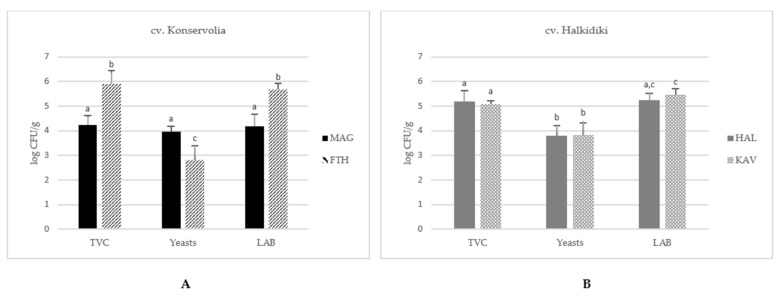
Figure 1 illustrates the mean population of TVC, LAB, and yeasts and molds in fermented table olives of Konservolia cultivar from the Magnesia and Fthiotida regions (Figure 1A) and Halkidiki cultivar from the Halkidiki and Kavala regions (Figure 1B). The specific population of the different microbial groups enumerated on each sample is shown at Table S1. TVC in natural fermented table olives of Konservolia variety exhibited an average value of 5.2 ± 0.9 log CFU/g, with the counts of olives from the Fthiotida region exhibiting about 2-log units higher value than the corresponding average population in olives harvested from Magnesia (p < 0.05) (Figure 1A). Similarly, the LAB population exhibited average values of 5.7 ± 0.2 log CFU/g and 4.2 ± 0.5 log CFU/g in table olives from the Fthiotida and Magnesia regions, respectively. By contrast, the yeast population in table olives from the Fthiotida was 2.8 ± 0.5 log CFU/g, about 1-log unit lower than the respective population in olives from the Magnesia region (p < 0.05).
Figure 1.
Microbial enumerations of total viable counts (TVC), lactic acid bacteria (LAB), and yeasts in fermented table olives of (A) cv. Konservolia from Magnesia (MAG) and Fthiotida (FTH) regions and (B) cv. Halkidiki from Kavala (KAV) and Halkidiki (HAL) regions. The results present average values ± SD. Different letters indicate statistically significant differences (p < 0.05).
In the case of Spanish-style fermented green olives of Halkidiki variety, no significant differences were observed in the microbial populations between samples from the Halkidiki and Kavala regions (p > 0.05) (Figure 1B). It has to be noted that, in all samples, Enterobacteriaceae population was lower than the detection limit of the method (<1 log CFU/g).
Regarding pH measurements, the pH values in the brine of olives did not exceed 4.30 (Table S1), a finding complying with the physicochemical characteristics for the safety of the final product [30]. More specifically, the pH value of brine samples from Magnesia (3.58 ± 0.01) was significantly lower (p < 0.05) than the pH of brine samples from the Fthiotida region (4.14 ± 0.14). On the other hand, brine samples from the Kavala and Halkidiki regions presented average similar pH values, i.e., 3.81 ± 0.04 and 3.79 ± 0.01, respectively. Moreover, the salt concentration in the brines of samples from Magnesia (10.3 ± 0.17% w/v) was significantly higher than the salt concentration in brine samples from Fthiotida (5.45 ± 0.78% w/v) (p < 0.05). Similarly, significant differences were also observed in salt concentration between brine samples from Kavala (5.75 ± 2.01% w/v) and Halkidiki (7.63 ± 0.13% w/v) (p < 0.05).
3.2. Sensory Evaluation of Fermented Table Olives
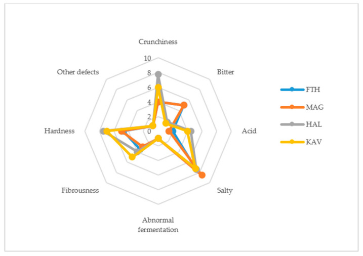
The scores of sensory attributes evaluated for the fermented table olives are presented in Figure 2. Regarding the gustatory sensations, naturally fermented black olives of cv. Konservolia from Fthiotida and Magnesia were perceived to be bitterer than green olives of cv. Halkidiki. Black olives from Magnesia received the highest score for saltiness by the panelists. Concerning acidity, green olives from Halkidiki and Kavala received higher scores compared to black olives from Fthiotida and Magnesia that developed a milder taste. No off odors indicating abnormal fermentation (i.e., butyric, putrid fermentation or zapateria) or other defects were detected by the panelists in any of the table olive samples. Moreover, Spanish-style fermented green olives of cv. Halkidiki from Halkidiki and Kavala received higher scores for the kinesthetic sensations of hardness, fibrousness, and crunchiness compared to naturally fermented black olives of cv. Konservolia from Fthiotida and Magnesia.
Figure 2.
Spider graph showing the sensory profiles (original scores) for the diverse fermented table olives samples. FTH (origin, Fthiotida; cultivar, Konservolia), MAG (Magnesia; Konservolia), HAL (Halkidiki; Halkidiki), KAV (Kavala; Halkidiki).
3.3. Bacterial Community Profiling
The NGS of 16S rRNA amplified from total DNA extracted from the surface of olive samples was applied to monitor the bacterial relative abundancies. The metataxonomic analysis in cv. Konservolia and cv. Halkidiki table olives revealed a complex bacterial microbiota that consisted of thirteen and eleven families, respectively. In brief, differences in bacterial families (Figure 3A) and species (data not shown) were observed based on olive varieties and were significantly higher (p < 0.05) in cv. Halkidiki than in cv. Konservolia table olives. In the case of cv. Halkidiki table olives, bacterial families were significantly higher in olives from the Halkidiki region than from the Kavala region (p < 0.05) (Figure 3B). Similarly, significantly higher bacterial communities were observed in table olives from the Magnesia region than from the Fthiotida region (Figure 3C).
Figure 3.
Alpha-diversity boxplots for table olive’s bacterial families of (A) cultivar Halkidiki and Konservolia, (B) cultivar Halkidiki from Halkidiki (A_1) and Kavala (B_2) regions, and (C) cultivar Konservolia from Magnesia (C_3) and Fthiotida (C_4) regions based on observed and Simpson indices.
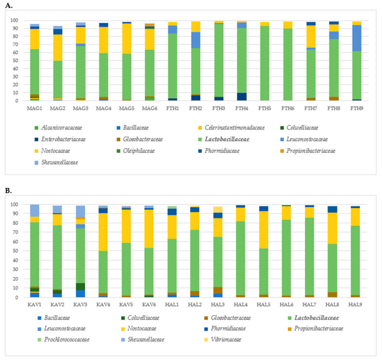
In Figure 4, the OTUs at family level on the olive surface of cv. Konservolia (Figure 4A) and cv. Halkidiki (Figure 4B) samples, representing at least 1% of the total sequence reads in each sample, are displayed. Lactobacillaceae was the predominant bacterial family identified across all olive samples of cv. Konservolia and cv. Halkidiki from both geographical regions (Figure 4A,B). The whole set of identifications at family, genus, and species level is shown as Supplementary Material (Table S2A–C).
Figure 4.
Relative abundance of total observed bacterial families on table olives of (A) cv. Konservolia originating from the regions of Magnesia (MAG) and Fthiotida (FTH) and (B) cv. Halkidiki originating from the regions of Kavala (KAV) and Halkidiki (HAL). Only families above 1% occurrence are reported.
Lactobacillus was the most common detected genus in all cases, followed by Pediococcus in samples of cv. Konservolia and samples from the Halkidiki region. In brief, the species Lactobacillus acidipiscis, Lactobacillus coryniformis, Lactobacillus paracollinoides, Lactobacillus parafarraginis, Lactobacillus harbinensis, Lactobacillus kisonensis, Pediococcus parvulus, and Pediococcus ethanolidurans were identified (Table S2C). Furthermore, Nostocaceae was the second most common family found in samples from Magnesia, Kavala and Halkidiki, whereas Leuconostocaceae was the second abundant family detected in samples from the Fthiotida region. Concerning the rest of the detected bacteria, in olives from Magnesia, Shewanellaceae (including Shewanella), Propionibacteriaceae (including Propionibacterium), and Gloeobacteraceae were also detected, with the remaining families being present at lower proportions (<2%) (Table S2B). Similarly, Nostocaceae, Enterobacteriaceae, Gloeobacteraceae, and Phormidiaceae were in samples from the Fthiotida region (Figure 4A). Moreover, other families contributing to the bacterial consortium in olives from Kavala were Shewanellaceae (including Shewanella), Bacillaceae, Colwelliaceae, Gloeobacteraceae, and Propionibacteriaceae, while Leuconostocaceae was identified only in one sample (Figure 4B). On the other hand, Phormidiaceae, Vibrionaceae, Gloeobacteraceae, Prochlorococaceae, and Bacillaceae were also detected on table olives from the Halkidiki region (Figure 4B).
3.4. Yeast Community Profiling
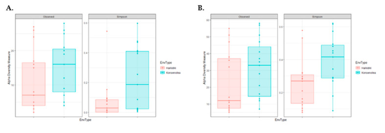
The yeast community of olive samples was revealed by NGS of the ITS region of yeast rDNA amplified from total DNA extracted from the surface of fermented table olive samples. The metataxonomic analysis in cv. Konservolia and cv. Halkidiki table olives revealed a complex yeast microbiota. In brief, differences in yeast families and genera were observed based on olive varieties and were significantly higher (p < 0.05) in cv. Konservolia than cv. Halkidiki table olives (Figure 5). However, no significant differences were observed between the different geographical regions for both cultivars (data not shown). The detected families at relative abundance >1% of the total sequence reads in each olive sample are presented in Figure 6, while the whole set of identifications at family, genus, and species level is shown as Supplementary Material (Table S3A–C).
Figure 5.
Alpha-diversity boxplots for table olives yeasts families (A) and species (B) of cultivar Halkidiki and Konservolia based on observed and Simpson indices.
Figure 6.
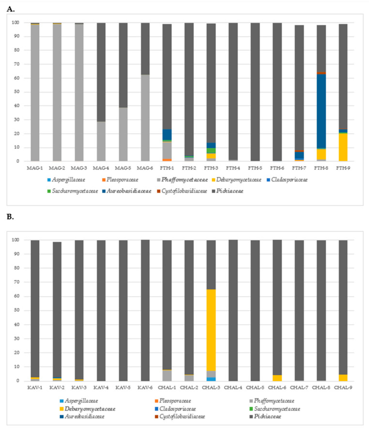
Relative abundance of total observed yeast families on table olives of (A) cv. Konservolia originating from the regions of Magnesia (MAG) and Fthiotida (FTH) and (B) cv. Halkidiki originating from the regions of Kavala (KAV) and Halkidiki (HAL). Only families above 1% occurrence are reported.
Pichiaceae was mainly detected at highest relative abundance in green, Spanish-style fermented olives cv. Halkidiki from both geographical regions (Figure 6B) and the majority of olive samples cv. Konservolia (Figure 6A). In the case of Konservolia olives from Magnesia, Phaffomycetaceae was the dominant family in four samples, followed by Pichiaceae that dominated in two samples, while the remaining families were present at very low proportions (<1%) (Figure 6A). In the latter case, the most detected species were Wickerhamomyces anomalus, Pichia membranifaciens, and Wickerhamomyces sydowiorum (Table S3C). Similarly, Pichiaceae was the predominant family across eight out of nine samples, followed by Aureobasidiaceae that dominated the yeast community in one sample in olives from Fthiotida. The rest of the families, i.e., Debaryomycetaceae and Phaffomycetaceae, were present at very low proportions (<1%) (Figure 6A). In brief, Pichia manshurica, Brettanomyces custersianus, Pichia membranifaciens, Aureobasidium pullulans, Schwanniomyces etchelsii, and Wickerhamomyces anomalus were characterized at species level (Table S3C). On the other hand, for cv. Halkidiki olives from Kavala, beyond Pichiaceae which was the most detected family, the rest of the families were detected in low relative percentages (< 1%) (Figure 6B). In brief, the yeast microbiota was dominated by Pichia (including Pichia manshurica) and Brettanomyces (including Brettanomyces custersianus) (Table S3B,C). In the case of olives from the Halkidiki region, the microbiota of one sample out of six was dominated by Debaryomycetaceae, while the majority of them were dominated by Pichiaceae. Pichia (including Pichia manshurica and Pichia membranifaciens) was the dominant genus detected in eight out of nine samples, while Schwanniomyces (i.e., Schwanniomyces etchelsii) dominated the ninth sample followed by Ogataea, Pichia, and Penicillium (Table S3B,C).
3.5. Cultivar and Geographical Discrimination of Table Olives by Multivariate Analysis
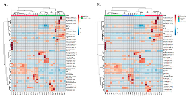
A dual hierarchal dendrogram (heatmap) was utilized to display the data obtained from this study (microbiological, physicochemical, sensory, and bacterial and yeast species—level OTUs) with clustering related to the different olive samples. Based on the clustering evident in Figure 7, there appears to be a clear distinction between samples based on cultivar (Figure 7A) and geographical origin (Figure 7B) classes.
Figure 7.
Hierarchically clustered heatmap of microbiological, physicochemical, organoleptic, and species level operational taxonomic units (OTUs) of bacteria and yeast communities data of table olive samples based on (A) the cultivar and (B) the geographical origin of the samples. The sample codes are indicated in Table 1.
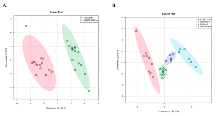
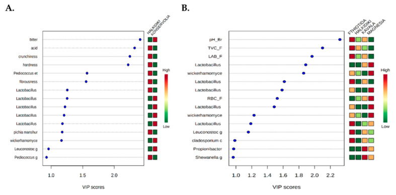
Furthermore, PLS-DA analysis effectively discriminated olive samples based on cultivar (Figure 8A) and geographical origin (Figure 8B) classes with no overlapping. However, a statistically significant difference (p < 0.001) was confirmed only for the discrimination of olives based on geographical origin. In this case, according to the VIP values (>1), pH_Br, TVC-F and LAB_F and Lactobacillus paracollinoides, Lactobacillus coryniformis, Leuconostoc, and Cladosporium cladosporioides were highly associated with the Fthiotida region (Figure 9B). Similarly, Lactobacillus acidipiscis, Wickerhamomyces anomalus, Lactobacillus suebicus, RBC_F, Lactobacillus vaccinostercus, and Wickerhamomyces sydowiorum were highly associated (VIP value > 1) with the Magnesia region and Propionibacterium and Shewanella with the Kavala region (Figure 9B).
Figure 8.
Partial least squares discriminant analysis (PLS-DA) clustering depending on (A) cultivar and (B) geographical origin of the olive samples.
Figure 9.
Most influential parameters of the olive samples based on the VIP scores from the PLS-DA analysis at (A) cultivar and (B) geographical origin levels.
4. Discussion
The effect of cultivar and geographical origin on the microbiota of the fermented table olives was assessed in this research. For this purpose, the bacterial and yeast diversity of fermented table olives of two main Greek varieties collected from different regions, i.e., black olives, cv. Konservolia, from Magnesia and Fthiotida and green olives, cv. Halkidiki, from Kavala and Halkidiki was evaluated using metataxonomics in parallel with the classical microbiological approach and taking into account physicochemical and organoleptic characteristics. The characterization of the microbial communities of Greek table olives aims at a comprehensive analysis of their microbial ecology and contributes to the exploitation of their microbial fingerprint based on cultivar and area of origin.
PLS-DA analysis indicated a satisfactory discrimination among the different geographical regions without overlapping between the cases, with the pH value and the TVC and LAB counts representing the most discriminative parameters.
LAB was the predominant microbial population in black Greek-style fermented olives cv. Konservolia from Fthiotida and in green Spanish-style fermented olives cv. Halkidiki from both geographical regions. The dominance of LAB is rather typical for Spanish-style processing and has been previously observed by other researchers [15,31]. This observation is also in line with previous findings for Greek table olives and is attributed to the low salt level (6–7%) used by the Greek industry in the brine during the period of active fermentation to ensure the dominance of LAB and therefore improve the preservation and sensory characteristics of the final product [6,32]. It is well documented that yeast development is favored against LAB by high salt concentrations, the presence of phenolic compounds, and low pH levels [33,34,35]. Low pH values and high salt concentrations were also measured in the case of black olives from the Magnesia region, where LAB and yeasts were detected at similar levels. The involvement of yeasts is particularly important in natural olives, when fruits are not lye-treated and phenolic compounds partly inhibit LAB development [36]. Similar yeast populations, i.e., 4.7 log cfu/g and 4 log cfu/mL were previously enumerated in Greek black dry-salted olives (cv. Thassos) with ~7.5% NaCl [37] and black table olives of cv. Hojiblanca with 4% NaCl and 0.3% acetic acid [38], respectively.
The metataxonomic analysis employed herein highlighted differences in bacterial and yeast ecology both at cultivar and geographical origin levels. Lactobacillaceae was the dominant family identified in olive samples from both cultivars, indicating that these were all lactic acid fermentations, which was also verified by the classical microbiological analysis. The significant role of this microbial group in olive fermentations has been extensively reviewed by Hurtado et al. [39], and it is commonly found in the microbiota of fermented green and black olives using both classical microbiological and metagenomics analyses [7,15,18,40,41,42]. NGS highlighted relevant differences in the occurrence of different Lactobacillus species, depending on the cultivar. According to multivariate analysis, the most discriminative species were Lactobacillus acidipiscis, Wickerhamomyces anomalus, and Lactobacillus paracollinoides (VIP > 1.6). In a recent study, the presence of Lactobacillus was also highly influential for the differentiation of Greek-style fermented olives cv. Kalamata from different geographic regions [20]. Furthermore, the species Lactobacillus paracollinoides was identified as responsible for the discrimination of Spanish-style green olive fermentations among different patios [22]. Moreover, L. harbinensis was found to colonize only the surface of green Spanish-style fermented table olives cv. Halkidiki, while L. vaccinostercus/L. suebicus, described by Abriouel et al. [13] were detected only on the surface of black naturally fermented table olives cv. Konservolia underlining the impact of cultivar in microbial diversity. In the present study, the occurrence of L. harbinensis at fermented table olives was revealed for the first time. L. harbinensis is a halotolerant species often isolated from fermented vegetables and dairy products [43]. However, the detection of L. coryniformis has also been reported previously in green table olive fermentations [44,45] and in black olives packed in modified atmosphere conditions [46]. In addition, L. acidipiscis was detected in green olives cv. Halkidiki from the Kavala region and in black olives cv. Konservolia from the Magnesia region, reinforcing the importance of regional characteristics (e.g., climatic conditions) in microbial diversity. Likewise, L. paracollinoides was detected in black table olives from Fthiotida and in one sample of green olives from Kavala. The occurrence of L. paracollinoides in table olive fermentations has been reported previously [13,22]. Moreover, pediococci were also detected in olives of both cultivars, with a higher abundance in green olives from Halkidiki where in some samples, they dominated over the Lactobacillus population. The dominant species were Pediococcus parvulus, detected in olives from both cultivars and Pediococcus ethanolidurans found in higher abundance in olives from Halkidiki than Konservolia variety, as it was detected at low relative abundance only in olives from Magnesia. In earlier studies, Pediococcus ethanolidurans was also isolated from black [46] and green [16] olive fermentations, while P. parvulus was found to be the dominant species in green table olives [47]. Regarding the rest of the LAB, the high relative abundance of Leuconostoc genus in black olives cv. Konservolia from the Fthiotida region in combination with the low salt concentration of these samples is in accordance with previous findings that observed a high occurrence of these heterofermentative cocci in fermentations carried out in brine with a low salt concentration [45].
An unusual finding of the present study was the detection of cyanobacteria in the microbiota of fermented table olives of Konservolia and Halkidiki cultivars, represented mainly by Nostocaceae family, followed by Phormidiaceae and Gloeobacteraceae with relative lower abundances. Cyanobacteria are ubiquitously present in soil and marine environments, and some species can survive harsh environmental conditions, including environments with high salt concentrations [48]. Their presence has been highlighted in earlier studies conducted on table olives [49] and olive-mill wastewater [50]; however, it should be carefully evaluated due to emerging human health issues related to this bacterial group [51,52,53].
Moreover, Enterobacteriaceae was detected in black naturally fermented olives cv. Konservolia only in some samples from the Fthiotida region at low relative abundances, although its presence was not confirmed by the classical microbiological methods. This could be attributed either to the amplification of DNA from dead bacteria or to the low detection limit of the plate counting method [49]. The presence of this family in the fermentation of table olives is rather habitual, with a well-known negative contribution in the quality of the final product [54].
Similarly, yeast diversity on olive surfaces was determined by targeting the ITS region of the nuclear ribosomal DNA, a widely accepted standard procedure for yeast identification not only in fermented table olives [19,20] but also in other food fermentations [55]. According to the results, the yeast microbiota of olive samples of both cultivars was less diverse compared to bacteria, a finding in accordance with the results obtained previously regarding fermented natural black olives cv. Kalamata [20].
Pichiaceae was the dominant family identified in green olives cv. Halkidiki from both regions, confirming its ability to colonize the surface of table olives [8]. Specifically, green olives from Kavala showed a homogeneous yeast population where Pichiaceae family prevailed in all samples. The species Pichia manshurica, Brettanomyces custersianus, Pichia membranifaciens, Schwanniomyces etchelsii, and Ogataea candida boidinii were the most common species detected. These results are in agreement with a recent work, where Pichia manshurica, Pichia membranifaciens, and Schwanniomyces etchellsii were found among the yeast species at the final stage of Spanish-style green olive fermentation [22]. The low occurrence (<1%) of Saccharomyces in the observed yeast consortium is of importance, as this genus has been highly associated with olive fermentation [9]. This finding is consistent with the results of biofilm community formed on the surface of plastic vessels used in Spanish-style green olive fermentation cv. Halkidiki [56] and middle stage of Spanish-style fermentation [22]. On the other hand, Brettanomyces are usually associated with the fermentation of alcoholic beverages like beer and wine having a controversial role from spoilage organisms to contributors to industrial fermentations. However, Brettanomyces was also recently detected in black olives cv. Kalamata at low levels [20], while Brettanomyces custersianus and D. bruxellensis have been isolated in the past from olives [57] and Greek-style black olives [58], respectively. It has to be noted that differences were observed among the dominant yeast families in black natural olives cv. Konservolia between the samples from the different geographical regions. The dominant yeast families identified in samples from Magnesia were Phaffomycetaceae (mainly Wickerhamomyces anomalus), followed by Pichiaceae (mainly Pichia membranifaciens). On the other hand, Pichiaceae was identified as the dominant yeast family in most of the black natural olives cv. Konservolia from the Fthiotida region, followed by Aureobasidiaceae. The prevalence of W. anomalus in the yeast consortium was probably attributed to its tolerance to diverse stress factors such as low pH and high salt concentration, characteristics found in the brines of the samples from Magnesia. Earlier studies have confirmed its presence in natural black olives of cv. Konservolia [59,60]. W. anomalus has been reported to exhibit β-glucosidase activity and produce antioxidant compounds and killer toxins against human pathogens and spoilage microorganisms [61,62], properties that may improve the quality of the final product both from nutritional and safety aspects. Pichia manshurica, Brettanomyces, and Pichia membranifaciens were also isolated recently from natural black olive fermentations of Konservolia and Kalamata cultivars [20,59,60], while Aureobasidium pullulans has been previously detected on first stages of cv. Konservolia olive fermentation [59] and Kalamata black olive natural fermentations [63]. P. membranifaciens has shown strain-specific killer activity against spoilage yeasts, thus preventing food spoilage [64].
5. Conclusions
In conclusion, discriminative analysis was performed to detect biogeographic patterns of the microbial populations along with physicochemical and organoleptic characteristics of Greek fermented table olives belonging to Konservolia and Halkidiki varieties. The diversity of the microbial community of olives from different regions was evaluated by metataxonomic analysis. The results obtained reveal the complex structure of the microbiota in these fermentations and point the microbial key taxa that may be linked to specific geographic areas. However, further studies are needed to enhance our knowledge of the microbial ecology of Greek table olives and probably enable the design of new strategies to improve their quality and safety.
Acknowledgments
The authors would like to thank the companies Georgoudis S.A. and Konstantopoulos S.A. for providing the samples of cv. Konservolia and cv. Halkidiki table olives.
Supplementary Materials
The following are available online at https://www.mdpi.com/2076-2607/8/8/1241/s1, Table S1: Microbiological counts of TVC, yeasts, LAB, and Enterobacteriaceae from olives (O) and pH and salt concentration (%, w/v) in brines (B) of Greek-style fermented cv. Konservolia black olives from the Magnesia (MAG) and Fthiotida (FTH) regions and of Spanish-style fermented cv. Halkidiki green olives from the Kavala (KAV) and Halkidiki (HAL) regions. Table S2A: Abundance of bacterial families identified in olive samples from the Magnesia (MAG), Fthiotida (FTH), Kavala (KAV), and Halkidiki (HAL) regions, Table S2B: Abundance of bacterial genera identified in olive samples from the Magnesia (MAG), Fthiotida (FTH), Kavala (KAV), and Halkidiki (HAL) regions, Table S2C: Abundance of bacterial species identified in olive samples from the Magnesia (MAG), Fthiotida (FTH), Kavala (KAV), and Halkidiki (HAL) regions, Table S3A: Abundance of yeast families identified in olive samples from the Magnesia (MAG), Fthiotida (FTH), Kavala (KAV), and Halkidiki (HAL) regions, Table S3B: Abundance of yeast genera identified in olive samples from the Magnesia (MAG), Fthiotida (FTH), Kavala (KAV), and Halkidiki (HAL) regions, Table S3C: Abundance of yeast species identified in olive samples from the Magnesia (MAG), Fthiotida (FTH), Kavala (KAV), and Halkidiki (HAL) regions.
Author Contributions
Conceptualization, C.C.T.; Methodology, A.G. and K.A.; Formal analysis, A.I.D.; Data curation, E.M. and K.A.; Writing—original draft preparation, K.A.; Writing—review and editing, A.I.D. and A.A.A.; Visualization, G.-J.E.N.; Supervision, C.C.T.; Project administration, C.C.T.; Funding acquisition, C.C.T. All authors have read and agreed to the published version of the manuscript.
Funding
This research has been financed by Greek national funds through the Public Investments Program (PIP) of General Secretariat for Research & Technology (GSRT), under the Emblematic Action “The Olive Road” (project code: 2018SE01300000).
Conflicts of Interest
The authors declare no conflict of interest. The funders had no role in the design of the study; in the collection, analyses, or interpretation of data; in the writing of the manuscript, or in the decision to publish the results.
References
- 1.Conte P., Fadda C., Del Caro A., Urgeghe P.P., Piga A. Table Olives: An overview on effects of processing on nutritional and sensory quality. Foods. 2020;9:514. doi: 10.3390/foods9040514. [DOI] [PMC free article] [PubMed] [Google Scholar]
- 2.IOC, International Olive Council World Table Olive Figures. [(accessed on 24 March 2020)];2020 Available online: https://www.internationaloliveoil.org/wp-content/uploads/2020/01/OT-W901-29-11-2019-P.pdf.
- 3.DOEPEL, Interprofessional Association for Table Olives. [(accessed on 24 March 2020)];2020 Available online: https://olivetreeroute.gr/wp-content/uploads/Studies_Publications_017a.pdf.
- 4.Panagou E.Z., Tassou C.C., Katsaboxakis K.Z. Induced lactic acid fermentation of untreated green olives of the Conservolea cultivar by Lactobacillus pentosus. J. Sci. Food Agric. 2003;83:667–674. doi: 10.1002/jsfa.1336. [DOI] [Google Scholar]
- 5.Kailis S., Harris D. Producing Table Olives. Landlinks Press; Collingwood, Australia: 2007. [Google Scholar]
- 6.Tassou C.C., Panagou E.Z., Katsaboxakis K.Z. Microbiological and physicochemical changes of naturally black olives fermented at different temperatures and NaCl levels in the brines. Food Microbiol. 2002;19:605–615. doi: 10.1006/fmic.2002.0480. [DOI] [Google Scholar]
- 7.Perpetuini G., Prete R., García-González N., Khairul Alam M., Corsetti A. Table olives. More than a fermented food. Foods. 2020;9:178. doi: 10.3390/foods9020178. [DOI] [PMC free article] [PubMed] [Google Scholar]
- 8.Nychas G.J.E., Panagou E.Z., Parker M.L., Waldron K.W., Tassou C.C. Microbial colonization of naturally black olives during fermentation and associated biochemical activities in the cover brines. Lett. Appl. Microbiol. 2002;34:173–177. doi: 10.1046/j.1472-765x.2002.01077.x. [DOI] [PubMed] [Google Scholar]
- 9.Arroyo-López F.N., Querol A., Bautista-Gallego J., Garrido-Fernández A. Role of yeasts in table olive production. Int. J. Food Microbiol. 2008;128:189–196. doi: 10.1016/j.ijfoodmicro.2008.08.018. [DOI] [PubMed] [Google Scholar]
- 10.Doulgeraki A., Pramateftaki P., Argyri A., Nychas G.J.E., Tassou C., Panagou E. Molecular characterization of lactic acid bacteria isolated from industrially fermented Greek table olives. LWT. 2013;50:353–356. doi: 10.1016/j.lwt.2012.07.003. [DOI] [Google Scholar]
- 11.Botta C., Cocolin L. Microbial dynamics and biodiversity in table olive fermentation: Culture-dependent and-independent approaches. Front. Microbiol. 2012;3:245. doi: 10.3389/fmicb.2012.00245. [DOI] [PMC free article] [PubMed] [Google Scholar]
- 12.Ercolini D. High-throughput sequencing and metagenomics: Moving forward in the culture-independent analysis of food microbial ecology. Appl. Environ. Microbiol. 2013;79:3148–3155. doi: 10.1128/AEM.00256-13. [DOI] [PMC free article] [PubMed] [Google Scholar]
- 13.Abriouel H., Benomar N., Lucas R., Gálvez A. Culture-independent study of the diversity of microbial populations in brines during fermentation of naturally fermented Aloreña green table olives. Int. J. Food Microbiol. 2011;144:487–496. doi: 10.1016/j.ijfoodmicro.2010.11.006. [DOI] [PubMed] [Google Scholar]
- 14.Randazzo C.L., Ribbera A., Pitino I., Romeo F.V., Caggia C. Diversity of bacterial population of table olives assessed by PCR-DGGE analysis. Food Microbiol. 2012;32:87–96. doi: 10.1016/j.fm.2012.04.013. [DOI] [PubMed] [Google Scholar]
- 15.Cocolin L., Alessandria V., Botta C., Gorra R., De Fillipis F., Ercolini D., Rantsiou K. NaOH-debittering induces changes in bacterial ecology during table olives fermentation. PLoS ONE. 2013;8:e69074. doi: 10.1371/journal.pone.0069074. [DOI] [PMC free article] [PubMed] [Google Scholar]
- 16.Lucena-Padrós H., Caballero-Guerrero B., Maldonado-Barragán A., Ruiz-Barba J.L. Genetic diversity and dynamics of bacterial and yeast strains associated to Spanish-style green table olive fermentations in large manufacturing companies. Int. J. Food Microbiol. 2014;190:72–78. doi: 10.1016/j.ijfoodmicro.2014.07.035. [DOI] [PubMed] [Google Scholar]
- 17.Lucena-Padrós H., Jiménez E., Maldonado-Barragán A., Rodríguez J.M., Ruiz-Barba J.L. PCR-DGGE assessment of the bacterial diversity in Spanish-style green table olive fermentations. Int. J. Food Microbiol. 2015;205:47–53. doi: 10.1016/j.ijfoodmicro.2015.03.033. [DOI] [PubMed] [Google Scholar]
- 18.Randazzo C.L., Todaro A., Pino A., Pitino I., Corona O., Caggia C. Microbiota and metabolome during controlled and spontaneous fermentation of Nocellara Etnea table olives. Food Microbiol. 2017;65:136–148. doi: 10.1016/j.fm.2017.01.022. [DOI] [PubMed] [Google Scholar]
- 19.Arroyo-López F.N., Medina E., Ruiz-Bellido M.A., Romero-Gil V., Montes-Borrego M., Landa B.B. Enhancement of the knowledge on fungal communities in directly brined Aloreña de Málaga green olive fermentations by metabarcoding analysis. PLoS ONE. 2016;11:e0163135. doi: 10.1371/journal.pone.0163135. [DOI] [PMC free article] [PubMed] [Google Scholar]
- 20.Kazou M., Tzamourani A., Panagou E.Z., Tsakalidou E. Unraveling the microbiota of natural black cv. Kalamata fermented olives through 16S and ITS metataxonomic analysis. Microorganisms. 2020;8:672. doi: 10.3390/microorganisms8050672. [DOI] [PMC free article] [PubMed] [Google Scholar]
- 21.Martiny J.B.H., Bohannan B.J.M., Brown J.H., Colwell R.K., Fuhrman J.A., Green J.L., Horner-Devine M.C., Kane M., Krumins J.A., Kuske C.R., et al. Microbial biogeography: Putting microorganisms on the map. Nat. Rev. Microbiol. 2006;4:102–112. doi: 10.1038/nrmicro1341. [DOI] [PubMed] [Google Scholar]
- 22.Lucena-Padrós H., Ruiz-Barba J.L. Microbial biogeography of Spanish-style green olive fermentations in the province of Seville, Spain. Food Microbiol. 2019;82:259–268. doi: 10.1016/j.fm.2019.02.004. [DOI] [PubMed] [Google Scholar]
- 23.Argyri A.A., Nisiotou A.A., Mallouchos A., Panagou E.Z., Tassou C.C. Performance of two potential probiotic Lactobacillus strains from the olive microbiota as starters in the fermentation of heat shocked green olives. Int. J. Food Microbiol. 2014;171:68–76. doi: 10.1016/j.ijfoodmicro.2013.11.003. [DOI] [PubMed] [Google Scholar]
- 24.Garrido-Fernández A., Fernández-Díez M.J., Adams R.M. Table Olives: Production and Processing. 1st ed. Chapman and Hall; London, UK: 1997. [Google Scholar]
- 25.IOC, International Olive Council . Sensory Analysis of Table Olives. International Olive Council; Madrid, Spain: 2011. Document COI/OT/MO No 1/Rev. 2. [Google Scholar]
- 26.Dowd S.E., Callaway T.R., Wolcott R.D., Sun Y., McKeehan T., Hagevoort R.G., Edrington T.S. Evaluation of the bacterial diversity in the feces of cattle using 16S rDNA bacterial tag-encoded FLX amplicon pyrosequencing (bTEFAP) BMC Microbiol. 2008;8:1–8. doi: 10.1186/1471-2180-8-125. [DOI] [PMC free article] [PubMed] [Google Scholar]
- 27.McMurdie P.J., Holmes S. Phyloseq: An R package for reproducible interactive analysis and graphics of microbiome census data. PLoS ONE. 2013;8:e61217. doi: 10.1371/journal.pone.0061217. [DOI] [PMC free article] [PubMed] [Google Scholar]
- 28.Gromski P.S., Muhamadali H., Ellis D.I., Xu Y., Correa E., Turner M.L., Goodacre R. A tutorial review: Metabolomics and partial least squares-discrimination analysis—A marriage of convenience or a shotgun wedding. Anal. Chim. Acta. 2015;879:10–23. doi: 10.1016/j.aca.2015.02.012. [DOI] [PubMed] [Google Scholar]
- 29.Chong J., Wishart D.S., Xia J. Using MetaboAnalyst 4.0 for comprehensive and interactive metabolomics data analysis. Curr. Protoc. Bioinform. 2019;68:e86. doi: 10.1002/cpbi.86. [DOI] [PubMed] [Google Scholar]
- 30.IOOC, International Olive Oil Council . Trade Standard Applying to Table Olives. International Olive Oil Council; Madrid, Spain: 2004. [Google Scholar]
- 31.Blana V.A., Grounta A., Tassou C.C., Nychas G.J.E., Panagou E.Z. Inoculated fermentation of green olives with potential probiotic Lactobacillus pentosus and Lactobacillus plantarum starter cultures isolated from industrially fermented olives. Food Microbiol. 2014;38:208–218. doi: 10.1016/j.fm.2013.09.007. [DOI] [PubMed] [Google Scholar]
- 32.Panagou E.Z., Schillinger U., Franz C.M.A.P., Nychas G.J.E. Microbiological and biochemical profile of cv Conservolea naturally black olives during controlled fermentation with selected strains of lactic acid bacteria. Food Microbiol. 2008;25:348–358. doi: 10.1016/j.fm.2007.10.005. [DOI] [PubMed] [Google Scholar]
- 33.Bautista Gallego J., Arroyo-Lopez F.N., Romero-Gil V., Rodriguez-Gomez F., Garcia-Garcia P., Garrido-Fernandez A. Fermentation profile of green Spanish-style Manzanilla olives according to NaCl content in brine. Food Microbiol. 2015;49:56–64. doi: 10.1016/j.fm.2015.01.012. [DOI] [PubMed] [Google Scholar]
- 34.Benítez-Cabello A., Romero-Gil V., Rodríguez-Gómez F., Garrido-Fernández A., Jiménez-Díaz R., Arroyo-López F.N. Evaluation and identification of poly-microbial biofilms on natural green Gordal table olives. Antonie Van Leeuwenhoek. 2015;108:597–610. doi: 10.1007/s10482-015-0515-2. [DOI] [PubMed] [Google Scholar]
- 35.Porru C., Rodriguez-Gomez F., Benitez-Cabello A., Jimenez-Diaz R., Zara G., Budroni M., Mannazzu I., Arroyo-Lopez F.N. Genotyping, identification and multifunctional features of yeasts associated to Bosana naturally table olive fermentations. Food Microbiol. 2018;69:33–42. doi: 10.1016/j.fm.2017.07.010. [DOI] [PubMed] [Google Scholar]
- 36.Arroyo-López F.N., Romero-Gil V., Bautista-Gallego J., Rodriguez-Gómez F., Jiménez-Díaz R., García-García P., Querol A., Garrido-Fernandez A. Yeasts in table olive processing: Desirable or spoilage microorganisms. Int. J. Food Microbiol. 2012;160:42–49. doi: 10.1016/j.ijfoodmicro.2012.08.003. [DOI] [PubMed] [Google Scholar]
- 37.Panagou E.Z. Greek dry-salted olives: Monitoring the dry-salting process and subsequent physico-chemical and microbiological profile during storage under different packing conditions at 4 and 20 °C. LWT-Food Sci. Technol. 2006;39:323–330. doi: 10.1016/j.lwt.2005.02.017. [DOI] [Google Scholar]
- 38.Arroyo Lopez F.N., Duran Quintana M.C., Ruiz-Barba J.L., Querol A., Garrido-Fernandez A. Use of molecular methods for the identification of yeast associated with table olives. Food Microbiol. 2006;23:791–796. doi: 10.1016/j.fm.2006.02.008. [DOI] [PubMed] [Google Scholar]
- 39.Hurtado A., Reguant A., Bordons A., Rozès N. Lactic acid bacteria from fermented table olives. Food Microbiol. 2012;31:1–8. doi: 10.1016/j.fm.2012.01.006. [DOI] [PubMed] [Google Scholar]
- 40.De Angelis M., Campanella D., Cosmai L., Summo C., Rizzello C.G., Caponio F. Microbiota and metabolome of un-started and started Greek-type fermentation of Bella di Cerignola table olives. Food Microbiol. 2015;52:18–30. doi: 10.1016/j.fm.2015.06.002. [DOI] [PubMed] [Google Scholar]
- 41.Medina E., Ruiz-Bellido M.A., Romero-Gil V., Rodríguez-Gómez F., Montes-Borrego M., Landa B.B., Arroyo-López F.N. Assessment of the bacterial community in directly brined Aloreña de Málaga table olive fermentations by metagenetic analysis. Int. J. Food Microbiol. 2016;236:47–55. doi: 10.1016/j.ijfoodmicro.2016.07.014. [DOI] [PubMed] [Google Scholar]
- 42.Rodríguez-Gómez F., Ruiz-Bellido M.Á., Romero-Gil V., Benítez-Cabello A., Garrido-Fernández A., Arroyo-López F.N. Microbiological and physicochemical changes in natural green heat-shocked Aloreña de Málaga table olives. Front. Microbiol. 2017;8:2209. doi: 10.3389/fmicb.2017.02209. [DOI] [PMC free article] [PubMed] [Google Scholar]
- 43.Miyamoto M., Seto Y., Hai Hao D., Teshima T., Bo Sun Y., Kabuki T., Bing Yao L., Nakajima H. Lactobacillus harbinensis sp. nov., consisted of strains isolated from traditional fermented vegetables ‘Suan cai’ in Harbin, Northeastern China and Lactobacillus perolens DSM 12745. Syst. Appl. Microbiol. 2005;15:688–694. doi: 10.1016/j.syapm.2005.04.001. [DOI] [PubMed] [Google Scholar]
- 44.Aponte M., Blaiotta G., La Croce F., Mazzaglia A., Farina V., Settanni L., Moschetti G. Use of selected autochthonous lactic acid bacteria for Spanish-style table olive fermentation. Food Microbiol. 2012;30:8–16. doi: 10.1016/j.fm.2011.10.005. [DOI] [PubMed] [Google Scholar]
- 45.De Bellis P., Valerio F., Sisto A., Lonigro S.L., Lavermicocca P. Probiotic table olives: Microbial populations adhering on olive surface in fermentation sets inoculated with the probiotic strain Lactobacillus paracasei IMPC2.1 in an industrial plant. Int. J. Food Microbiol. 2010;140:6–13. doi: 10.1016/j.ijfoodmicro.2010.02.024. [DOI] [PubMed] [Google Scholar]
- 46.Doulgeraki A.I., Hondrodimou O., Iliopoulos V., Panagou E. Lactic acid bacteria heterogeneity during aerobic and modified atmosphere packaging storage of natural black Conservolea olives in polyethylene pouches. Food Control. 2012;26:49–57. doi: 10.1016/j.foodcont.2012.01.006. [DOI] [Google Scholar]
- 47.Franzetti L., Scarpellini M., Vecchio A., Planeta D. Microbiological and safety evaluation of green table olives marketed in Italy. Ann. Microbiol. 2011;61:843–851. doi: 10.1007/s13213-011-0205-x. [DOI] [Google Scholar]
- 48.Billi D., Friedmann E.I., Helm R.F., Potts M. Gene transfer to the desiccation-tolerant cyanobacterium Chroococcidiopsis. J. Bacteriol. 2001;183:2298–2305. doi: 10.1128/JB.183.7.2298-2305.2001. [DOI] [PMC free article] [PubMed] [Google Scholar]
- 49.Zinno P., Guantario B., Perozzi G., Pastore G., Devirgillis C. Impact of NaCl reduction on lactic acid bacteria during fermentation of Nocellara del Belice table olives. Food Microbiol. 2017;63:239–247. doi: 10.1016/j.fm.2016.12.001. [DOI] [PubMed] [Google Scholar]
- 50.Tsiamis G., Tzagkaraki G., Chamalaki A., Xypteras N., Andersen G., Vayenas D., Bourtzis K. Olive-mill wastewater bacterial communities display a cultivar specific profile. Curr. Microbiol. 2012;64:197–203. doi: 10.1007/s00284-011-0049-4. [DOI] [PubMed] [Google Scholar]
- 51.He X., Liu Y.L., Conklin A., Westrick J., Weavers L.K., Dionysiou D.D., Lenhart J.J., Mouser P.J., Szlag D., Walker H.W. Toxic cyanobacteria and drinking water: Impacts, detection and treatment. Harmful Algae. 2016;54:174–193. doi: 10.1016/j.hal.2016.01.001. [DOI] [PubMed] [Google Scholar]
- 52.Gutierrez-Praena D., Jos A., Pichardo S., Moreno I.M., Camean A.M. Presence and bioaccumulation of microcystins and cylindrospermopsin in food and the effectiveness of some cooking techniques at decreasing their concentrations: A review. Food Chem. Toxicol. 2013;53:139–152. doi: 10.1016/j.fct.2012.10.062. [DOI] [PubMed] [Google Scholar]
- 53.Manganelli M., Scardala S., Stefanelli M., Palazzo F., Funari E., Vichi S., Buratti F.M., Testai E. Emerging health issues of cyanobacterial blooms. Ann. Ist. Super. Sanità. 2012;48:415–428. doi: 10.4415/ANN_12_04_09. [DOI] [PubMed] [Google Scholar]
- 54.Sánchez-Gómez A.H., García-García P., Rejano Navarro L. Elaboration of table olives. Grasas Y Aceites. 2006;57:86–94. doi: 10.3989/gya.2006.v57.i1.24. [DOI] [Google Scholar]
- 55.De Fillipis F., Parente E., Ercolini D. Metagenomics insights into food fermentations. Microb. Biotechnol. 2017;10:91–102. doi: 10.1111/1751-7915.12421. [DOI] [PMC free article] [PubMed] [Google Scholar]
- 56.Grounta A., Doulgeraki A.I., Panagou E.Z. Quantification and characterization of microbial biofilm community attached on the surface of fermentation vessels used in green table olive processing. Int. J. Food Microbiol. 2015;203:41–48. doi: 10.1016/j.ijfoodmicro.2015.03.001. [DOI] [PubMed] [Google Scholar]
- 57.Crauwels S., Zhu B., Steensels J., Busschaert P., De Samblanx G., Marchal K., Willems K.A., Verstrepen K.V., Lievens B. Assessing genetic diversity among Brettanomyces yeasts by DNA fingerprinting and whole-genome sequencing. Appl. Environ. Microbiol. 2014;80:4398–4413. doi: 10.1128/AEM.00601-14. [DOI] [PMC free article] [PubMed] [Google Scholar]
- 58.Kotzekidou P. Identification of yeasts from black olives in rapid system microtitre plates. Food Microbiol. 1997;14:609–616. doi: 10.1006/fmic.1997.0133. [DOI] [Google Scholar]
- 59.Nisiotou A.A., Chorianopoulos N., Nychas G.J.E., Panagou E.Z. Yeast heterogeneity during spontaneous fermentation of black Conservolea olives in different brine solutions. J. Appl. Microbiol. 2010;108:396–405. doi: 10.1111/j.1365-2672.2009.04424.x. [DOI] [PubMed] [Google Scholar]
- 60.Bleve G., Tufariello M., Durante M., Grieco F., Ramires F.A., Mita G., Tasioula-Margari M., Logrieco A.F. Physico-chemical characterization of natural fermentation process of Conservolea and Kalamata table olives and development of a protocol for the pre-selection of fermentation starters. Food Microbiol. 2015;46:368–382. doi: 10.1016/j.fm.2014.08.021. [DOI] [PubMed] [Google Scholar]
- 61.Hernández A., Martin A., Aranda E., Pérez-Nevado F., Córdoba M.G. Identification and characterization of yeast isolated from the elaboration of seasoned green table olives. Food Microbiol. 2007;24:346–351. doi: 10.1016/j.fm.2006.07.022. [DOI] [PubMed] [Google Scholar]
- 62.Gazi M.R., Hoshikuma A., Kanda K., Murata A., Kato F. Detection of free radical scavenging activity in yeast culture. Bull. Fac. Agric. -Saga Univ. (Jpn.) 2001;86:67–74. [Google Scholar]
- 63.Bonatsou S., Paramithiotis S., Panagou E.Z. Evolution of yeast consortia during the fermentation of Kalamata natural black olives upon two initial acidification treatments. Front. Microbiol. 2018;8:2673. doi: 10.3389/fmicb.2017.02673. [DOI] [PMC free article] [PubMed] [Google Scholar]
- 64.Santos A., Marquina D., Leal J.A., Peinado J.M. (1→6)-β-D-Glucan as cell wall receptor for Pichia membranifaciens killer toxin. Appl. Environ. Microbiol. 2000;66:1809–1813. doi: 10.1128/AEM.66.5.1809-1813.2000. [DOI] [PMC free article] [PubMed] [Google Scholar]
Associated Data
This section collects any data citations, data availability statements, or supplementary materials included in this article.


