Abstract
Background
Lipedema is often unrecognized or misdiagnosed; despite an estimated prevalence of 10% in the overall female population, its cause is still unknown. There is increasing awareness of this condition, but its differential diagnosis can still be challenging. In this article, we summarize current hypotheses on its pathogenesis and the recommendations of current guidelines for its diagnosis and treatment.
Methods
This review is based on publications about lipedema that were retrieved by a selective search in the MEDLINE, Web of Science, and Cochrane Library databases.
Results
The pathophysiology of lipedema remains unclear. The putative causes that have been proposed include altered adipogenesis, microangiopathy, and disturbed lymphatic microcirculation. No specific biomarker has yet been found, and the diagnosis is currently made on clinical grounds alone. Ancillary tests are used only to rule out competing diagnoses. The state of the evidence on treatment is poor. Treatment generally consists of complex decongestive therapy. In observational studies, liposuction for the permanent reduction of adipose tissue has been found to relieve symptoms to a significant extent, with only rare complications. The statutory health-insurance carriers in Germany do not yet regularly cover the cost of the procedure; studies of high methodological quality will be needed before this is the case.
Conclusion
The diagnosis of lipedema remains a challenge because of the heterogeneous presentation of the condition and the current lack of objective measuring instruments to characterize it. This review provides a guide to its diagnosis and treatment in an interdisciplinary setting. Research in this area should focus on the elucidation of the pathophysiology of lipedema and the development of a specific biomarker for it.
cme plus
This article has been certified by the North Rhine Academy of Continuing Medical Education. The test questions for this article can be found at http://daebl.de/RY95. This unit can be processed for CME credit until 31 May 2021.
Participation is possible only via the Internet at cme.aerztebatt.de.
Lipedema is a chronic condition that is currently thought to be progressive as well. It mainly affects women, male sufferers having been described in only a few case reports (1) (e1, e2). Its progressive nature, though not yet unequivocally demonstrated, is assumed on the basis of clinical experience. Epidemiologic estimates from the sparse available data suggest an approximately 10% prevalence in the overall female population (2, 3, e3– e6).
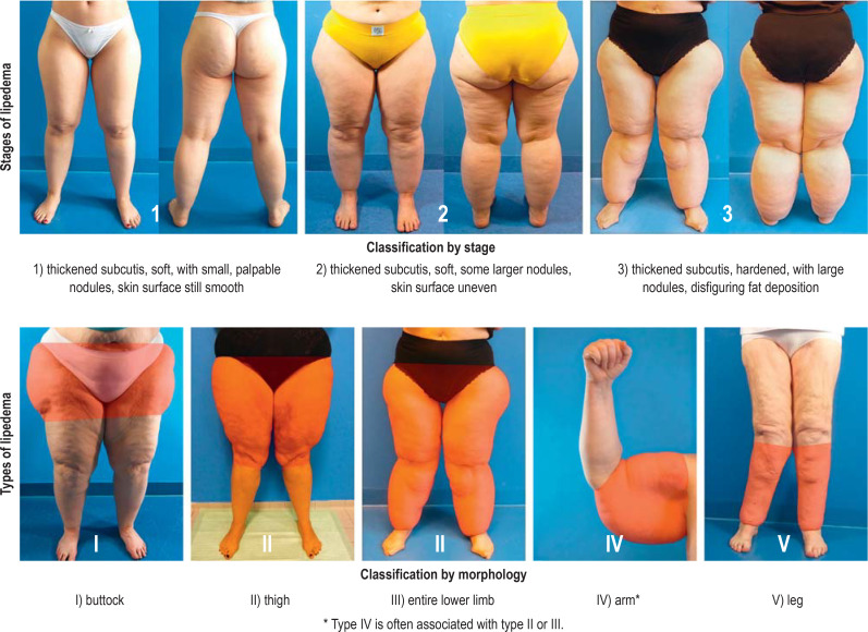
The initial manifestations of lipedema often arise in phases of hormonal change (puberty, pregnancy, menopause). Its hallmark is a disproportionate distribution of body fat on the extremities, while the trunk remains slim. Hands and feet are not involved. (4) (Figure).
Figure 1.
the staging and typological classification of lipedema
Aside from the circumscribed, bilaterally symmetrical, localized increase of the subcutaneous fatty tissue of the limbs, lipedema has the typical clinical manifestations listed in the Box (5). Three clinical stages have been described through which the disease progresses (figure 1) (6).
Although Allan und Hines (7) described lipedema as early as 1940, the condition attracted little attention for many years. Even now that awareness of it has been heightened by frequent discussion in the news media (e7), there remains a great deal of uncertainty as to how it can be correctly diagnosed. The diagnosis is only rarely made on the patient’s first contact with a physician (e8), and there is often a delay of several years before specific treatment is initiated (8).
Current research focuses on the pathophysiology of lipedema and on the development of tools to facilitate its correct diagnosis and the exclusion of competing diagnoses. In this review, we present the current state of knowledge of, and hypotheses about, the etiology and pathogenesis of lipedema. We also hope to increase physicians’ awareness of the urgency of early diagnosis and promptly initiated treatment.
Method
We selectively searched for publications about lipedema in the MEDLINE (via PubMed), Web of Science, and Cochrane Library databases using the key words “Lipödem,” “lipedema,” “lipoedema,” and “multiple symmetric lipomatosis,” and we carried out a supplementary search among the references of these publications. We included articles that were published in English or German up to February 2020.
Pathophysiology
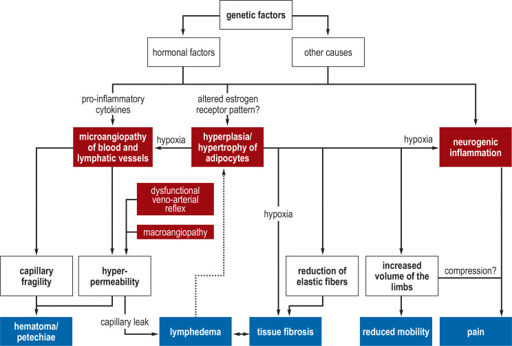
The cause of lipedema is still unexplained. There are various hypotheses about its pathophysiology (figure 2).
Figure 2.
Hypotheses about pathogenesis
?Note: the etiology of lipedema has not yet been conclusively determined.
The figure depicts a number of possible hypotheses about its pathogenesis.
As the condition has repeatedly been described in familial clusters, a genetic predisposition is assumed (9, e1, e9). As many as 60% of patients have an affected first-degree relative (3, 10, e9, e10). Analyses of familial clusters suggest an autosomal dominant inheritance pattern with incomplete penetrance (11, 12, e11).
As lipedema usually first manifests itself in periods of hormonal change, it is generally thought to be estrogen-mediated (13). Despite the autosomal dominant inheritance pattern suggested by pedigree analyses, it has been proposed that the disorder results from a polygenically mediated change in the pattern of distribution of alpha- and beta-estrogen receptors (ER) in the white fatty tissue of affected areas (ER-α expression ↓, ER-ß expression ↑) (13, 14, e12).
It is not yet entirely clear whether, in lipedema, the subcutaneous fat cells become more numerous (hyperplasia) (15– 17, e13, e14) or merely larger in size (hypertrophy) (15, e15).
Cytobiological and protein-expression studies on lipo-aspirates taken from lipedema patients suggest that the disorder mainly arises through changes in the initial steps of cell differentiation in adipogenesis (15, 16, 18– 20).
Another pathophysiological hypothesis involves primary microvascular dysfunction in the lymphatic and blood capillaries (21, 22). This, in turn, is thought to be due to a hypoxic stimulus brought about by excessive expansion of adipose tissue, leading to endothelial dysfunction, and thereby to increased angiogenesis; alternatively, it may be due to a mechanical disturbance of lymph drainage (13, 17, 23, e16, e17). Capillary damage is also a proposed cause of the observed increased tendency to form hematomas and petechiae (21, 24).
Increased capillary permeability leads to shifting of protein into the extracellular compartment (“capillary leak”) and thereby to tissue edema. At first, the additional fluid entering the interstitial space can be compensated for by increased lymph drainage. As the disorder progresses, however, the capacity of the draining lymphatic vessels is exceeded, and high-volume insufficiency (e18) results, while the larger lymphatic vessels remain intact (e9, e19, e20). Quantitative lymphatic scintigraphy has revealed early and, in part, stage-dependent disturbances of lymphatic transport capacity (e21, e22), as well as initially increased lymphatic transport (e23).
The effect of capillary hyperpermability is increased by pathological abnormalities in large blood vessels. Stiffness of the aorta, which has been described in patients with lymphedema, might promote premature vascular remodeling and local hypertension (13, e16). Moreover, there is also dysregulation of the veno-arterial reflex (VAR), which protects the capillary bed from locally elevated hydrostatic pressure by constriction of the arterioles (17). This, combined with the capillary leak due to microangiopathy, promotes the formation of edema and hematoma.
The increased perception of pain that typifies lipedema has been attributed to dysregulation of locoregional sensory nerve fibers through an inflammatory mechanism. This hypothesis is based on single case reports; there are no valid data showing a significant increase of pro-inflammatory markers in patients with lipedema (15, e24– 25). Disordered pain perception seems unlikely to be due to mechanical compression of nerve fibers by the expanding mass of fatty tissue and tissue edema, as there is no such disturbance in other types of lipohypertrophy or lymphedema (10).
The advanced stages of lipedema are associated with various sequelae. A fluid load exceeding the capacity of the lymphatic system can cause secondary lymphedema (“lipo-lymphedema”) in any stage of the disease (12). Mechanical irritation from large fatty deposits near the joints can macerate the skin; such deposits on the thighs and around the knee joints can also interfere with normal gait and cause secondary arthritis (5). Further secondary effects include the emotional disturbance and lessened self-esteem that result from an appearance that falls short of the contemporary ideal of beauty (e7, e26).
Diagnostic evaluation
The diagnosis is generally made on clinical grounds after the exclusion of competing diagnoses. As the presenting manifestations of lipedema are heterogeneous, the diagnosis should be confirmed by an experienced lymphologist in doubtful cases. The basic diagnostic evaluation consists of history-taking, inspection, and palpation, with particular attention to the manifestations listed in the Box. The clinical constellation of the major manifestations of the disorder appearing together—tissue tenderness, a feeling of tightness, and an excessive tendency toward hematoma formation, with worsening symptoms over the course of the day, in a patient with a bilaterally symmetrical, disproportionate proliferation of fatty tissue on the limbs but not on the hands/feet—points toward the diagnosis of lipedema. Thus, the history obtained from the patient is a major factor in the establishment of the correct diagnosis.
BOX. Clinical criteria for the diagnosis of lipedema.
bilateral, symmetrical, disproportionate fatty tissue hypertrophy on the limbs
sparing of the hands and feet (cuff phenomenon)
approximately 30% involvement of the arms
negative Stemmer sign*
a feeling of heaviness and tension in the affected limbs
pain on pressure and touch
marked tendency to form hematomas
stable limb circumference with weight reduction or caloric restriction
worsening of symptoms over the course of the day
telangiectases and visible vascular markings around fat deposits
hypothermia of the skin
*positive Stemmer sign (in case of secondary lymphedema): the skin fold between the second and third toe is thickened and cannot be lifted
Persons suffering from lipedema often have a positive family history of the disorder. The physician taking the history of the present illness must also ask, in particular, about the time of onset of the initial manifestations and progression in the intervening time.
The onset of lipedema is typically triggered by hormonal changes (puberty, pregnancy, menopause); this helps in the differentiation of lipedema from simple obesity. The distinction can be difficult to draw, as these entities often appear together and the clinical picture can vary (table) (25). Even in an obese person, however, the characteristic symptoms of pain, a feeling of tightness, and a tendency toward bruising (hematoma formation) indicate that lipedema is present as well (box). Sometimes lipedema is unmasked only after successful bariatric surgery for obesity, when, after marked weight loss, a persistent abnormal pattern of fat distribution reveals itself that is typical of lipedema (26, e27).
Table. The differential diagnosis of lipedema (modified from [5]).
| Lipedema | Lipohypertrophy | Obesity | Lymphedema | |
| Sex | female | female/male | female/male | female/male |
| Family history | ++ | (+) | +++ | primary ++ secondary Ø |
| Symmetry | +++ | (+) | +++ | (+) |
| Swollen feet | Ø | (+) | (+) | +++ |
| Increased fatty tissue | +++ | +++ | +++ | (+) |
| Disproportion | +++ | +++ | (+) | + |
| Edema | depending on stage Ø/+++ |
Ø | (+) | +++ |
| Tenderness | +++ | Ø | Ø | Ø |
| Hematoma tendency | +++ | (+) | Ø | Ø |
| Influence of diet | (+) | Ø | +++ | Ø |
+ to +++ present, (+) possible, Ø absent
The physician taking the history must also routinely inquire about the commonly associated psychiatric comorbidities, so that early treatment of these can be initiated where necessary (e28).
Clinical examination
The three stages of the disease are characterized by progressive changes in the structure of the skin surface (stage I, smooth; stage II, uneven or corrugated; stage 3, markedly thickened and indurated) and in the findings on palpation:
stage I: small nodules, reversible edema
stage II: walnut-sized nodules, reversible or irreversible edema
stage III: disfiguring fat deposits, macronodular changes, with accompanying lymphedema, potentially Stemmer sign positive (e29).
The symptoms and subjective degree of suffering are not necessarily correlated with the disease stage (5).
Standardized anthropometric measurements should be a part of routine clinical follow-up, both to assess the spontaneous course of the disorder and to monitor its response to treatment: body weight, body-mass index (BMI), waist-to-hip ratio (WHR), waist-to-height ratio (WHtR), and the circumference and volume of the limbs. The BMI is of limited utility in distinguishing lipedema from obesity (11, 25, e24).
Moreover, pain perception should be assessed at regular intervals, with, e.g., the Visual Analog Scale (VAS) and the Schmeller questionnaire (e30). An index of daily activity should also be documented, e.g., by the step-counting app of the patient’s mobile telephone (5).
The tissue tenderness that is characteristic of lipedema can be checked with the pinch test, which is often felt as very unpleasant in the affected areas but causes no pain elsewhere. Increased capillary fragility manifests itself in spontaneous hematoma formation. There is no need, in routine clinical practice, to document this further with any special measuring instruments or stress tests (e31– e33).
Laboratory tests
Renal and hepatic dysfunction, hypothyroidism (possibly subclinical), pathological lipid profiles, and insulin resistance should be ruled out by laboratory testing. Any hormonal or edema-promoting disturbances that are found should be treated, although no evidence yet indicates a benefit of such treatment with respect to the severity or course of lipedema (1).
Ancillary diagnostic testing
Diagnostic procedures that require special equipment are used only to rule out competing elements of the differential diagnosis; they play no established role in the routine evaluation of lipedema (3, 12, 27, e34, e35).
The skin and subcutaneous tissue can be studied qualitatively and quantitatively with ultrasonography (e36– e38), computed tomography (e39, e40), or magnetic resonance imaging (e41, e42).
Structural and functional evaluation of the lymphatic system with tests such as indirect lymphography (22, e43, e44), fluorescence microlymphography (21, e45), functional lymphatic scintigraphy (22, e9, e19, e21, e23, e46), and magnetic resonance lymphangiography (e47) does not reveal any specific or pathognomonic findings of lipedema.
Other diagnostic methods, such as dual-energy X-ray absorptiometry (DEXA) (e48) or bio-impedance analysis (e49), are used only to answer certain specific questions that may arise.
Treatment
Conservative management
Ever since lipedema was first described, the consensus medical recommendation has been that patients should be advised to accept the condition and modify their mode of living accordingly. This remains true today, despite the availability of treatments that can bring relief (7). To prevent frustration, the physician must inform the patient that the main goal of conservative treatment is to relieve symptoms, not to improve the appearance of the extremities (17). No causally directed treatment for lipedema has yet been described.
The initiation, extent, and duration of treatment should be agreed on with the patient, in consideration of the individual degree of suffering caused by the disease. The classic components of conservative management are the following:
manual lymph drainage, on a regular basis if necessary
appropriate compression therapy with custom-made, flat-knitted compressive clothing (compression classes II–III)
physiotherapy and exercise therapy
psychosocial therapy
dietary counseling and weight management
patient education on self-management.
Although conservative management brings about only a small reduction in tissue volume—5–10% in various studies, including one randomized, controlled trial—it does lessen tenderness (pain on pressure) and feelings of tightness in the limbs (10, 24, 28, 29, e17, e50). A further goal of treatment is to prevent secondary complications, such as skin lesions in advanced disease (11).
Reports that several weeks of inpatient treatment can be beneficial (24, 28, 29, e17) do not imply any long-term benefit from outpatient treatment. In fact, there is hardly any evidence for the efficacy of conservative outpatient treatment “under the conditions of normal, everyday life,” and the authors therefore do not think conservative management can be considered the gold standard of treatment. Nor does any convincing evidence suggest that classic conservative management prevents the progression of the disease.
Patient education
Patients should be comprehensively informed about the nature of the disease and the fact that it is chronic. They should be told in a “non-ideological” way about all of the treatment options and about the ways they themselves can actively influence the disease. They should also be offered the option of professional help in coping emotionally with the disease. The pros and cons of confronting the patient with the diagnosis are discussed in detail by De la Torre et al. (25). As lipedema is a chronic, progressive condition, the patient should be given adequate informational material as soon as the diagnosis is made, along with contact data for the relevant self-help organizations. If necessary, the patient should also be educated about complex decongestive therapy (30).
Weight control
Patients with lipedema are at increased risk of developing morbid obesity (25); conversely, overweight worsens the manifestations of lipedema (11). The pathological subcutaneous fat in lipedema is considered to be diet-resistant (e51), but weight normalization can nevertheless improve symptoms (e52). Obesity should be treated if necessary, as recommended in current guidelines.
Dietary modification
There is no specific, evidence-based diet for patients with lipedema, as no randomized and controlled trials on this topic have been published. Current dietary approaches generally rely on empirical data and are designed to lower body weight through hypocaloric nutrition (e52), inhibition of systemic inflammation with anti-oxidative and anti-inflammatory components (e53– e55), and fluid removal (e54). Because many patients with lipedema also suffer from eating disorders (12, 25), dietary modification should be carried out under the care of a psychologist wherever possible (5).
Complex decongestive therapy
Manual lymph drainage (MLD), compression therapy, exercise therapy, and skin care are the pillars of complex decongestive therapy (1, 24, 28, e17).
As for the use of intermittent pneumatic compression devices (IPC), 30 minutes of intermittent compression in addition to 30 minutes of MLD was not found to have any convincing, synergistic, beneficial effect on the symptoms of lipedema in a randomized trial carried out in the inpatient setting (28). When used in ambulatory care, however, intermittent compression may lessen the frequency of MLD and lessen both tissue tension and the patient’s symptoms. Only mild pressure should be applied in the supplementary use of IPC to treat lipedema, in order not to bring about the collapse of the superficial lymphatic vessels, with ensuing tissue damage (28).
Exercise therapy should be tailored to the patient’s individual needs and disease stage. In general, the beneficial types of sport are those typified by controlled, cyclical walking or running movements that activate the calf-muscle pump but do not cause any excessive tissue trauma (e32, e56). As the pressure gradient under water helps lessen edema, swimming, aqua-jogging, and aqua-gymnastics are recommended; exercise under water also puts less stress on the joints in overweight patients.
Patients who do not benefit from outpatient treatment can be hospitalized in specialized lymphological units for further care.
Surgery
If the symptoms persist and impair the patient’s quality of life despite appropriate conservative management, the potential indication for liposuction should be evaluated (5). Its therapeutic benefit has not yet been evaluated in any randomized, controlled trials.
Lymph-sparing liposuction
In five observational studies of liposuction for the lasting reduction of fatty tissue, with follow-up for up to eight years, significant relief of symptoms was found (31– 35, e57, e58). Surgery brought about improvement both in subjective criteria (pain perception, feeling of tightness, tendency to form hematomas, quality of life) and in objectively measured variables, such as leg circumference and the frequency and extent of conservative treatment. Complication rates were low and corresponded to the reported rates after liposuction in larger cohorts of patients who did not have lipedema (1% hemorrhage, 4% erysipelas, 4.5 % wound infection).
The available evidence in favor of liposuction for lipedema still does not document its efficacy clearly enough to justify its inclusion in the German health insurers’ catalog of regularly reimbursable procedures; whether it can be reimbursed must be decided in each individual case (36). Its long-term therapeutic benefit is now being investigated in a prospective, randomized multicenter trial sponsored by the German Joint Federal Committee (Gemeinsamer Bundesausschuss, G-BA) (e59). For the time being, this treatment is only selectively reimbursed by the statutory health-insurance carriers after individual case assessment, and it is thus mainly available to patients who have adequate financial resources to pay for it themselves.
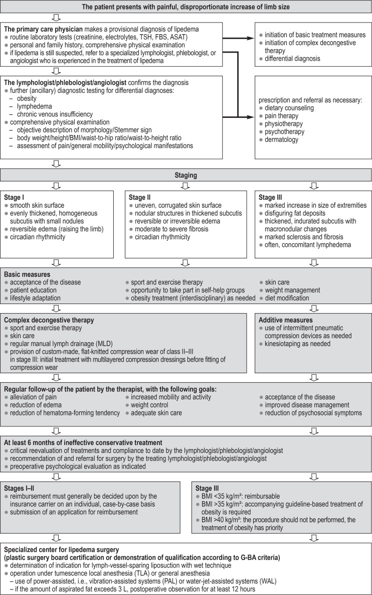
Liposuction is, however, reimbursable as of January 2020 and until 31 December 2024 for patients with stage III lipedema who meet certain further conditions. Six months of prior conservative treatment are a prerequisite, and reimbursement further depends, to a great extent, on the patient’s BMI (figure 3) (e60). Yet the BMI is of only limited utility for deciding on the indication for surgery, particularly in stage III patients who may have advanced fibrotic tissue changes in the involved areas of subcutaneous fat (e61). The patient self-help organizations have complained that these patients are receiving inadequate care (e62).
Figure 3.
Treatment algorithm interdisciplinary treatment of patients with lipedema in Germany
Patients in any stage of the disease whose weight exceeds 120 kg or whose BMI exceeds 32 kg/m² should be treated for obesity in conformity with current guidelines before the potential indication for liposuction is considered (5, 37). Liposuction should be performed with wet technique to spare the lymphatic vessels (33, 38– 40, e63– e65). Patients from whom more than 3 L of pure adipose tissue have been aspirated should remain under qualified postoperative care for at least 12 hours after the procedure. The surgical techniques described in the literature differ from one another in many ways, but it is generally recommended that liposuction should be performed in multiple sittings, rather than a single sitting (40).
Surgical debulking
In highly advanced stages of the disease, with accompanying lymphedema, the involved tissue is so fibrotic that liposuction cannot adequately reduce its volume. In such cases, open surgical debulking (dermato-fibro-lipectomy) may be indicated (e66).
Key Messages.
It is not yet clear whether lipedema should be best defined as a primary lipodystrophy (pathological adipogenesis) or as a primary microangiopathy of small blood and lymphatic vessels. No specific biomarker is yet available.
Its estimated prevalence in the overall female population is 10%. The costs engendered by the treatment of lipedema are difficult to calculate, as it remains unclear what percentage of the affected persons need to be treated.
The diseases is diagnosed on clinical grounds, on the basis of its main manifestations: pain, a feeling of tension, and increased tendency to form hematomas in the affected areas. Ancillary diagnostic testing is recommended mainly to rule out competing diagnoses.
Treatment is symptomatically oriented and based on complex decongestive therapy. Conservative treatment can lessen the painful feeling of tension and pressure, the tendency to form hematomas, and the sequelae of the disease.
If conservative treatment is unsuccessful, lymph-sparing liposuction can be considered as a means of permanently reducing fatty tissue mass. Only low-level evidence supports this procedure to date; the long-term outcome of treatment is to be studied in a prospective interventional trial commissioned by the German Joint Federal Committee (Gemeinsamer Bundesausschuss, G-BA).
Questions for the article in issue 22–23/2020: Lipedema – Pathogenesis, Diagnosis and Treatment Options.
CME credit for this unit can be obtained via cme.aerzteblatt.de until 31. 5. 2021.
Only one answer is possible per question. Please select the answer that is most appropriate.
Question 1
What is the estimated prevalence of lipedema in the female population?
3%
6%
10%
12%
15%
Question 2
Which of the following is a risk factor associated with the development of lipedema?
a carbohydrate-rich diet
smoking
lack of exercise
prolonged standing
positive family history
Question 3
Which of the following is a typical manifestation of lipedema?
a feeling of tension in the affected limb
hypertension
body-mass index >28
ankle-to-arm index <0.75
ecessively warm skin
Question 4
Which of the following features is characteristic of lipedema?
improvement of symptoms over the course of the day
sparing of the hands and feet
insensitivity to pressure
knee arthritis
mild redness of the skin
Question 5
Which of the following is a feature of stage I lipedema?
positive Stemmer sign
irreversible edema
subcuticular induration
a smooth skin surface
concomitant lymphedema
Question 6
What disease should be ruled out by laboratory testing in the differential diagnosis of lipedema?
PCO syndrome
gout
hypothyroidism
lysosoma storage disease
celiac disease
Question 7
Which of the following is a central element of conservative treatment?
Kneipp baths
manual lymph drainage
hypercaloric diet
restricted fluid intake
vibration training
Question 8
Which of the following is a typical finding in stage III lymphedema?
small subuticular nodules
moderate increase in size
skin eruption on the calves
moderate fibrosis
e) disfiguring fat deposits/tissue overhangs
Question 9
What type of sport is especially recommended for persons with lipedema?
aqua-gymnastics
power sports
badminton
rock climbing
beach volleyball
Question 10
What surgical procedure is used to treat severe lipedema?
lymphovenous anastomosis
Roux-en-Y gastric bypass
liposuction
femoropopliteal bypass
debridement
Acknowledgments
Translated from the original German by Ethan Taub, M.D.
Footnotes
Conflict of interest statement
Dr. Klein-Weigel has served as a paid medicolegal expert for the Berlin Social Court (Sozialgericht Berlin) in cases related to the topic of this article.
The other authors state that they have no conflict of interest.
References
- 1.Földi M, Földi E, Strößenreuther R, Kubik S. Urban & Fischer. München, Germany: 2012. Lipedema Földi‘s textbook of lymphology: for physicians and lymphedema therapists 3rd Edition; pp. 364–369. [Google Scholar]
- 2.Meier-Vollrath I, Schneider W, Schmeller W. Lipödem: Verbesserte Lebensqualität durch Therapiekombination. Dtsch Arztebl. 2005;102:A1061–A-1067. [Google Scholar]
- 3.Forner-Cordero I, Szolnoky G, Forner-Cordero A, Kemeny L. Lipedema: an overview of its clinical manifestations, diagnosis and treatment of the disproportional fatty deposition syndrome—systematic review. Clin Obes. 2012;2:86–95. doi: 10.1111/j.1758-8111.2012.00045.x. [DOI] [PubMed] [Google Scholar]
- 4.Peled AW, Kappos EA. Lipedema: diagnostic and management challenges. Int J Womens Health. 2016;8:389–395. doi: 10.2147/IJWH.S106227. [DOI] [PMC free article] [PubMed] [Google Scholar]
- 5.Deutsche Gesellschaft für Phlebologie D. S1-Leitlinie Lipödem. AWMF. 2015 [Google Scholar]
- 6.Buck DW 2nd, Herbst KL. Lipedema: arelatively common disease with extremely common misconceptions. Plast Reconstr Surg Glob Open. 2016;4 doi: 10.1097/GOX.0000000000001043. [DOI] [PMC free article] [PubMed] [Google Scholar]
- 7.Allen EV, Hines EA. Lipedema of the legs: a syndrome characterized by fat legs and orthostatic edema. Proc Staff Mayo Clinic. 1940:184–187. [Google Scholar]
- 8.Bauer AT, von Lukowicz D, Lossagk K, et al. New insights on lipedema: the enigmatic disease of the peripheral fat. Plast Reconstr Surg. 2019;144:1475–1484. doi: 10.1097/PRS.0000000000006280. [DOI] [PubMed] [Google Scholar]
- 9.Fife CE, Maus EA, Carter MJ. Lipedema: a frequently misdiagnosed and misunderstood fatty deposition syndrome. Adv Skin Wound Care. 2010;23:81–92. doi: 10.1097/01.ASW.0000363503.92360.91. quiz 3-4. [DOI] [PubMed] [Google Scholar]
- 10.Langendoen SI, Habbema L, Nijsten TE, Neumann HA. Lipoedema: from clinical presentation to therapy A review of the literature. Br J Dermatol. 2009;161:980–986. doi: 10.1111/j.1365-2133.2009.09413.x. [DOI] [PubMed] [Google Scholar]
- 11.Child AH, Gordon KD, Sharpe P, et al. Lipedema: an inherited condition. Am J Med Genet A. 2010;152a:970–976. doi: 10.1002/ajmg.a.33313. [DOI] [PubMed] [Google Scholar]
- 12.Herbst KL. Rare adipose disorders (RADs) masquerading as obesity. Acta Pharmacol Sin. 2012;33:155–172. doi: 10.1038/aps.2011.153. [DOI] [PMC free article] [PubMed] [Google Scholar]
- 13.Szel E, Kemeny L, Groma G, Szolnoky G. Pathophysiological dilemmas of lipedema. Med Hypotheses. 2014;83:599–606. doi: 10.1016/j.mehy.2014.08.011. [DOI] [PubMed] [Google Scholar]
- 14.Wiedner M, Aghajanzadeh D, Richter DF. Lipedema—basics and current hypothesis of pathomechanism. Handchir Mikrochir Plast Chir. 2018;50:380–385. doi: 10.1055/a-0767-6842. [DOI] [PubMed] [Google Scholar]
- 15.Suga H, Araki J, Aoi N, Kato H, Higashino T, Yoshimura K. Adipose tissue remodeling in lipedema: adipocyte death and concurrent regeneration. J Cutan Pathol. 2009;36:1293–1298. doi: 10.1111/j.1600-0560.2009.01256.x. [DOI] [PubMed] [Google Scholar]
- 16.Priglinger E, Wurzer C, Steffenhagen C, et al. The adipose tissue-derived stromal vascular fraction cells from lipedema patients:aAre they different? Cytotherapy. 2017;19:849–860. doi: 10.1016/j.jcyt.2017.03.073. [DOI] [PubMed] [Google Scholar]
- 17.Földi E, Földi M. Lipedema Foldi’s textbook of lymphology. In: Földi E, Földi M, editors. 2. Munich, Germany Elsevier: 2006. pp. 417–427. [Google Scholar]
- 18.Prantl L, Schreml J, Gehmert S, et al. Transcription profile in sporadic multiple symmetric lipomatosis reveals differential expression at the level of adipose tissue-derived stem cells. Plast Reconstr Surg. 2016;137:1181–1190. doi: 10.1097/PRS.0000000000002013. [DOI] [PubMed] [Google Scholar]
- 19.Bauer AT, von Lukowicz D, Lossagk K, et al. Adipose stem cells from lipedema and control adipose tissue respond differently to adipogenic stimulation in Vitro. Plast Reconstr Surg. 2019;144:623–632. doi: 10.1097/PRS.0000000000005918. [DOI] [PubMed] [Google Scholar]
- 20.Al-Ghadban S, Diaz ZT, Singer HJ, et al. Increase in leptin and PPAR-gamma gene expression in lipedema adipocytes differentiated in vitro from adipose-derived stem cells. Cells. 2020;9 doi: 10.3390/cells9020430. [DOI] [PMC free article] [PubMed] [Google Scholar]
- 21.Amann-Vesti BR, Franzeck UK, Bollinger A. Microlymphatic aneurysms in patients with lipedema. Lymphology. 2001;34:170–175. [PubMed] [Google Scholar]
- 22.Weissleder H, Brauer JW, Schuchhardt C, Herpertz U. Value of functional lymphoscintigraphy and indirect lymphangiography in lipedema syndrome. Z Lymphol. 1995;19:38–41. [PubMed] [Google Scholar]
- 23.AL-Ghadban S, Cromer W, Allen M, et al. Dilated blood and lymphatic microvessels, angiogenesis, increased macrophages, and adipocyte hypertrophy in lipedema thigh skin and fat tissue. J Obes 2019. 2019;10 doi: 10.1155/2019/8747461. [DOI] [PMC free article] [PubMed] [Google Scholar]
- 24.Szolnoky G, Nagy N, Kovacs RK, et al. Complex decongestive physiotherapy decreases capillary fragility in lipedema. Lymphology. 2008;41:161–166. [PubMed] [Google Scholar]
- 25.Torre YS, Wadeea R, Rosas V, Herbst KL. Lipedema: friend and foe. Horm Mol Biol Clin Investig. 2018;33:1–10. doi: 10.1515/hmbci-2017-0076. [DOI] [PMC free article] [PubMed] [Google Scholar]
- 26.Pouwels S, Huisman S, Smelt HJM, Said M, Smulders JF. Lipoedema in patients after bariatric surgery: report of two cases and review of literature. Clin Obes. 2018;8:147–150. doi: 10.1111/cob.12239. [DOI] [PubMed] [Google Scholar]
- 27.Schiltz D, Anker A, Ortner C, et al. Multiple symmetric lipomatosis: new classification system based on the largest german patient cohort. Plast Reconstr Surg Glob Open. 2018;6 doi: 10.1097/GOX.0000000000001722. [DOI] [PMC free article] [PubMed] [Google Scholar]
- 28.Szolnoky G, Borsos B, Barsony K, Balogh M, Kemeny L. Complete decongestive physiotherapy with and without pneumatic compression for treatment of lipedema: a pilot study. Lymphology. 2008;41:40–44. [PubMed] [Google Scholar]
- 29.Szolnoky G, Varga E, Varga M, Tuczai M, Dosa-Racz E, Kemeny L. Lymphedema treatment decreases pain intensity in lipedema. Lymphology. 2011;44:178–182. [PubMed] [Google Scholar]
- 30.Reich-Schupke S, Mohren E, Stucker M. Survey on the diagnostics and therapy of patients with lymphedema and lipedema. Hautarzt. 2018;69:471–477. doi: 10.1007/s00105-018-4151-4. [DOI] [PubMed] [Google Scholar]
- 31.Baumgartner A, Hueppe M, Schmeller W. Long-term benefit of liposuction in patients with lipoedema: a follow-up study after an average of 4 and 8 years. Br J Dermatol. 2016;174:1061–1067. doi: 10.1111/bjd.14289. [DOI] [PubMed] [Google Scholar]
- 32.Schmeller W, Hüppe M, Meier-Vollrath I. Tumescent liposuction in lipoedema yields good long-term results. Br J Dermatol. 2012;166:161–168. doi: 10.1111/j.1365-2133.2011.10566.x. [DOI] [PubMed] [Google Scholar]
- 33.Dadras M, Mallinger P, Corterier C, Theodosiadi S, Ghods M. Liposuction in the treatment of lipedema: alongitudinal study. Arch Plast Surg. 2017;44:324–331. doi: 10.5999/aps.2017.44.4.324. [DOI] [PMC free article] [PubMed] [Google Scholar]
- 34.Rapprich S, Dingler A, Podda M. Liposuction is an effective treatment for lipedema-results of a study with 25 patients. J Dtsch Dermatol Ges. 2011;9:33–40. doi: 10.1111/j.1610-0387.2010.07504.x. [DOI] [PubMed] [Google Scholar]
- 35.Wollina U, Heinig B. Treatment of lipedema by low-volume micro-cannular liposuction in tumescent anesthesia: results in 111 patients. Dermatol Ther. 2019 doi: 10.1111/dth.12820. e12820. [DOI] [PubMed] [Google Scholar]
- 36.Motamedi M, Herold C, Allert S. Kostenübernahmen beim Lipödem - was ist zu beachten? Handchir Mikrochir Plast Chir. 2019;51:139–143. doi: 10.1055/a-0826-4844. [DOI] [PubMed] [Google Scholar]
- 37.Bertsch T, Erbacher G. Lipödem - Mythen und Fakten Teil 3. Phlebologie. 2018;47:188–198. [Google Scholar]
- 38.Stutz JJ, Krahl D. Water jet-assisted liposuction for patients with lipoedema: histologic and immunohistologic analysis of the aspirates of 30 lipoedema patients. Aesthetic Plast Surg. 2009;33:153–162. doi: 10.1007/s00266-008-9214-y. [DOI] [PubMed] [Google Scholar]
- 39.Cornely M, Gensior M. Update Lipödem 2014: Kölner Lipödemstudie. LymphForsch. 2014;18:66–71. [Google Scholar]
- 40.Ghods M, Kruppa P. Surgical treatment of lipoedema. Handchir Mikrochir Plast Chir. 2018;50:400–411. doi: 10.1055/a-0767-6808. [DOI] [PubMed] [Google Scholar]
- E1.Wold LE, Hines EA Jr., Allen EV. Lipedema of the legs; a syndrome characterized by fat legs and edema. Ann Intern Med. 1951;34:1243–1250. doi: 10.7326/0003-4819-34-5-1243. [DOI] [PubMed] [Google Scholar]
- E2.Chen SG, Hsu SD, Chen TM, Wang HJ. Painful fat syndrome in a male patient. Br J Plast Surg. 2004;57:282–286. doi: 10.1016/j.bjps.2003.12.020. [DOI] [PubMed] [Google Scholar]
- E3.Herpertz U. Krankheitsspektrum des Lipödems an einer Lymphologischen Fachklinik - Erscheinungsformen, Mischbilder und Behandlungsmöglichkeiten. Vasomed. 1997;9:301–307. [Google Scholar]
- E4.Forner-Cordero I, Navarro-Monsoliu R, Langa J, Rel-Monzó P. Early or late diagnosis of lymphedema in our lymphedema unit. Eur J Lymphology Relat Probl. 2006;16:19–23. [Google Scholar]
- E5.Schook CC, Mulliken JB, Fishman SJ, Alomari AI, Grant FD, Greene AK. Differential diagnosis of lower extremity enlargement in pediatric patients referred with a diagnosis of lymphedema. Plast Reconstr Surg. 2011;127:1571–1581. doi: 10.1097/PRS.0b013e31820a64f3. [DOI] [PubMed] [Google Scholar]
- E6.Marshall M, Schwahn-Schreiber C. Prävalenz des Lipödems bei berufstätigen Frauen in Deutschland (Lipödem-3-Studie) Phlebologie. 2011;3:127–134. [Google Scholar]
- E7.Fetzer A, Fetzer S. Lipoedema UK big survey 2014 Research report. www.lipoedema.co.uk/wp-content/uploads/2016/04/UK-Big-Surey-version-web.pdf (last accessed on 5 May 2020) [Google Scholar]
- E8.Schubert N, Viethen H. Lipödem und Liplymphödem - Alles eine Frage des Lebensstils? Ergebnisse der ersten deutschlandweiten Online-Umfrage zur Auswirkung auf die Lebensqualität der Betroffenen. Teil 1: Hintergrund, Prävalenz, medizinisch-therapeutisch-fachliche Betreuung. LymphForsch. 2016;20:2–11. [Google Scholar]
- E9.Harwood CA, Bull RH, Evans J, Mortimer PS. Lymphatic and venous function in lipoedema. Br J Dermatol. 1996;134:1–6. [PubMed] [Google Scholar]
- E10.Gregl A. Lipedema. Z Lymphol. 1987;11:41–43. [PubMed] [Google Scholar]
- E11.Lindner A, Marbach F, Tschernitz S, et al. Calcyphosine-like (CAPSL) is regulated in multiple symmetric lipomatosis and is involved in adipogenesis. Sci Rep. 2019;9 doi: 10.1038/s41598-019-44382-1. [DOI] [PMC free article] [PubMed] [Google Scholar]
- E12.Gavin KM, Cooper EE, Hickner RC. Estrogen receptor protein content is different in abdominal than gluteal subcutaneous adipose tissue of overweight-to-obese premenopausal women. Metabolism. 2013;62:1180–1188. doi: 10.1016/j.metabol.2013.02.010. [DOI] [PubMed] [Google Scholar]
- E13.Schneble N, Wetzker R, Wollina U. Lipedema lack of evidence for the involvement of tyrosine kinases. J Biol Regul Homeost Agents. 2016;30:161–163. [PubMed] [Google Scholar]
- E14.Cornely M. Das Lipödem an Armen und Beinen: Teil 1: Pathophysiologie. Phlebologie. 2011;40:21–25. [Google Scholar]
- E15.Kaiserling K. Lehrbuch der Lymphologie. Stuttgart, New York Fischer: 2005. Morphologische Befunde beim Lymphödem, Lipödem, Lipolymphödem In: Földi E, Földi M, Kubik S (eds.): pp. 374–378. [Google Scholar]
- E16.Szolnoky G, Nemes A, Gavaller H, Forster T, Kemeny L. Lipedema is associated with increased aortic stiffness. Lymphology. 2012;45:71–79. [PubMed] [Google Scholar]
- E17.Siems W, Grune T, Voss P, Brenke R. Anti-fibrosclerotic effects of shock wave therapy in lipedema and cellulite. Biofactors. 2005;24:275–282. doi: 10.1002/biof.5520240132. [DOI] [PubMed] [Google Scholar]
- E18.Marsch W. Ist das Lipödem ein lymphologisches Krankheitsbild? Lymphologie. 2001;1:22–24. [Google Scholar]
- E19.Bilancini S, Lucchi M, Tucci S, Eleuteri P. Functional lymphatic alterations in patients suffering from lipedema. Angiology. 1995;46:333–339. doi: 10.1177/000331979504600408. [DOI] [PubMed] [Google Scholar]
- E20.van Geest A, Esten S, Cambier J, et al. Lymphatic disturbances in lipoedema. Phlebologie 2003. 2003:138–142. [Google Scholar]
- E21.Boursier V, Pecking A, Vignes S. Comparative analysis of lymphoscintigraphy between lipedema and lower limb lymphedema. J Mal Vasc. 2004;29:257–261. doi: 10.1016/s0398-0499(04)96770-4. [DOI] [PubMed] [Google Scholar]
- E22.Gould DJ, El-Sabawi B, Goel P, Badash I, Colletti P, Patel KM. Uncovering lymphatic transport abnormalities in patients with primary lipedema. J Reconstr Microsurg. 2020;36:136–141. doi: 10.1055/s-0039-1697904. [DOI] [PubMed] [Google Scholar]
- E23.Brauer W, Brauer V. Altersabhangigkeit des Lymphtransportes beim Lipödem und Lipolymphödem. LymphForsch. 2005;9:6–9. [Google Scholar]
- E24.Beltran K, Herbst KL. Differentiating lipedema and dercum‘s disease. Int J Obes (Lond) 2017;41:240–245. doi: 10.1038/ijo.2016.205. [DOI] [PubMed] [Google Scholar]
- E25.Shin BW, Sim YJ, Jeong HJ, Kim GC. Lipedema, a rare disease. Ann Rehabil Med. 2011;35:922–927. doi: 10.5535/arm.2011.35.6.922. [DOI] [PMC free article] [PubMed] [Google Scholar]
- E26.Dudek JE, Bialaszek W, Ostaszewski P, Smidt T. Depression and appearance-related distress in functioning with lipedema. Psychol Health Med. 2018;23:846–853. doi: 10.1080/13548506.2018.1459750. [DOI] [PubMed] [Google Scholar]
- E27.Bast JH, Ahmed L, Engdahl R. Lipedema in patients after bariatric surgery. Surg Obes Relat Dis. 2016;12:1131–1132. doi: 10.1016/j.soard.2016.04.013. [DOI] [PubMed] [Google Scholar]
- E28.Dudek JE, Bialaszek W, Ostaszewski P. Quality of life in women with lipoedema: a contextual behavioral approach. Qual Life Res. 2016;25:401–408. doi: 10.1007/s11136-015-1080-x. [DOI] [PubMed] [Google Scholar]
- E29.Wollina U, Heinig B. Differenzialdiagnostik von Lipödem und Lymphödem. Der Hautarzt. 2018;69:1039–1047. doi: 10.1007/s00105-018-4304-5. [DOI] [PubMed] [Google Scholar]
- E30.Schmeller W, Baumgartner A. Schmerzen beim Lipödem - Versuch einer Annäherung. LymphForsch. 2008;12:8–12. [Google Scholar]
- E31.Streeten DH. Idiopathic edema. Pathogenesis, clinical features, and treatment. Endocrinol Metab Clin North Am. 1995;24:531–547. [PubMed] [Google Scholar]
- E32.Szolnoky G. Bettany-Saltikov J, Paz-Lourido B. Lipedema Physical Therapy Perspectives in the 21st Century: Challenges and Possibilities. BoD - Books on Demand. 2012 [Google Scholar]
- E33.Szolnoky G. Differential Diagnosis: Lipedema In: Lee B-B, Rockson SG, Bergan J, (eds.): Lymphedema: a concise compendium of theory and practice. Cham: Springer International Publishing. 2018:239–249. [Google Scholar]
- E34.Coppel T. UK Best Practice Guidelines: The management of lipoedema. London: Wounds UK. 2017 [Google Scholar]
- E35.Halk A, Damstra R. First dutch guidelines on lipedema using the international classification of functioning, disability and health. Phlebology. 2017;32:152–159. doi: 10.1177/0268355516639421. [DOI] [PubMed] [Google Scholar]
- E36.Breu FX, Marshall M. Neue Ergebnisse der duplexsonographischen Diagnostik des Lip- und Lymphödems. Phlebologie. 2000;29:124–128. [Google Scholar]
- E37.Naouri M, Samimi M, Atlan M, et al. High-resolution cutaneous ultrasonography to differentiate lipoedema from lymphoedema. Br J Dermatol. 2010;163:296–301. doi: 10.1111/j.1365-2133.2010.09810.x. [DOI] [PubMed] [Google Scholar]
- E38.Iker E, Mayfield CK, Gould DJ, Patel KM. Characterizing lower extremity lymphedema and lipedema with cutaneous ultrasonography and an objective computer-assisted measurement of dermal echogenicity. Lymphat Res Biol. 2019;17:525–530. doi: 10.1089/lrb.2017.0090. [DOI] [PubMed] [Google Scholar]
- E39.Monnin-Delhom ED, Gallix BP, Achard C, Bruel JM, Janbon C. High resolution unenhanced computed tomography in patients with swollen legs. Lymphology. 2002;35:121–128. [PubMed] [Google Scholar]
- E40.Vaughan BF. CT of swollen legs. Clin Radiol. 1990;41:24–30. doi: 10.1016/s0009-9260(05)80927-4. [DOI] [PubMed] [Google Scholar]
- E41.Dimakakos PB, Stefanopoulos T, Antoniades P, Antoniou A, Gouliamos A, Rizos D. MRI and ultrasonographic findings in the investigation of lymphedema and lipedema. Int Surg. 1997;82:411–416. [PubMed] [Google Scholar]
- E42.Duewell S, Hagspiel KD, Zuber J, von Schulthess GK, Bollinger A, Fuchs WA. Swollen lower extremity: role of MR imaging. Radiology. 1992;184:227–231. doi: 10.1148/radiology.184.1.1609085. [DOI] [PubMed] [Google Scholar]
- E43.Partsch H, Stoberl C, Urbanek A, Wenzel-Hora BI. Clinical use of indirect lymphography in different forms of leg edema. Lymphology. 1988;21:152–160. [PubMed] [Google Scholar]
- E44.Tiedjen K-U, Schultz-Ehrenburg U. Berlin, Heidelberg Springer Berlin Heidelberg: 1985. Isotopenlymphographische Befunde beim Lipödem; pp. 432–438. [Google Scholar]
- E45.Bollinger A, Amann-Vesti BR. Fluorescence microlymphography: diagnostic potential in lymphedema and basis for the measurement of lymphatic pressure and flow velocity. Lymphology. 2007;40:52–62. [PubMed] [Google Scholar]
- E46.Brauer WJ, Weissleder H. Methodik und Ergebnisse der Funktionslymphszintigraphie: Erfahrungen bei 924 Patienten. Phlebologie. 2002;31:118–125. [Google Scholar]
- E47.Lohrmann C, Foeldi E, Langer M. MR imaging of the lymphatic system in patients with lipedema and lipo-lymphedema. Microvasc Res. 2009;77:335–339. doi: 10.1016/j.mvr.2009.01.005. [DOI] [PubMed] [Google Scholar]
- E48.Dietzel R, Reisshauer A, Jahr S, Calafiore D, Armbrecht G. Body composition in lipoedema of the legs using dual-energy X-ray absorptiometry: a case-control study. Br J Dermatol. 2015;173:594–596. doi: 10.1111/bjd.13697. [DOI] [PubMed] [Google Scholar]
- E49.Crescenzi R, Donahue PMC, Weakley S, Garza M, Donahue MJ, Herbst KL. Lipedema and dercum‘s disease: a new application of bioimpedance. Lymphat Res Biol. 2019;17:671–679. doi: 10.1089/lrb.2019.0011. [DOI] [PMC free article] [PubMed] [Google Scholar]
- E50.Deri G, Weissleder H. Vergleichende prä- und posttherapeutische Volumenmessungen in Beinsegmenten beim Lipödem. Lymph Forsch. 1997;1:35–37. [Google Scholar]
- E51.Warren AG, Janz BA, Borud LJ, Slavin SA. Evaluation and management of the fat leg syndrome. Plast Reconstr Surg. 2007;119:9e–15e. doi: 10.1097/01.prs.0000244909.82805.dc. [DOI] [PubMed] [Google Scholar]
- E52.Faerber G. Ernährungstherapie bei Lipödem und Adipositas - Ergebnisse eines leitliniengerechten Therapiekonzepts. Vasomed. 2017;29:176–177. [Google Scholar]
- E53.Li W, Li V, Hutnik M, Chiou A. Tumor angiogenesis as a target for dietary cancer prevention. J Oncol. 2012;2012 doi: 10.1155/2012/879623. 879623. [DOI] [PMC free article] [PubMed] [Google Scholar]
- E54.Coetzee O, Filatov D. Lipidema and lymphedema: the “leaky lymph,” weight loss resistance and the intestinal permeability connection. EC Nutrition. 2017;11:233–243. [Google Scholar]
- E55.Ehrlich C, Iker E, Herbst K, et al. Lymphedema and lipedema nutrition guide: foods, vitamins, minerals, and supplements. San Francisco, USA: Lymph Notes. 2016 [Google Scholar]
- E56.Burger R, Jung M, Becker J, et al. Wirkung von Aqua-Cycling als Bewegungstherapie bei der Diagnose Lipödem. Phlebologie. 2019;48:182–186. [Google Scholar]
- E57.Peled AW, Slavin SA, Brorson H. Long-term outcome after surgical treatment of lipedema. Ann Plast Surg. 2012;68:303–307. doi: 10.1097/SAP.0b013e318215791e. [DOI] [PubMed] [Google Scholar]
- E58.Cobos L, Herbst KL, Ussery C. Liposuction for Lipedema (Persistent Fat) in the US Improves Quality of Life. J Endocr Soc. 2019;3(Suppl 1) MON-116. [Google Scholar]
- E59.G-BA. Erprobungsstudie soll offene Frage des Nutzens der Liposuktion bei Lipödem beantworten: G-BA beauftragt wissenschaftliche Institution mit Studienbegleitung. www.g-ba.de/downloads/34-215-795/12_2019-04-18_Vergabe%20uwI_Liposuktion.pdf (last accessed on 1 August 2019) [Google Scholar]
- E60.G-BA. Methodenbewertung: Liposuktion wird befristet Kassenleistung bei Lipödem im Stadium III. www.g-ba.de/presse/pressemitteilungen/811/ (last accessed on 5 May 2020) [Google Scholar]
- E61.Herpertz U. Adipositas-Diagnostik in der Lymphologie - Warum der BMI bei Ödemen unsinnig sein kann! LymphForsch. 2009;13:34–7. [Google Scholar]
- E62.Lütz D. Gemeinsame Presseerklärung der organisierten Selbsthilfe von Frauen mit Lipödem vom 20.9.2019. www.lipoedem-fakten.de/app/download/6021579766/Gemeinsame+Presseerkl%C3%A4rung+Lip%C3%B6dembetroffener+vom+20.09.2019.pdf?t=1579150244 (last accessed on 22 March 2020) [Google Scholar]
- E63.Rapprich S, Baum S, Kaak I, Kottmann T, Podda M. Therapie des Lipödems mittels Liposuktion im Rahmen eines umfassenden Behandlungskonzeptes - Ergebnisse eigener Studien. Phlebologie. 2015;44:121–132. [Google Scholar]
- E64.Heck FC. Liposuktion beim Lipödem in WAL-Technik - Kreislaufstörungen sind kein Problem für die ambulante Vorgehensweise Auswertung von 326 Liposuktionen. Akt Dermatol. 2015;41 [Google Scholar]
- E65.Baumgartner A, Frambach Y. Liposuction and lipoedema. Phlebologie. 2016;45:47–53. [Google Scholar]
- E66.Wollina U, Heinig B, Schonlebe J, Nowak A. Debulking surgery for elephantiasis nostras with large ectatic podoplanin-negative lymphatic vessels in patients with lipo-lymphedema. Eplasty. 2014;14 [PMC free article] [PubMed] [Google Scholar]


