Abstract
Background: VIM (Verona Integron-encoded Metallo-beta-lactamase) is a member of the Metallo-Beta-Lactamases (MBLs), and is able to hydrolyze all beta-lactams antibiotics, except for monobactams, and including carbapenems. Here we characterize a VIM-producing IncA plasmid isolated from a clinical ST69 Escherichia coli strain from an Italian Long-Term Care Facility (LTCF) inpatient. Methods: An antimicrobial susceptibility test and conjugation assay were carried out, and the transferability of the blaVIM-type gene was confirmed in the transconjugant. Whole-genome sequencing (WGS) of the strain 550 was performed using the Sequel I platform. Genome assembly was performed using “Microbial Assembly”. Genomic analysis was conducted by uploading the contigs to ResFinder and PlasmidFinder databases. Results: Assembly resulted in three complete circular contigs: the chromosome (4,962,700 bp), an IncA plasmid (p550_IncA_VIM_1; 162,608 bp), harboring genes coding for aminoglycoside resistance (aac(6′)-Ib4, ant(3″)-Ia, aph(3″)-Ib, aph(3′)-XV, aph(6)-Id), beta-lactam resistance (blaSHV-12, blaVIM-1), macrolides resistance (mph(A)), phenicol resistance (catB2), quinolones resistance (qnrS1), sulphonamide resistance (sul1, sul2), and trimethoprim resistance (dfrA14), and an IncK/Z plasmid (p550_IncB_O_K_Z; 100,306 bp), free of antibiotic resistance genes. Conclusions: The increase in reports of IncA plasmids bearing different antimicrobial resistance genes highlights the overall important role of IncA plasmids in disseminating carbapenemase genes, with a preference for the blaVIM-1 gene in Italy.
Keywords: E. coli, blaVIM-1, IncA
1. Introduction
VIM (Verona Integron-encoded Metallo-beta-lactamase) is a member of the Metallo-beta-Lactamases (MBLs), and is able to hydrolyze all beta-lactams antibiotics, except for monobactams, but including carbapenems. The VIM family was first detected and described in Italy from a Pseudomonas aeruginosa (P. aeruginosa) strain isolated in 1997 [1]; to date, 69 different enzyme variants have been described and grouped in sub-lineages: VIM-1 like, VIM-2 like, and VIM-7 like [2]. Among MBLs, VIM enzymes are commonly detected in European/Mediterranean countries such as Italy and Greece. In particular, VIM-1 and VIM-4 are geographically predominant in Europe, VIM-2 is globally distributed in P. aeruginosa, VIM-3 is widespread in Taiwan, VIM-6 in Asia, and VIM-7 in USA [3]. The blaVIM-type genes are often inserted within class 1 integrons and located on plasmids playing a key role in the interspecies distribution of such determinants [4]. According to Matsumura et al. (2017), more than 20 different VIM-harboring integrons have been described [5]. Geographically, In87, In624, and In916 are spread mostly in European countries and associated with blaVIM-1; In339 is globally distributed and harbors blaVIM-2; In450 in Taiwan carrying blaVIM-3; and In496, harboring blaVIM-6, circulates across the Asian continent. Moreover, In916 has been identified in Escherichia coli (E. coli), Klebsiella pneumoniae (K. pneumoniae), Enterobacter cloacae (E. cloacae), Klebsiella aerogenes (K. aerogenes), and Klebsiella oxytoca (K. oxytoca); In339 has been reported in P. aeruginosa and Acinetobacter baumannii (A. baumannii); and In496 in P. aeruginosa and Pseudomonas putida (P. putida) [5]. The blaVIM genes are mainly reported in IncN [6,7], IncY [8], IncR [9], and IncA type plasmids [10]. The IncA group, once assigned to the IncA/C family, was later allocated to a separate incompatibility group, not yet well characterized and recently recognized as an alternative reservoir of carbapenemase genes in the Enterobacterales family [11,12]. Here we characterize a VIM-producing IncA plasmid isolated from a clinical ST69 E. coli strain from an Italian Long-Term Care Facility (LTCF) inpatient.
2. Materials and Methods
2.1. Case Presentation, Antimicrobial Susceptibility Test and Molecular Investigations
On the 2nd of May 2018, a rectal swab was collected from a female patient, resident in the GP1 ward of the rehabilitation center “Giovanni Paolo II” (Milan, Italy). The swab, collected as part of a carbapenemase passive surveillance screening, was sent to the Clinical Microbiology Laboratory of the ASP “Golgi-Redaelli” of Milan for initial phenotypic characterization. Preliminary investigation, through Vitek-2 (bioMérieux, Marcy-l’Étoile, France) and a phenotypic synergy test, revealed the presence of a carbapenemase-producing E. coli strain. The isolate was sent to the Microbiology Laboratory of University of Pavia and to the “Biomedical Center” in Plzen, Czech Republic, for further molecular investigation. The species identification was confirmed through MALDI-TOF MS (Matrix-Assisted Laser Desorption Ionization-Time of Flight Mass Spectrometry) using MALDI Biotyper software (Brucker Daltonics, Bremen, Germany). Carbapenemase production was confirmed by meropenem hydrolysis assay [13], and antimicrobial susceptibility profiles were obtained by Microscan AutoScan-4 (Beckman-Coulter, Brea, CA, USA) and interpreted in accordance with EUCAST 2020 clinical breakpoints (https://www.eucast.org/fileadmin/src/media/PDFs/EUCAST_files/Breakpoint_tables/v_10.0_Breakpoint_Tables.pdf). Fosfomycin, colistin, and meropenem minimum inhibitory concentrations (MICs) were confirmed through broth-microdilution. Production of class B, D, and A carbapenemases was evaluated using disk combination synergy tests with meropenem and EDTA, temocillin, and phenylboronic acid, as inhibitors [14,15,16], respectively. The presence of carbapenemase genes was confirmed by polymerase chain reaction (PCR) as described by Shirani et al. 2016 [17]. Phylogenetic groups were determined by a two-step triplex PCR as described by Clermont et al. 2000 [18].
2.2. Conjugation Assay
To test the transferability of the blaVIM-type gene, conjugation experiments were performed in Mueller Hilton (MH) broth (OXOID, Basingstoke, UK) using E. coli A15rRif as the recipient. Transconjugant selection was assessed on MH agar (OXOID, Basingstoke, UK) plates supplemented with rifampicin (100 mg/μL) (Sigma-Aldrich, St. Louis, MO, USA) and ampicillin (50 mg/μL) (Sigma-Aldrich, St. Louis, MO, USA). The presence of the blaVIM-type gene in the transconjugants was confirmed through PCR.
2.3. Whole-Genome Sequencing (WGS)
The E. coli strain designated as 550 was subjected to whole genome DNA extraction using the NucleoSpin Microbial DNA kit (Macherey-Nagel, Duren, Germany). The obtained DNA was sheared using Megaruptor 2 using the Hydropore-long (Diagenode). Library preparation of the sheared DNA was performed in accordance with the manufacturer’s recommendation for microbial multiplexing for the Express kit 2.0 (Pacific Biosciences, Menlo Park, CA, USA). No size selection was performed during library preparation. The constructed library was sequenced using long-reads sequencing technology on Sequel I (Pacific Biosciences, Menlo Park, CA, USA).
2.4. Whole-Genome-Sequencing-Data Analysis
Genome assembly was performed with minimum seed coverage of 30×, using the “Microbial Assembly” pipeline offered by “SMRT Link v8.0”. Antibiotic resistance genes, plasmid replicons, virulence factors, serotype, FimH type, FumC type, MLST, pMLST, and phage detection was obtained through uploading assembled contigs to ResFinder (https://cge.cbs.dtu.dk/services/ResFinder/) [19], PlasmidFinder (https://cge.cbs.dtu.dk/services/PlasmidFinder/) [20], VirulenceFinder (https://cge.cbs.dtu.dk/services/VirulenceFinder/) [21], VFDB: Virulence factors database (http://www.mgc.ac.cn/VFs/) [22], SeroTypeFinder 2.0 (https://cge.cbs.dtu.dk/services/SerotypeFinder/) [23], FimTyper 1.0 (https://cge.cbs.dtu.dk/services/FimTyper/) [24], CHTyper 1.0 (https://cge.cbs.dtu.dk/services/chtyper/) [25], MLST 2.0 (https://cge.cbs.dtu.dk/services/MLST/) [26], pMLST 2.0 (https://cge.cbs.dtu.dk/services/pMLST/) [20], and Phaster (https://phaster.ca) [27]. The genome was annotated by the NCBI Prokaryotic Genome Annotation Pipeline (PGAP). Plasmid comparisons were assessed through the Blast Ring Image Generator (BRIG) application (http://brig.sourceforge.net) and EasyFig (https://mjsull.github.io/Easyfig/) [28].
2.5. Nucleotide Accession Numbers
The chromosome sequence of 550 and the plasmid sequences of p550_IncA_VIM_1 and p550_IncB_O_K_Z were deposited in GenBank under the accession numbers CP058223, CP058224, and CP058225, respectively.
3. Results
The strain showed a multi-drug resistance (MDR) profile, expressing resistance against ampicillin, piperacillin, 3rd and 4th generation cephalosporins, ertapenem, gentamycin, tobramycin, and trimethoprim-sulfamethoxazole, yet remained susceptible to amikacin, colistin, fosfomycin, meropenem, imipenem, ciprofloxacin, and tigecycline (Table 1). The E. coli 550 strain was identified as an MBL producer by disk combination synergy test, and as blaVIM-1 positive through PCR and sequencing. A conjugation assay confirmed the transferability of the plasmid carrying the carbapenemase gene (Table 1).
Table 1.
Antimicrobial susceptibility profile of E. coli A15, E. coli 550, and the transconjugant A15*E. coli 550 MIC (minimum inhibitory concentration, mg/L).
| AMP | AMS | ATM | CTX | FEP | CAZ | CIP | CO | FOS | ETP | MP | IMP | CN | TOB | TGC | TMX | |
|---|---|---|---|---|---|---|---|---|---|---|---|---|---|---|---|---|
| E. coli A15 | 2 S | 2 S | ≤0.125 S | ≤0.068 S | ≤0.125 S | ≤0.25 S | ≤0.068 S | ≤0.5 S | ≤0.125 S | ≤0.032 S | ≤0.125 S | ≤0.125 S | ≤0.25 S | ≤0.125 S | ≤0.125 S | ≤0.068 S |
| E. coli 550 | >128 R | >128 R | 16 R | >8 R | 16 R | >16 R | 0.25 S | 0.25 S | 8 S | 1 R | 1 S | 2 S | 1 S | 4 R | 0.25 S | >4 R |
|
A15*
E. coli 550 |
128 R | 128 R | 16 R | >8 R | 8 R | >16 R | 0.25 S | 0.25 S | 8 S | 0.38 S | 1 S | 2 S | 0.5 S | 2 S | 0.12 5S | >4 R |
MIC, minimum inhibitory concentration: ampicillin; AMP, ampicillin/sulbactam; AMS, aztreonam; ATM, cefotaxime; CTX, cefepime; FEP, ceftazidime; CAZ, ciprofloxacin; CIP, colistin; CO, fosfomycin; FOS, ertapenem; ETP, meropenem; MP, imipenem; IMP, gentamycin; CN, tobramycin; TO, tigecycline; TIG; TMX, trimethoprim/sulfamethoxazole. R-resistant, S-susceptible.
Whole-genome sequencing revealed the presence of three contigs: a complete circular chromosome with a size of 4,962,700 bp, and two complete circular contigs corresponding to two different plasmids of 162,608 bp and 100,306 bp in size. The isolate belonged to the serotype O15:H18, uropathogenic sequence type ST69 (Achtman scheme), phylogenetic group D and CH-Type FumC35/FimH27 (fimbrial adhesion gene fimH with allele 27 and fumarate hydratase class II gene fumC with allele 35). Moreover, the strain’s chromosome harbored several virulence genes involved in: adhesion (hemorragic E. coli pilus, EaeH, Type I fimbriae), autotransporter (ag43: autoaggregation and flocculation of E. coli cells in static cultures [29], cah: carbonic anhydrase, air: enteroaggregative immunoglobulin repeat protein, vat: vacuolating autotransporter gene which contributes to uropathogenic E. coli (UPEC) fitness during systemic infection, invasion (ibeB and ibeC facilitates invasion of brain endothelial cells), iron uptake (chu: Hemin uptake, sit: Iron/manganese transport and Yersiniabactin siderophore), secretion (Type III secretion system), and toxin production (clyA: expressing the pore-forming hemolytic and cytotoxic cytolysin A). Additionally, the chromosome harbored an antibiotic resistance gene (mdf(A)) coding for macrolides resistance and two phages free of antibiotic resistance genes (Table 2).
Table 2.
Antibiotic resistance genes and virulence determinants detected on the chromosome and the plasmid of the isolate 550.
| Position | Antibiotic Resistance | Adhesion | Autotransporter | Invasion | Iron Uptake | Secretion | Toxin |
|---|---|---|---|---|---|---|---|
| Chromosome | mdf(A) | Pilus, EaeH, Type I fimbriae | ag43, cah, air, vat | ibeB, ibeC | chu, sit | Type III Secretion System | clyA |
| p550_IncA_VIM_1 | aac(6′)-lb4, ant(3″)-Ia, aph(3″)-lb, aph(3′)-XV, aph(6)-ld, bla SHV-12 , bla VIM-1 , mph(A), catB2, qnrS1, sul1, sul2, dfrA14 |
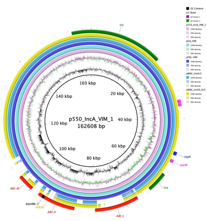
The second contig belonged to an IncA plasmid (p550_IncA_VIM_1: 162,608 bp) and pMLST IncA/C 12. The plasmid harbored genes coding for aminoglycoside resistance (aac(6’)-Ib4, ant(3″)-Ia, aph(3″)-Ib, aph(3’)-XV, aph(6)-Id), β-lactam resistance (blaSHV-12, blaVIM-1), macrolides resistance (mph(A)), phenicol resistance (catB2), quinolones resistance (qnrS1), sulphonamide resistance (sul1, sul2), and trimethoprim resistance (dfrA14) genes. p550_IncA_VIM_1 shared high identity scores with the pGA_VIM plasmid (182,016 bp) collected from a clinical ST12 E. coli strain in Italy (MN783743.2 [10]; 100% sequence and identity), pFDL-VIM plasmid (164,903 bp) isolated from a clinical K. oxytoca in Italy (MN783744.1 [10]; sequence coverage 100%, sequence identity 99.99%), pIBAC_IncA/C plasmid (145,294 bp) isolated from a clinical Citrobacter freundii (C. freundii) isolated in Italy (MH594477.1 [11]; 84% sequence coverage, 99.99% sequence identity), and pIBAC_Incx3_A/C plasmid (192,802 bp) isolated from a clinical C. freundii in Italy (MH594478.1 [11]; 87% sequence coverage and 100% sequence identity) (Figure 1). p550_IncA_VIM_1 backbone harbored conjugal transfer genes (tra), a replication initiation gene (repA), maintenance and stability genes (parA, parM), genes coding for mercuric uptake system (mer), and toxin/antitoxin system (HigB/HipA). Moreover, the plasmid contained three antimicrobial resistance islands (ARIs); ARI-I (12,914 bp) flanked by IS26 on both ends in opposite orientation and harbored sul-2, aph(6)-Id and qnrS1 genes. When blasted, ARI-I showed perfect identity with pKC-BO-N1-VIM found in a Kluyvera cryocrescens collected from a rectal swab from Italy (MG228427.1; query cover 100%, identity 100%). ARI-II (11,263 bp), flanked by an IS4321R and an IntI1 in the same orientation, and harbored an In916 integron with a cassette containing sul2, catB2, ant(3″)-Ia, aph(3′)-XV, aac(6′)-Ib4 and blaVIM-1 genes. When blasted, the region shared high identity score with pKC-BO-N1-VIM (MG228427.1; query cover 100%, identity 100%). Moreover, a mercury uptake system region separated the two ARIs. ARI-III, of 4895 bp, contained just the blaSHV-12 gene, flanked by an IS6-like element and an IS26 in opposite orientation. This region revealed high identities with pFDL-VIM found in K. oxytoca from Italy (MN783744.1; query cover 100%, identity 100%).
Figure 1.
Circular map of p550_IncA_VIM_1 against pGA_VIM (pink), pFDL-VIM (turquoise), pIBAC_IncA/C (violet), and pIBAC_Incx3_A/C (yellow). At the outer curved segments; red, yellow, black, green, purple and blue corresponds to ARIs, In916, blaVIM-1, tra region, maintenance and stability region, and repA.
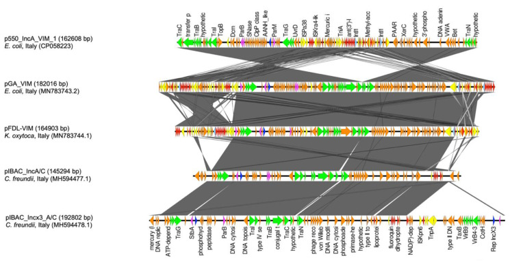
p550_IncA_VIM_1 shared most of the IncA backbone with pGA_VIM, pFDL-VIM, pIBAC_IncA/C, and pIBAC_Incx3_A/C. Nevertheless, regarding ARIs, p550_IncA_VIM_1 and pGA_VIM shared most of the region; both plasmids shared the ARI-I region but in the opposite direction. Conversely, pGA_VIM harbored two copies of ARI-II while sharing one copy with p550_IncA_VIM_1 but in the opposite orientation. Moreover, the ARI-III region was also shared by both plasmids in opposite directions except for the IS26. In pGA_VIM, the “mercury system region” directly flanked the first copy of the ARI-II region, whereas in p550_IncA_VIM_1 it separated ARI-I and ARI-II, maintaining the same orientation. The orientation of the mercury system region and the opposite orientation of ARI-II in p550_IncA_VIM_1 suggests a recombination event responsible for the genetic restructuring of this region (Figure 2).
Figure 2.
Genetic linear map of p550_IncA_VIM_1, pGA_VIM, pFDL-VIM, pIBAC_IncA/C, and pIBAC_Incx3_A/C. Replicons, partitioning genes, mobile elements, conjugal transfer genes, antibiotic resistance, and other remaining genes are designated by blue, purple, yellow, green, red, and orange, respectively. Gray shaded area shows nucleotide similarity.
The third contig belonged to an IncK/Z plasmid (p550_Inc_B_O_K_Z; 100306 bp) and did not harbor any antibiotic resistance gene.
4. Discussion
Here, we report the first detection of a VIM-producing ST69 E. coli strain in Italy. E. coli ST69 is a member of the extraintestinal pathogenic E. coli (ExPEC) group, mostly involved in urinary tract infections (UTIs). It was first detected in 1999, in a study conducted in California, among 255 E. coli collected from urine of women with UTIs [30]. The ST69 lineage is part of the phylogenetic group D and belongs to the clonal group A (CGA) [31]. This group reported several serotypes such as O11, O15, O86, O125ab, and O25b [32]. ST69 has been associated with high virulence and pathogenicity, due to several virulence genes content coding for adhesins, toxins, autotransporters, and siderophores [33]. It has often been associated with trimethoprim-sulphametoxazole resistance and to the expression of CTX-M and TEM type enzymes. Recently, sporadic cases of ST69 E. coli, expressing several hydrolyzing enzymes, such as KPC-3 [34], NDM-1 [35] and mcr-1, have been reported [36]. Moreover, Fibke et al. identified a possible link between acquisition/infection with ST69 and travel histories. Additionally, the consumption of high-risk foods such as raw meat or vegetables, undercooked eggs, and seafood could play a role in the acquisition/infection with ST69 [37]. These data support the definition of ST69 as a high-risk clone, with increased ability for antimicrobial resistance genes acquisition. Moreover, the presence of type 1 fimbriae (fimH27), has been correlated with persisting colonization and bacteremia in patients [38].
VIM-type enzymes are widely detected in Italy and in Europe [39,40], harbored predominantly on IncN plasmids [6,7]. The expression of a VIM-1 enzyme by a high-risk ST69 clone, can represent a challenge, limiting the therapeutic options. In particular, the spread of VIM enzymes reduces the efficacy of the recently introduced therapeutic options such as ceftazidime-avibactam (CAZ-AVI). According to Arcari et al. the rise of MBL-producer circulation could be facilitated by extending the CAZ-AVI usage for treatment of KPC-producing-strains [10].
IncA plasmids are highly conjugative plasmids which are not yet well characterized. Recently, studies have suggested that IncA plasmids could act as carbapenemase reservoirs in different Enterobacterales species [6,11,41]. Few reports of complete closed IncA plasmids have been reported in the NCBI database. Nevertheless, from the plasmids discussed in Figure 2, IncA plasmids maintained a relatively stable backbone. The aforementioned ability could be explained by the presence of different integrative hotspot regions in the IncA plasmid backbone, particularly IS26, IS6, and IntI1 elements, as described by Johnson et al. 2012 [42].
In conclusion, the high identity with other IncA plasmids highlights the predisposition of the IncA group to acquire several antimicrobial resistance genes. These data emphasize the overall important role of IncA plasmids in disseminating carbapenemase genes and, in particular, the blaVIM-1 in Italy [10]. The ability of these plasmids to accumulate different antibiotic resistant determinants in a high-risk clone, poses a health threat that might be difficult to control.
Author Contributions
I.B., V.M.M. and R.M. played an important role in interpreting the results and writing the manuscript. A.P., A.M., E.F., J.H. and R.M. helped to acquire data. I.B. and V.M.M. carried out experimental work. I.B. supervised the experiments and revised the final manuscript, which was approved by all authors. All authors have read and agreed to the final version of the manuscript.
Funding
The study was supported by the research project grant 17-29239A provided by Czech Health Research Council, by the Charles University Research Fund PROGRES (project number Q39), by the National Sustainability Program I (NPU I) Nr. LO1503 and by the project Nr. CZ.02.1.01/0.0/0.0/16_019/0000787 “Fighting Infectious Diseases”, provided by the Ministry of Education Youth and Sports of the Czech Republic. The study was also supported by “Fondo Ricerca & Giovani 2018”, University of Pavia, Italy.
Conflicts of Interest
The authors declare no conflict of interest. The funders had no role in the design of the study; in the collection, analyses, or interpretation of data; in the writing of the manuscript, or in the decision to publish the results.
References
- 1.Lauretti L., Riccio M.L., Mazzariol A., Cornaglia G., Amicosante G., Fontana R., Rossolini G.M. Cloning and characterization of blaVIM, a new integron-borne metallo-beta-lactamase gene from a Pseudomonas aeruginosa clinical isolate. Antimicrob. Agents Chemother. 1999;43:1584–1590. doi: 10.1128/AAC.43.7.1584. [DOI] [PMC free article] [PubMed] [Google Scholar]
- 2.Mojica M.F., Bonomo R.A., Fast W. B1-Metallo-β-Lactamases: Where Do We Stand? Curr. Drug Targets. 2016;17:1029–1050. doi: 10.2174/1389450116666151001105622. [DOI] [PMC free article] [PubMed] [Google Scholar]
- 3.Hong D.J., Bae I.K., Jang I.H., Jeong S.H., Kang H.K., Lee K. Epidemiology and Characteristics of Metallo-β-Lactamase-Producing Pseudomonas aeruginosa. Infect. Chemother. 2015;47:81–97. doi: 10.3947/ic.2015.47.2.81. [DOI] [PMC free article] [PubMed] [Google Scholar]
- 4.Mathers A.J., Peirano G., Pitout J.D. The role of epidemic resistance plasmids and international high-risk clones in the spread of multidrug-resistant Enterobact. Clin. Microbiol. Rev. 2015;28:565–591. doi: 10.1128/CMR.00116-14. [DOI] [PMC free article] [PubMed] [Google Scholar]
- 5.Matsumura Y., Peirano G., Devinney R., Bradford P.A., Motyl M.R., Adams M.D., Chen L., Kreiswirth B., Johann D., Pitout D. Genomic epidemiology of global VIM-producing Enterobact. J. Antimicrob. Chemother. 2017;72:2249–2258. doi: 10.1093/jac/dkx148. [DOI] [PMC free article] [PubMed] [Google Scholar]
- 6.Carattoli A., Aschbacher R., March A., Larcher C., Livermore D.M., Woodford N. Complete nucleotide sequence of the IncN plasmid pKOX105 encoding VIM-1, QnrS1 and SHV-12 proteins in Enterobacteriaceae from Bolzano, Italy compared with IncN plasmids encoding KPC enzymes in the USA. J. Antimicrob. Chemother. 2010;65:2070–2075. doi: 10.1093/jac/dkq269. [DOI] [PubMed] [Google Scholar]
- 7.Miriagou V., Papagiannitsis C.C., Kotsakis S.D., Loli A., Tzelepi E., Legakis N.J., Tzouvelekis L.S. Sequence of pNL194, a 79.3-kilobase IncN plasmid carrying the blaVIM-1 metallo-beta-lactamase gene in Klebsiella pneumoniae. Antimicrob. Agents Chemother. 2010;54:4497–4502. doi: 10.1128/AAC.00665-10. [DOI] [PMC free article] [PubMed] [Google Scholar]
- 8.Roschanski N., Guenther S., Vu T.T.T., Fischer J., Semmler T., Huehn S., Alter T., Roesler U. VIM-1 carbapenemase-producing Escherichia coli isolated from retail seafood, Germany 2016. Euro. Surveill. 2017;22:17-00032. doi: 10.2807/1560-7917.ES.2017.22.43.17-00032. [DOI] [PMC free article] [PubMed] [Google Scholar]
- 9.Drieux L., Decré D., Frangeul L., Arlet G., Jarlier V., Sougakoff W. Complete nucleotide sequence of the large conjugative pTC2 multireplicon plasmid encoding the VIM-1 metallo-β-lactamase. J. Antimicrob. Chemother. 2013;68:97–100. doi: 10.1093/jac/dks367. [DOI] [PubMed] [Google Scholar]
- 10.Arcari G., Di Lella F.M., Bibbolino G., Mengoni F., Beccaccioli M., Antonelli G., Faino L., Carattoli A. A Multispecies Cluster of VIM-1 Carbapenemase-Producing Enterobacterales Linked by a Novel, Highly Conjugative, and Broad-Host-Range IncA Plasmid Forebodes the Reemergence of VIM-1. Antimicrob. Agents Chemother. 2020;64:e02435-19. doi: 10.1128/AAC.02435-19. [DOI] [PMC free article] [PubMed] [Google Scholar]
- 11.Bitar I., Caltagirone M., Villa L., Mattioni M.V., Nucleo E., Sarti M., Migliavacca R., Carattoli A. Interplay among IncA and blaKPC-Carrying Plasmids in Citrobacter freundii. Antimicrob. Agents Chemother. 2019;63:e02609-18. doi: 10.1128/AAC.02609-18. [DOI] [PMC free article] [PubMed] [Google Scholar]
- 12.Gaibani P., Ambretti S., Scaltriti E., Cordovana M., Berlingeri A., Pongolini S., Landini M.P., Re M.C. A novel IncA plasmid carrying blaVIM-1 in a Kluyvera cryocrescens strain. J. Antimicrob. Chemother. 2018;73:3206–3208. doi: 10.1093/jac/dky304. [DOI] [PubMed] [Google Scholar]
- 13.Rotova V., Papagiannitsis C.C., Skalova A., Chudejova K., Hrabak J. Comparison of imipenem and meropenem antibiotics for the MALDI-TOF MS detection of carbapenemase activity. J. Microbiol. Methods. 2017;137:30–33. doi: 10.1016/j.mimet.2017.04.003. [DOI] [PubMed] [Google Scholar]
- 14.Lee K., Lim Y.S., Yong D., Yum J.H., Chong Y. Evaluation of the hodge test and the imipenem-EDTA double-disk synergy test for differentiating Metallo-β-Lactamase-producing isolates of Pseudomonas spp. and Acinetobacter spp. J. Clin. Microbiol. 2003;41:4623–4629. doi: 10.1128/JCM.41.10.4623-4629.2003. [DOI] [PMC free article] [PubMed] [Google Scholar]
- 15.Doi Y., Potoski B.A., Adams-Haduch J.M., Sidjabat H.E., Pasculle A.W., Paterson D.L. Simple disk-based method for detection of Klebsiella pneumoniae Carbapenemase-type -lactamase by use of a boronic acid compound. J. Clin. Microbiol. 2008;46:4083–4086. doi: 10.1128/JCM.01408-08. [DOI] [PMC free article] [PubMed] [Google Scholar]
- 16.Glupczynski Y., Huang T.D., Bouchahrouf W., Castro R.R.D., Bauraing C., Geérard M., Verbruggen A., Deplano A., Denis O., Bogaerts P. Rapid emergence and spread of OXA-48-producing carbapenem-resistant Enterobacteriaceae isolates in Belgian hospitals. Int. J. Antimicrob. Agents. 2012;39:168–172. doi: 10.1016/j.ijantimicag.2011.10.005. [DOI] [PubMed] [Google Scholar]
- 17.Shirani K., Ataei B., Roshandel F. Antibiotic resistance pattern and evaluation of metallo-beta lactamase genes (VIM and IMP) in Pseudomonas aeruginosa strains producing MBL enzyme, isolated from patients with secondary immunodeficiency. Adv. Biomed. Res. 2016;5:124. doi: 10.4103/2277-9175.186986. [DOI] [PMC free article] [PubMed] [Google Scholar]
- 18.Clermont O., Christenson J.K., Denamur E., Gordon D.M. The Clermont Escherichia coli phylo-typing method revisited: Improvement of specificity and detection of new phylo-groups. Environ. Microbiol. Rep. 2013;5:58–65. doi: 10.1111/1758-2229.12019. [DOI] [PubMed] [Google Scholar]
- 19.Zankari E., Hasman H., Cosentino S., Vestergaard M., Rasmussen S., Lund O., Aarestrup F.M., Voldby Larsen M. Identification of acquired antimicrobial resistance genes. J. Antimicrob. Chemother. 2012;67:2640–2644. doi: 10.1093/jac/dks261. [DOI] [PMC free article] [PubMed] [Google Scholar]
- 20.Carattoli A., Zankari E., García-Fernández A., Voldby L.M., Lund O., Villa L., Aarestrup F.M., Hasman H. In silico detection and typing of plasmids using PlasmidFinder and plasmid multilocus sequence typing. Antimicrob. Agents Chemother. 2014;58:3895–3903. doi: 10.1128/AAC.02412-14. [DOI] [PMC free article] [PubMed] [Google Scholar]
- 21.Joensen K.G., Scheutz F., Lund O., Hasman H., Kaas R.S., Nielsen E.M., Aarestrup F.M. Real-time whole-genome sequencing for routine typing, surveillance, and outbreak detection of verotoxigenic Escherichia coli. J. Clin. Microbiol. 2014;52:1501–1510. doi: 10.1128/JCM.03617-13. [DOI] [PMC free article] [PubMed] [Google Scholar]
- 22.Liu B., Zheng D., Jin Q., Chen L., Yang J. VFDB 2019: A comparative pathogenomic platform with an interactive web interface. Nucleic Acids Res. 2019;47:D687–D692. doi: 10.1093/nar/gky1080. [DOI] [PMC free article] [PubMed] [Google Scholar]
- 23.Joensen K.G., Tetzschner A.M., Iguchi A., Aarestrup F.M., Scheutz F. Rapid and easy in silico serotyping of Escherichia coli using whole genome sequencing (WCS) data. J. Clin. Microbiol. 2015;53:2410–2426. doi: 10.1128/JCM.00008-15. [DOI] [PMC free article] [PubMed] [Google Scholar]
- 24.Roer L., Tchesnokova V., Allesøe R., Muradova M., Chattopadhyay S., Ahrenfeldt J., Thomsen M.C.F., Lund O., Hansen F., Hammerum A.M., et al. Development of a Web Tool for Escherichia coli Subtyping Based on fimH Alleles. J. Clin. Microbiol. 2017;55:2538–2543. doi: 10.1128/JCM.00737-17. [DOI] [PMC free article] [PubMed] [Google Scholar]
- 25.Roer L., Johannesen T.B., Hansen F., Stegger M., Tchesnokova V., Sokurenko E., Garibay N., Allesøe R., Thomsen M.C.F., Lund O., et al. CHTyper, a Web Tool for Subtyping of Extraintestinal Pathogenic Escherichia coli Based on the fumC and fimH Alleles. J. Clin. Microbiol. 2018;56:e00063-18. doi: 10.1128/JCM.00063-18. [DOI] [PMC free article] [PubMed] [Google Scholar]
- 26.Larsen M.V., Cosentino S., Rasmussen S., Friis C., Hasman H., Marvig R.L., Jelsbak L., Sicheritz-Pontén T., Ussery D.W., Aarestrup F.M., et al. Multilocus sequence typing of total-genome-sequenced bacteria. J. Clin. Microbiol. 2012;50:1355–1361. doi: 10.1128/JCM.06094-11. [DOI] [PMC free article] [PubMed] [Google Scholar]
- 27.Arndt D., Grant J.R., Marcu A., Sajed T., Pon A., Liang Y., Wishart D.S. PHASTER: A better, faster version of the PHAST phage search tool. Nucleic Acids Res. 2016;44:W16–W21. doi: 10.1093/nar/gkw387. [DOI] [PMC free article] [PubMed] [Google Scholar]
- 28.Sullivan M.J., Petty N.K., Beatson S.A. Easyfig: A genome comparison visualizer. Bioinformatics. 2011;27:1009–1010. doi: 10.1093/bioinformatics/btr039. [DOI] [PMC free article] [PubMed] [Google Scholar]
- 29.Kjaergaard K., Schembri M.A., Hasman H., Klemm P. Antigen 43 from Escherichia coli induces inter- and intraspecies cell aggregation and changes in colony morphology of Pseudomonas Fluoresc. J. Bacteriol. 2000;182:4789–4796. doi: 10.1128/JB.182.17.4789-4796.2000. [DOI] [PMC free article] [PubMed] [Google Scholar]
- 30.Manges A.R., Johnson J.R., Foxman B., O’Bryan T.T., Fullerton K.E., Riley L.W. Widespread distribution of urinary tract infections caused by a multidrug-resistant Escherichia coli clonal group. N. Engl. J. Med. 2001;345:1007–1013. doi: 10.1056/NEJMoa011265. [DOI] [PubMed] [Google Scholar]
- 31.Johnson J.R., Magistro G., Clabots C., Stephen P., Amee M., Paul T., Sören S. Contribution of yersiniabactin to the virulence of an Escherichia coli sequence type 69 (“clonal group A”) cystitis isolate in murine models of urinary tract infection and sepsis. Microb. Pathog. 2018;120:128–131. doi: 10.1016/j.micpath.2018.04.048. [DOI] [PubMed] [Google Scholar]
- 32.Riley L.W. Pandemic lineages of extraintestinal pathogenic Escherichia coli. Clin. Microbiol. Infect. 2014;20:380–390. doi: 10.1111/1469-0691.12646. [DOI] [PubMed] [Google Scholar]
- 33.Alghoribi M.F., Gibreel T.M., Dodgson A.R., Beatson S.A., Upton M. Galleria mellonella infection model demonstrates high lethality of ST69 and ST127 uropathogenic E. coli. PLoS ONE. 2014;9:e101547. doi: 10.1371/journal.pone.0101547. [DOI] [PMC free article] [PubMed] [Google Scholar]
- 34.Giacobbe D.R., Del Bono V., Coppo E., Marchese A., Viscoli C. Emergence of a KPC-3-Producing Escherichia coli ST69 as a Cause of Bloodstream Infections in Italy. Microb. Drug Resist. 2015;21:342–344. doi: 10.1089/mdr.2014.0230. [DOI] [PubMed] [Google Scholar]
- 35.Abd E.I., Ghany M., Sharaf H., Al-Agamy M.H., Shibl A., Hill-Cawthorne G.A., Hong P.Y. Genomic characterization of NDM-1 and 5, and OXA-181 carbapenemases in uropathogenic Escherichia coli isolates from Riyadh, Saudi Arabia. PLoS ONE. 2018;13:e0201613. doi: 10.1371/journal.pone.0201613. [DOI] [PMC free article] [PubMed] [Google Scholar]
- 36.Hammad A.M., Hoffmann M., Gonzalez-Escalona N., Nasser H.A., Yao K., Koenig S., Allué-Guardia A., Eppinger M. Genomic features of colistin resistant Escherichia coli ST69 strain harboring mcr-1 on IncHI2 plasmid from raw milk cheese in Egypt. Infect. Genet. Evol. 2019;73:126–131. doi: 10.1016/j.meegid.2019.04.021. [DOI] [PubMed] [Google Scholar]
- 37.Fibke C.D., Croxen M.A., Geum H.M., Glass M., Wong E., Avery B.P., Daignault D., Mulvey M.R., Reid-Smith R.J., Parmley E.J., et al. Genomic Epidemiology of Major Extraintestinal Pathogenic Escherichia coli Lineages Causing Urinary Tract Infections in Young Women Across Canada. Open Forum. Infect. Dis. 2019;6:ofz431. doi: 10.1093/ofid/ofz431. [DOI] [PMC free article] [PubMed] [Google Scholar]
- 38.Boll E.J., Overballe-Petersen S., Hasman H., Roer L., Ng K., Scheutz F., Hammerum A.M., Dungu A., Hansen F., Johannesen T.B., et al. Emergence of Enteroaggregative Escherichia coli within the ST131 Lineage as a Cause of Extraintestinal Infections. mBio. 2020;11:e00353-20. doi: 10.1128/mBio.00353-20. [DOI] [PMC free article] [PubMed] [Google Scholar]
- 39.Loconsole D., Accogli M., De Robertis A.L., Capozzo L., Bianco A., Morea A., Mallamaci R., Quarto M., Parisi A., Chironna M. Emerging high-risk ST101 and ST307 carbapenem-resistant Klebsiella pneumoniae clones from bloodstream infections in Southern Italy. Ann. Clin. Microbiol. Antimicrob. 2020;19:24. doi: 10.1186/s12941-020-00366-y. [DOI] [PMC free article] [PubMed] [Google Scholar]
- 40.Flores C., Bianco K., de Filippis I., Clementino M.M., Romão C.M.C. Genetic Relatedness of NDM-Producing Klebsiella pneumoniae Co-occurring VIM, KPC, and OXA-48 Enzymes from Surveillance Cultures from an Intensive Care Unit. Microb. Drug Resist. 2020 doi: 10.1089/mdr.2019.0483. [DOI] [PubMed] [Google Scholar]
- 41.Caltagirone M., Bitar I., Piazza A., Spalla M., Nucleo E., Navarra A., Migliavacca R. Detection of an IncA/C plasmid encoding VIM-4 and CMY-4 β-lactamases in Klebsiella oxytoca and Citrobacter koseri from an inpatient in a cardiac rehabilitation unit. New Microbiol. 2015;38:387–392. [PubMed] [Google Scholar]
- 42.Johnson T.J., Lang K.S. IncA/C plasmids: An emerging threat to human and animal health? Mob. Genet. Elem. 2012;2:55–58. doi: 10.4161/mge.19626. [DOI] [PMC free article] [PubMed] [Google Scholar]


