Abstract
Multiple tissue samples were obtained during emergent abdominal surgery in 4 patients with coronavirus disease 2019 (COVID-19) to examine for tissue involvement by severe acute respiratory syndrome coronavirus 2 (SARS-CoV-2). The first patient underwent a laparoscopic cholecystectomy for gallbladder empyema and died from severe respiratory failure. The second patient with Crohn disease underwent emergent laparotomy for a perforation in the terminal ileum and recovered. The third patient underwent an open appendectomy and recovered. The fourth patient underwent emergent laparotomy for a perforated peptic ulcer and died from sepsis. Although the SARS-CoV-2 RNA was found in the feces of 3 patients and in the duodenal wall of the patient with perforated peptic ulcer, real time reverse transcriptase polymerase chain reaction (RT-PCR) examination of abdominal fluid was negative for the virus. The RT-PCR did not detect viral RNA in the wall of small intestine, appendix, gallbladder, bile, liver, and urine. Visceral fat (omentum) and abdominal subcutaneous fat of 4 patients were also not infected with the SARS-CoV-2. Although this limited experience did not show direct involvement of abdominal fluid and omentum, assessment in large series is suggested to provide answers about the safety of abdominal surgery in patients with COVID-19.
Keywords: adipose, Coronavirus, COVID-19, laparoscopy, laparotomy, obesity, SARS-CoV-2
Many questions remain unanswered about the involvement of different body organs and tissues by the severe acute respiratory syndrome coronavirus 2 (SARS-CoV-2) causing novel coronavirus disease 2019 (COVID-19).1–4
First, are the abdominal cavity and organs directly infected with the virus? This has important implications in abdominal surgery. Concerns about aerosolization have been raised that during laparotomy or at the time of the evacuation of abdominal gas and smoke during laparoscopy contamination of the operative field and personnel may occur.5,6 Although there are some reports indicating that smoke generated during surgery may act as bio-aerosols that can contain the hepatitis B virus, human immunodeficiency virus, and human papillomavirus,7–10 there is no documented report on transmission of viral diseases through this route. Currently, there is also no evidence to confirm this effect is seen with SARS-CoV-2. Nonetheless, some surgeons have hypothesized that having a relatively stagnant heated volume of gas in the abdominal cavity during laparoscopic surgery may create a concentrated aerosolization of the virus with theoretical risk of contamination during evacuation of pneumoperitoneum.5,6
Second, is the adipose tissue directly infected with the virus? Recent data indicate that patients with obesity and morbid obesity are disproportionately affected with a severe form of COVID-19.11,12 Obesity-related comorbidities including metabolic, cardiovascular, thromboembolic, and pulmonary diseases, a persistent pro-inflammatory state, impaired immunity, and restrictive changes to the mechanics of the lungs and chest wall may contribute to this observation. An alternative hypothesis to explain the pathogenic role of obesity in the severity of COVID-19 infection would be the possibility of direct infection of adipocytes by the SARS-CoV-2 and a subsequent exaggerated inflammatory response.13,14 The SARS-CoV-2 uses the angiotensin-converting enzyme 2 (ACE2) as a cell surface receptor to invade host cells, and theoretically tissues with greater expression of ACE2 would be a potential target for the SARS-CoV-2.15–16 In addition to lung, the ACE2 is expressed in a wide variety of human tissues. Evidences suggest that the expression of ACE2 in adipose tissue (including subcutaneous and visceral) is higher than its expression in lung.17 Infection of adipose tissue by other infectious agents have been reported,18 and likewise the possible infection of adipose tissue by SARS-CoV-2 may have significant clinical implication in patients with obesity.
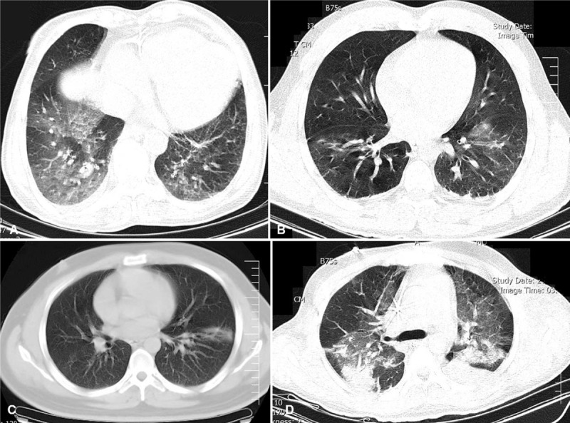
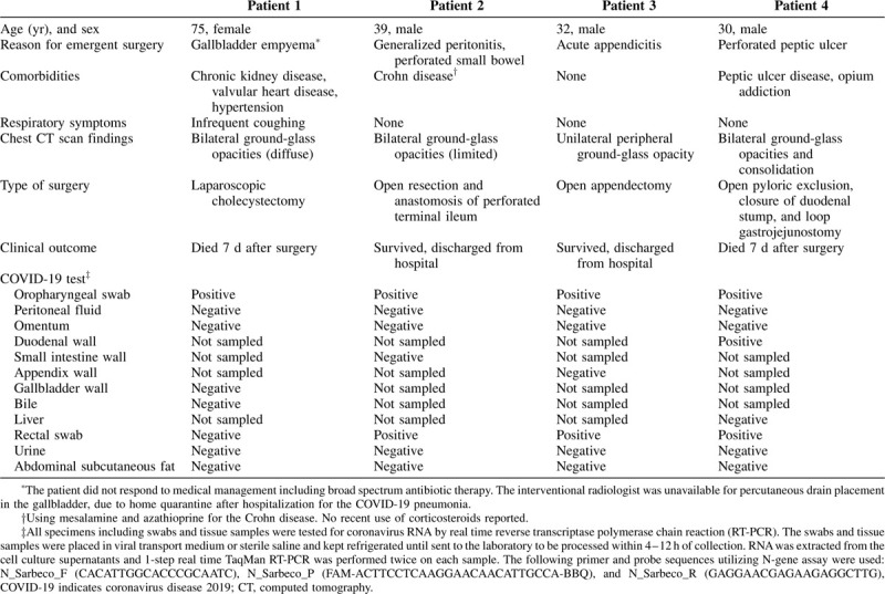
To examine the involvement by the SARS-CoV-2, multiple clinical specimens were obtained during the emergent abdominal surgery in 4 COVID-19 patients between March 27 and May 11, 2020. Diagnosis of COVID-19 was confirmed by preoperative chest CT scan (Fig. 1) and real time reverse transcriptase polymerase chain reaction (RT-PCR) testing of oropharyngeal swab (Table 1).
FIGURE 1.

Chest CT scan of COVID-19 pneumonia before emergent abdominal operations. A, Patient 1: Bilateral diffuse ground-glass opacities before laparoscopic cholecystectomy. B, Patient 2: Bilateral limited ground-glass opacities before exploratory laparotomy for perforated ileum. C, Patient 3: Peripheral ground-glass opacity of left lung before open appendectomy. D, Patient 4: Bilateral diffuse ground-glass opacities and consolidation before exploratory laparotomy for perforated peptic ulcer. COVID-19 indicates coronavirus disease 2019; CT, computed tomography.
TABLE 1.
Demographic, Clinical, and Laboratory Characteristics of 4 Patients With COVID-19 Pneumonia Who Underwent Emergent Abdominal Surgery

The first patient was a 75-year-old woman with abdominal pain who was admitted with a diagnosis of acute cholecystitis for nonoperative management. Her only respiratory symptom was infrequent coughing. After unsuccessful nonoperative management for 48 hours, she was scheduled for laparoscopic cholecystectomy. Preoperative chest computed tomography (CT) scan showed bilateral diffuse ground-glass opacities (Fig. 1A), and gallbladder empyema was found intraoperatively. During the operation, she developed transient hypoxemia and cardiac dysrhythmia. In her postoperative course in the intensive care unit, serial abdominal examinations, surgical drain output, and liver function tests remained unremarkable. The patient died from severe respiratory failure on postoperative day 7.
The second patient was a 39-year-old man with a 10-year history of Crohn disease, managed with medications, who was admitted to the hospital with acute abdominal pain without any respiratory symptoms. He had history of a previous small bowel perforation 4 years prior. CT scan showed free abdominal air and pulmonary involvement (Fig. 1B). The intraoperative finding was purulent peritonitis secondary to a small sealed perforation in the terminal ileum. After resection and primary anastomosis of the small bowel, he was discharged home on postoperative day 5. Histopathological examination reported a perforated active ulcer with no granulomatous and malignant changes.
The third patient was a 32-year-old man who was admitted with abdominal pain, nausea, and vomiting for 4 days, and fever for 1 day before admission. He did not have any respiratory complaints. CT scan showed acute appendicitis and limited COVID-19 pneumonia (Fig. 1C). He underwent an open appendectomy. The intraoperative finding was a locally perforated and sealed appendicitis, and the patient was discharged home on postoperative day 4.
The fourth patient was a 30-year-old man with massive hematemesis and epigastric abdominal pain who was admitted for emergent endoscopic gastroduodenoscopy. In his history, he had surgical repair of a perforated peptic ulcer 3 years prior. Although he did not have any respiratory symptoms, oropharyngeal swab was sent at the time of admission which was positive for SARS-CoV-2. He underwent multiple endoscopic interventions to control the bleeding from a large peptic ulcer. Seven days after admission, a CT scan showed diffuse COVID-19 pneumonia (Fig. 1D), free abdominal air, and leakage of luminal contrast in the pyloric area. He underwent an emergent laparotomy which showed diffuse purulent peritonitis from a very large unrepairable perforation (sealed by the omentum and liver) in the pyloric region. Closure of the stomach and duodenum on both sides of the hole and a loop gastrojejunostomy were performed. He died from severe sepsis on postoperative day 7.
Although the SARS-CoV-2 RNA was found in the feces of the last 3 patients and in the duodenal wall of the patient with perforated peptic ulcer, RT-PCR examination of abdominal fluid was negative for the virus (Table 1). Although these patients had localized or diffuse purulent peritonitis at the time of their operations, there was no gross contamination of the peritoneal cavity with gastrointestinal contents. The RT-PCR did not detect viral RNA in the wall of the small intestine, appendix, gallbladder, bile, liver, and urine. Visceral fat (omentum) and abdominal subcutaneous fat of 4 patients were also not infected with the SARS-CoV-2.
Small case series have shown an unexpected high mortality rate in surgical cases who developed COVID-19 pneumonia.19,20 Our patient was the second patient with gallbladder disease reported in the literature who died after laparoscopic cholecystectomy with COVID-19 pneumonia.20 Unlike the other case who developed COVID-19 pneumonia 2-weeks after cholecystectomy, the current case had COVID-19 at the time of surgery. It is unknown that whether the surgical stress and postoperative physiological changes can exacerbate the COVID-19 infection and worsen the outcomes of surgical patients.
As the first report in the surgical literature, lack of involvement of abdominal fluid, omentum, liver, gallbladder, and bile by the SARS-CoV-2 in these 4 COVID-19 cases can be reassuring, given the current concerns around the potential hazard of infected surgical fields in abdominal surgery such as gastric and small bowel operations, appendectomy, and cholecystectomy. Furthermore, operating on these 4 cases, who did not have prominent respiratory signs and symptoms before surgery, highlights the importance of preoperative screening tests in asymptomatic patients. Depending on availability of tests and local protocols, chest CT scan, RT-PCR, and immunoassays for COVID-19 can be considered before surgery.
This study is limited by reporting only 4 patients and lack of molecular data from sequential sampling and collecting of multiple specimens from each tissue. Some clinical specimens were actually sampled from only 1 patient. The presence of viremia and extent of viral load at the time of sampling in our patients were unknown. Furthermore, the accuracy of molecular tests for measurement of viral RNA in tissue samples has not been characterized. Viral culture was also not performed.
Although our findings indicate no direct involvement of abdominal fluid and fat tissues, careful sampling and assessment in large series is suggested to provide answers about the safety of abdominal surgery in patients with COVID-19 and to identify the possible role of adipose tissue in the pathogenesis of COVID-19 infection. This study does not provide evidence to suggest that severity of COVID-19 infection in patients with obesity is the direct result of infection of adipocytes by the SARS-CoV-2.
Footnotes
Author contributions: Dr. SS had full access to all of the data in the study and takes responsibility for the integrity of the data. Concept and design: all authors. Acquisition and interpretation of data: SS, HK, NMA, AD, MRH, and BNH. Drafting of the manuscript: AA. Critical revision of the manuscript: All authors.
All authors have completed and submitted the ICMJE Form for Disclosure of Potential Conflicts of Interest. None was directly related to the study.
The authors report no conflicts of interest.
REFERENCES
- 1.Wang W, Xu Y, Gao R, et al. Detection of SARS-CoV-2 in different types of clinical specimens. JAMA 2020; 323:1843–1844. [DOI] [PMC free article] [PubMed] [Google Scholar]
- 2.Zhang W, Du RH, Li B, et al. Molecular and serological investigation of 2019-nCoV infected patients: implication of multiple shedding routes. Emerg Microbes Infect 2020; 9:386–389. [DOI] [PMC free article] [PubMed] [Google Scholar]
- 3.Pan Y, Zhang D, Yang P, et al. Viral load of SARS-CoV-2 in clinical samples. Lancet Infect Dis 2020; 20:411–412. [DOI] [PMC free article] [PubMed] [Google Scholar]
- 4. Qiu L, Liu X, Xiao M, et al. SARS-CoV-2 is not detectable in the vaginal fluid of women with severe COVID-19 infection. Clin Infect Dis. 2020 Apr 2:ciaa375. doi: 10.1093/cid/ciaa375. Epub ahead of print. PMID: 32241022; PMCID: PMC7184332. [DOI] [PMC free article] [PubMed] [Google Scholar]
- 5.Francis N, Dort J, Cho E, et al. SAGES and EAES recommendations for minimally invasive surgery during COVID-19 pandemic. Surg Endosc 2020; 34:2327–2331. [DOI] [PMC free article] [PubMed] [Google Scholar]
- 6.Vigneswaran Y, Prachand VN, Posner MC, et al. What is the appropriate use of laparoscopy over open procedures in the current COVID-19 climate? J Gastrointest Surg 2020; 1–6. [DOI] [PMC free article] [PubMed] [Google Scholar]
- 7.Kwak HD, Kim SH, Seo YS, et al. Detecting hepatitis B virus in surgical smoke emitted during laparoscopic surgery. Occup Environ Med 2016; 73:857–863. [DOI] [PubMed] [Google Scholar]
- 8.Alp E, Bijl D, Bleichrodt RP, et al. Surgical smoke and infection control. J Hosp Infect 2006; 62:1–5. [DOI] [PubMed] [Google Scholar]
- 9.Eubanks S, Newman L, Lucas G. Reduction of HIV transmission during laparoscopic procedures. Surg Laparosc Endosc 1993; 3:2–5. [PubMed] [Google Scholar]
- 10.Gloster HM, Jr, Roenigk RK. Risk of acquiring human papillomavirus from the plume produced by the carbon dioxide laser in the treatment of warts. J Am Acad Dermatol 1995; 32:436–441. [DOI] [PubMed] [Google Scholar]
- 11.Simonnet A, Chetboun M, Poissy J, et al. High prevalence of obesity in severe acute respiratory syndrome coronavirus-2 (SARS-CoV-2) requiring invasive mechanical ventilation. Obesity (Silver Spring) 2020; 28:1195–1199. [DOI] [PMC free article] [PubMed] [Google Scholar]
- 12.Kass DA, Duggal P, Cingolani O. Obesity could shift severe COVID-19 disease to younger ages. Lancet 2020; 395:1544–1545. [DOI] [PMC free article] [PubMed] [Google Scholar]
- 13.Kassir R. Risk of COVID-19 for patients with obesity. Obes Rev 2020; 21:e13034. [DOI] [PMC free article] [PubMed] [Google Scholar]
- 14.Kruglikov IL, Scherer PE. The role of adipocytes and adipocyte-like cells in the severity of COVID-19 infections. Obesity (Silver Spring) 2020; 28:1187–1190. [DOI] [PMC free article] [PubMed] [Google Scholar]
- 15.Wrapp D, Wang N, Corbett KS, et al. Cryo-EM structure of the 2019-nCoV spike in the prefusion conformation. Science 2020; 367:1260–1263. [DOI] [PMC free article] [PubMed] [Google Scholar]
- 16.Zou X, Chen K, Zou J, et al. Single-cell RNA-seq data analysis on the receptor ACE2 expression reveals the potential risk of different human organs vulnerable to 2019-nCoV infection. Front Med 2020; 14:185–192. [DOI] [PMC free article] [PubMed] [Google Scholar]
- 17.Li MY, Li L, Zhang Y, et al. Expression of the SARS-CoV-2 cell receptor gene ACE2 in a wide variety of human tissues. Infect Dis Poverty 2020; 9:45. [DOI] [PMC free article] [PubMed] [Google Scholar]
- 18.Desruisseaux MS, Nagajyothi, Trujillo ME, et al. Adipocyte, adipose tissue, and infectious disease. Infect Immun 2007; 75:1066–1078. [DOI] [PMC free article] [PubMed] [Google Scholar]
- 19.Lei S, Jiang F, Su W, et al. Clinical characteristics and outcomes of patients undergoing surgeries during the incubation period of COVID-19 infection. EClinicalMedicine 2020; 21:100331. [DOI] [PMC free article] [PubMed] [Google Scholar]
- 20.Aminian A, Safari S, Razeghian-Jahromi A, et al. COVID-19 outbreak and surgical practice: unexpected fatality in perioperative period. Ann Surg 2020; 272:e27–e29. [DOI] [PMC free article] [PubMed] [Google Scholar]


