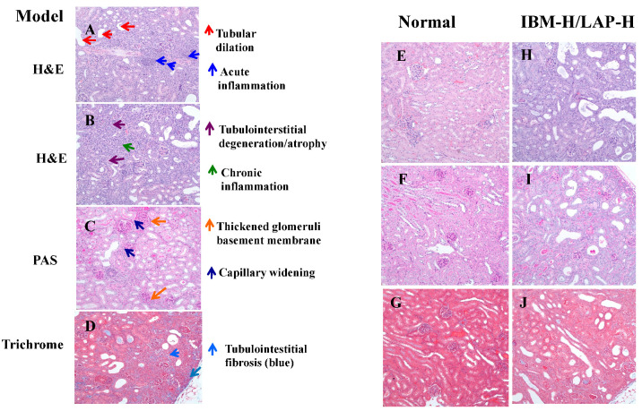
Figure 2.
Histopathological evaluation of kidney lesions. Representative histopathology slides from the adenine-induced CKD mice showing tubular dilation and acute inflammation (A), tubulointerstitial degeneration/atrophy and chronic inflammation (B), thickened glomeruli basement membrane and capillary widening (C), and tubulointerstitial fibrosis (D). The representative histology slides from the normal mice showing no lesions (E–G), and the representative histology slides from the effective treatment mice showing reduced lesions (H–J).

