Sir,
Solitary pelvic ectopic kidney is quite rare, seen in 1 in 22,000 autopsies.[1] To the best of our knowledge, there are only three case reports of solitary pelvic kidney from India in the literature.[2,3] When renal anomalies coexisted with limb defects, it is known as acrorenal syndrome. The term was coined by Curran et al.[4] in 1972. Since then, the term is often used but still lacks proper definition as several patterns of anomalies are being described. The commonly observed limb anomalies include syndactyly, oligodactyly, polydactyly, ectrodactyly, or brachydactyly of the carpal and tarsal bones. Renal anomalies commonly observed include unilateral renal agenesis, bilateral renal hypoplasia, ureteric hypoplasia, hydroureteronephrosis, and duplication abnormalities. Anomalies may be seen in other areas, such as oromandibular area, lungs, trachea, testes, vas deferens, uterus, heart, skin, spine, and ocular area. We describe uncommon case of solitary ectopic pelvic kidney presenting as rare variant of acrorenal syndrome. No such case is reported from India.
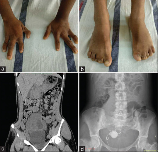
A 16-year-old female presented to us with lower abdominal pain, decreased urine output, and shortness of breath for 2 days. On physical examination, she had oligodactyly in both hands, syndactyly of the feet [Figure 1a and b]. A tender mass was palpable in the suprapubic region. There was no visual, ocular, hearing, ear anomaly, or any other facial dysmorphism. Cardiovascular, respiratory, and central nervous system examination was within normal limit. There was no history of delay in developmental milestones. Laboratory parameters showed hemoglobin 10.9 g/dl, total leucocyte count 9100/mm3, platelet count 2.6 × 10⁹/L, blood urea 223 mg/dl, serum creatinine 12.9 mg/dl, metabolic acidosis, microhematuria, and pus cells in urine but sterile urine culture. Emergency ultrasonography showed ectopic pelvic kidney with prominent pelvicalyceal system and multiple calculi. The patient received two hemodialysis sessions. Subsequently, computed tomography (CT) scan revealed single ectopic kidney in pelvis with moderate hydronephrosis and 24 × 18 mm calculi in renal pelvis and other calculi in inferior calyx [Figure 1c]. The patient received two hemodialysis sessions followed by DJ stenting [Figure 1d], following which the patient had improvement in urine output and creatinine came down to normal level obviating the need for further dialysis.
Figure 1.

(a) Bilateral hands showing oligodactyly. (b) Bilateral foot showing syndactyly. (c) CT abdomen showing large solitary ectopic pelvic kidney with hydronephrosis. (d) Plain X-ray KUB showing DJ stent in situ and renal calculi
Defect in acrorenal syndrome may be due to teratogenic, chromosomal, or dysplastic disorders.[5] The mode of inheritance can be autosomal recessive, autosomal dominant, or sporadic. The index case is likely sporadic mutation as there was no similar family history. Limb and renal anomalies coexistence is due to similar time period of development at 4 to 12 weeks of embryonic period.[6] There are various acrorenal syndromes described in literature-(a) Acro-renal-ocular syndrome which is characterized by optic nerve coloboma, radial ray anomalies of hand and various renal anomalies, (b) Townes–Brock syndrome characterized by thumb anomaly, ear defect with conductive hearing loss, imperforate anus and renal anomalies, (c) Pallister–Hall syndrome which includes limb anomalies, imperforate anus, renal and genitor-urinary anomalies, (d) Hajdu–Cheney syndrome characterized by coarse face, short neck, short stature dental anomalies, acroosteolysis, heart defect, hepatosplenomegaly, hydrocephalus and cleft palate in association with renal anomalies, (e) acro-renal-mandibular syndrome with characteristic unilateral renal agenesis, electrodactyly and hypoplastic mandible, (f) Fraser syndrome associated with unilateral renal agenesis, cryptopthalmos, syndactyly, and genital anomalies, (g) short-rib polydactyly syndrome associated with polydactyly, syndactyly, short limbs, dwarfism, cystic dysplasia, and hypoplastic ureter, (h) other miscellaneous syndromes like VACTERL, atrioacrorenal syndrome, MURCS (Mullerian duct aplasia, renal aplasia, and cervico-thoracic somite dysplasia) syndrome, and Poland's syndrome.[7] The key message of the letter is that limb defects can be indirect indicator of serious renal anomalies. General practitioners and pediatricians should be aware of the possible renal anomalies in a child with limb defects. Hence, screening ultrasonography of whole abdomen should be done early in a child with limb anomalies.
Declaration of patient consent
The authors certify that they have obtained all appropriate patient consent forms. In the form the patient(s) has/have given his/her/their consent for his/her/their images and other clinical information to be reported in the journal. The patients understand that their names and initials will not be published and due efforts will be made to conceal their identity, but anonymity cannot be guaranteed.
Financial support and sponsorship
Nil.
Conflicts of interest
There are no conflicts of interest.
References
- 1.Bergman RA, Afifi AK, Miyauchi R. Illustrated Encyclopedia of human anatomic variation. opus IV: Organ system: Urinary system: Kidneys, ureters, bladders and urethra. [Last accessed on 2019 May 20]. Available from: http://www.anatomyatlases.org/AnatomicVariants/Organ System/Text/UrinarySystem.shtml .
- 2.Bhowmik D, Tiwari SC, Gupta S, Agarwal SK, Dash SC. Chronic renal failure in a patient with solitary pelvic ectopic kidney and ipsilateral ectopic testes. Indian J Nephrol. 2001;11:68–9. [Google Scholar]
- 3.Neki NS, Amritpal S, Gagandeep SS, Puneet BS, Amanpreet K, Taranjit S. Solitary pelvic kidney. Int J Med Health Sci. 2017;6:127–9. [Google Scholar]
- 4.Curran AS, Curran JP. Associated acral and renal malformations: A new syndrome? Pediatrics. 1972;49:716–25. [PubMed] [Google Scholar]
- 5.Rascher W, Rosch WH. Congenital abnormalities of the urinary tract. In: Davison AM, editor. Oxford Textbook of Clinical Nephrology. New Delhi: Oxford University Press; 2005. pp. 2471–94. [Google Scholar]
- 6.Krzemieln G, Blaim MR, Kostro I, Wojnar J, Karpińska M, Sekowska R. Urological anomalies in children with renal agenesis or multicystic dysplastic kidney. J Appl Genet. 2006;47:171–6. doi: 10.1007/BF03194618. [DOI] [PubMed] [Google Scholar]
- 7.Natarajan G, Jeyachandran D, Subramaniyan B, Thanigachalam D, Rajagopalan A. Congenital anomalies of kidney and hand: A review. Clin Kidney J. 2013;6:144–9. doi: 10.1093/ckj/sfs186. [DOI] [PMC free article] [PubMed] [Google Scholar]


