PLOS ONE logoLink to PLOS ONE
. 2020 Sep 3;15(9):e0238177. doi: 10.1371/journal.pone.0238177

Renal hyperfiltration as a risk factor for chronic kidney disease: A health checkup cohort study

Se Won Oh 1, Ji Hyun Yang 1, Myung-Gyu Kim 1, Won Yong Cho 1, Sang Kyung Jo 1,*
Editor: Tatsuo Shimosawa2
PMCID: PMC7470278  PMID: 32881893

Abstract

Introduction

Renal hyperfiltration (RHF) has been found to be an independent predictor of adverse cardiovascular outcome. However, it remains uncertain whether it is precursor of chronic kidney disease (CKD) in a healthy population.

Materials and methods

To determine relative risks and identify the predictor of incident proteinuria and decline of estimated glomerular filtration rate (eGFR) in subjects with RHF. A total of 55,992 subjects aged ≥20 years who underwent health check-up during 2004–2017 were included. Among them, 16,946 subjects who completed at least two health checkups were analyzed.

Results

A total of 949 (5.6%) subjects developed proteinuria and 98 (0.6%) subjects showed ≥ 30% of eGFR decline. The risk of incident proteinuria was significantly higher in those with RHF (RR: 1.644; 95% CI: 1.064–2.541). Those with RHF showed 8.720 fold (95% CI: 4.205–18.081) increased risk for ≥30% decline. ESR, CRP, and monocyte count showed reversed J shaped curve according to the increase of eGFR. The adjusted mean of monocyte count was significantly higher in participants with eGFR ≥90ml/min/1.73m2 or < 60ml/min/1.73m2 compared to that in patients with eGFR 75-89ml/min/1.73m2. Compared to subjects with the lowest tertile of monocyte and no RHF, those with the highest tertile of monocyte count in the RHF group had 3.314-fold (95% CI: 1.893–5.802) higher risk of incident proteinuria and 3.822-fold (95% CI, 1.327–11.006) risk of 30% eGFR decline.

Conclusions

RHF had significantly increased risk of developing proteinuria and CKD in healthy subjects. Higher monocyte count might be used as a predictor of CKD in subjects with RHF.

Introduction

Renal hyperfiltration (RHF) is an absolute increase of glomerular filtration rate (GFR). It can occur physiologically after consuming high protein meals or during pregnancy [1]. However, increased whole kidney GFR is well known to precede the onset of albuminuria and progressive decline of GFR in type 1 diabetes mellitus. Several cross-sectional studies have demonstrated that RHF is also associated with hypertension, obesity, prediabetes, and smoking [27]. RHF has recently been found to be an independent predictor of adverse cardiovascular outcome or all-cause mortality [8, 9]. However, it still remains uncertain whether it is a precursor of chronic kidney disease (CKD) in apparently healthy population. Factors that can differentiate physiologic vs. pathologic increase of GFR also remain unknown. Given the irreversible nature of CKD associated with cardiovascular risks, identifying patients who are in the very early stage of CKD have a paramount importance in developing possible preventive and therapeutic strategies. Inflammation plays an important role in the loss of renal function in CKD. Previous studies have demonstrated the association between monocyte count and incident CKD or progression to end stage renal disease (ESRD) in a large cohort of veterans with significant comorbidities [10]. Monocytes are cells of the innate immune system with heterogenous phenotypes.

The objective of this longitudinal analysis of apparently healthy subjects with repeat voluntary health check-ups was to determine relative risks of incident proteinuria and decline of eGFR in subjects with RHF. Results showed that RHF might represent an early stage of CKD. This study also showed that higher monocyte count could predict the development of proteinuria and more rapid decline of eGFR.

Materials and methods

Participants

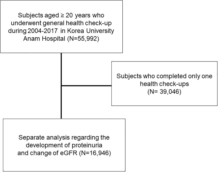
A total of 55,992 subjects aged ≥ 20 years who underwent general health check-up during 2004–2017 in Korea University Anam Hospital were included. Among them, 16,946 subjects who completed at least two health check-ups regarding the development of proteinuria and change of eGFR were subjected to a separate analysis for (Fig 1). This study was conducted in accordance with the Declaration of Helsinki. It was approved by the Institutional Review Board (IRB) of Korea University Anam Hospital Clinical Trial Center (IRB No. 2019AN0181). Institutional review board approved that informed consent is not necessary because this is a retrospective study.

Fig 1. Selection of study population.

Fig 1

Measurements

After an 8-hour fast, blood samples were collected year-round and immediately processed, refrigerated, and transported in cold storage to the laboratory for analysis within 12 hours. The measurement of serum creatinine was performed using a Toshiba Neo (Toshiba Medical System Co, Otawara, Japan) from 2004 to Nov. 2012 and a Beckman Coulter AU5811, 5821 (Diamond Diagnostics, Holliston, MA, USA) from Dec. 2012 to Nov. 2018. Serum creatinine level was measured using the Jaffe kinetic method (Jan. 2004- Nov. 2012, CLINIMATE Creatinine sekisui, Sekisui Medical Co. Ltd., Tokyo, Japan; Dec. 2012- Nov. 2018; Beckman Creatinine, Beckman Coulter, Inc., USA). GFR was estimated using the Modification of Diet in Renal Disease equation. Urine protein was measured by dipstick urinalysis. Results are reported using a semiquantitative scale from negative to 4+. Body fat and lean body mass percent were measured using bioelectrical impedance analysis (InBody770, InBody Co, Ltd., Seoul, Korea).

Definitions

We divided participants into 10-year age groups. RHF was defined as eGFR above age- and sex-specific 97.5th percentile (S1 Fig) [11]. Hypertension (HTN) was defined as the presence of either (i) systolic blood pressure (SBP) ≥ 140 mmHg or diastolic blood pressure (DBP) ≥ 90 mmHg or (ii) diagnostic code of HTN. Diabetes mellitus (DM) was defined as participants who fulfilled at least one of the following three criteria: (i) fasting blood sugar (FBS) ≥ 126 mg/dL; (ii) HbA1c ≥ 6.5%; (iii) diagnostic code of diabetes. Body mass index (BMI) was calculated on the basis of weight and height (kg/m2). Proteinuria was defined as dipstick urinalysis above 1+. Decline of renal function was defined by 30% and 40% of decrease at follow-up examination compared to baseline eGFR. Malignancy was defined as the presence of “C” code in electronic medical record. Coronary artery disease was defined as participants who underwent coronary angiography.

Statistical analysis

All analyses were performed using SPSS software (SPSS version 25.0, Chicago, IL, USA). Data are presented as mean ± standard deviation (SD) for continuous variables and as percentage for categorical variables. Differences were analyzed using Chi-square test for categorical variables and analysis of variance for continuous variables. Analysis of covariance was used to adjust independent factors related to monocyte count and post-hoc analysis was used to correct for multiple comparisons. Risks and 95% confidence intervals (95% CIs) of proteinuria and decline of eGFR were calculated using cox regression analysis. A P-value < 0.05 was considered statistically significant.

Results

Baseline characteristics of participants with RHF

The mean age of all participants was 46.8 ± 12.4 years. Of them, 52.5% were men. RHF was identified in 1,511 (2.69%) subjects. Participants with RHF were slightly younger. The proportion of BMI > 30 kg/m2 or < 20 kg/m2 was also significantly higher in those with RHF. SBP, FBS level, and the prevalence of DM were significantly higher while BUN, albumin, high density lipoprotein (HDL)-cholesterol levels were significantly lower in the RHF group. Significantly higher percentage of participants with RHF showed dipstick positive proteinuria and elevated levels of inflammatory markers including CRP, ESR, and monocyte count (Table 1).

Table 1. Characteristics of patients with renal hyperfiltration (RHF).

No RHF (N = 54,481) RHF(N = 1,511) P
Age (years) 46.8±12.4 46.2±12.7 0.040
Men (%) 28,587 (52.5) 765 (50.6) 0.157
BMI (kg/m2) <0.001
<20 6,322 (11.6) 245 (16.4)
20–24 29,729 (54.7) 776 (52.1)
25–29 16,177 (29.8) 394 (26.4)
≥30 2,143 (3.9) 75 (5.0)
SBP (mmHg) 115.7±14.0 115.0±15.3 0.064
≥130 11,450 (21.0) 371 (24.6) 0.001
eGFR (ml/min/1.73m2) 86.5±13.7 126.5±14.1 <0.001
BUN (mg/dL) 13.1±3.7 11.6±3.3 <0.001
Proteinuria (%) 3,120 (5.8) 114 (7.6) 0.003
Hematuria (%) 4,428 (8.2) 127 (8.4) 0.716
Hb (g/dL) 14.3±1.6 13.9±1.7 <0.001
WBC (/mm3) 5.99±2.05 6.04±1.78 0.286
Monocyte count (/mm3) 0.40±0.15 0.43±0.17 <0.001
ESR (mm/hr) 8.4±8.2 10.5±11.6 <0.001
CRP (mg/L) 1.7±5.4 2.6±10.3 0.006
AST (IU/L) 25.1±17.2 28.7±34.2 <0.001
ALT(IU/L) 25.5±25.1 28.0±24.8 <0.001
ALP(IU/L) 59.5±20.2 67.1±27.8 <0.001
GGT(IU/L) 36.6±55.0 47.4±98.0 <0.001
Bilirubin (mg/dL) 0.8±0.4 0.8±0.5 0.682
FBS (mg/dL) 95.6±20.8 101.2±32.0 <0.001
Albumin (g/dL) 4.5±0.3 4.4±0.3 <0.001
Triglyceride (mg/dL) 129.7±91.6 129.1±105.2 0.818
HDL cholesterol (mg/dL) 52.7±12.8 51.6±12.9 0.001
HTN (%) 8,802 (16.2) 255 (16.9) 0.456
DM (%) 3,219 (5.9) 164 (10.9) <0.001
CAD (%) 1,840 (3.4) 38 (2.5) 0.066
Cancer (%) 2,759 (5.1) 71 (4.7) 0.523

BMI, body mass index; SBP, systolic blood pressure; eGFR, estimated glomerular filtration rate; BUN, blood urea nitrogen; Hb, hemoglobin; WBC, white blood cell; ESR, erythrocyte sedimentation rate; CRP, C-reactive protein; AST, aspartate transaminase; ALT, aspartate aminotransferase; ALP, alkaline phosphatase; GGT, gamma-glutamyltransferase; FBS, fasting blood sugar; HDL, high density lipoprotein; hypertension, HTN; diabetes mellitus, DM; CAD, coronary artery disease.

Factors associated with incident proteinuria and eGFR decline

In the analysis of 16,946 participants with repeat health check-ups, 949 (5.6%) subjects newly developed proteinuria during a median follow-up period of 46.0 [24.0–77.0] months. Younger age, higher systolic BP (> 130 mmHg), presence of diabetes, higher monocyte counts, lower eGFR (< 60 ml/min/1.73m2), and RHF were independently associated with the development of proteinuria (P < 0.001). Out of 16,946 subjects, 98 (0.59%) participants developed ≥ 30% of eGFR decline. Older age, lower hemoglobin, higher monocyte counts, lower eGFR, and RHF were significantly associated with ≥ 30% of eGFR decline.

RHF predicts incident proteinuria and decline of eGFR

The risk of incident proteinuria was significantly higher in subjects with RHF after adjusting by age, sex, and eGFR (RR: 1.680; 95% CI: 1.100–2.568). Even after adjusting multiple factors, RHF had a 1.566-fold (95% CI: 1.013–2.420 folds) increase risk of developing incident proteinuria (Table 2).

Table 2. Risks of renal hyperfiltration (RHF) for the development of proteinuria and decline of eGFR.

Model 1* Model2
RR 95% CI P RR 95% CI P
Development of proteinuria
RHF 1.680 1.100–2.568 0.016 1.566* 1.013–2.420 0.044
30% decline in eGFR
RHF 3.265 1.446–7.372 0.004 8.720 4.205–18.081 <0.001
40% decline in eGFR
RHF 7.962 1.421–44.607 0.018 7.948 2.094–30.169 0.002

* Risks are adjusted by age, sex, and estimated glomerular filtration rate

**Risk is adjusted by age, sex, systolic blood pressure, body mass index, estimated glomerular filtration rate, hemoglobin, monocyte count, aspartate transaminase, aspartate aminotransferase, total cholesterol, diabetes, hypertension, and malignancy.

Risk is adjusted by age, sex, systolic blood pressure, body mass index, estimated glomerular filtration rate, fasting glucose, hemoglobin, monocyte count, aspartate transaminase, aspartate aminotransferase, alkaline phosphatase, total cholesterol, diabetes, hypertension, coronary artery disease, and malignancy.

Among participants developed ≥ 30% decline of eGFR, 31 (8.4%) subjects were in the RHF group while 67 (0.4%, p < 0.001) subjects were not. RHF was significantly associated with 30% decline in eGFR adjusted by age, sex, and eGFR (RR: 3.265; 95% CI: 1.446–7.372). Multivariate analysis showed that RHF had a 8.720-fold (95% CI: 4.205–18.081) increased risk for developing ≥ 30% decline in eGFR. More than 40% decline of eGFR was observed in 32 (0.2%) participants: 10 (2.7%) in the RHF group and 22 (0.1%) in the no-RHF group (p < 0.001). The relative risk of developing ≥ 40% decline of eGFR was also 7.948-fold (95% CI: 2.094–30.169) higher in the RHF group (Table 2).

Inflammatory markers, monocytes, and RHF

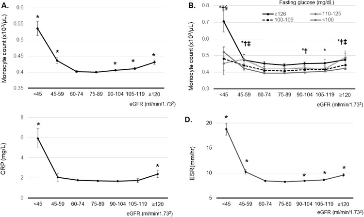
A previous report has demonstrated that higher monocyte count can predict the development and progression of CKD10. Thus, we examined associations between monocyte count and different eGFR ranges. The lowest adjusted means of monocyte count and ESR were noted in participants with GFR 75–89 ml/min/1.73m2. The lowest adjusted mean of CRP was observed in those with eGFR of 90–105 ml/min/1.73m2. Monocyte count showed a reversed J-shape curve. The adjusted mean of monocyte count was significantly higher in participants with eGFR ≥ 90 ml/min ml/min/1.73m2 or eGFR < 60 ml/min ml/min/1.73m2 compared to that in patients with eGFR 75–89 ml/min/1.73m2 (Fig 2A). This reverse J-shape association was independent of fasting blood glucose level. Adjusted mean monocyte count was higher in participants with eGFR <60 ml/min/1.73m2 compared to that in patients with eGFR of 75–89 ml/min/1.73m2 with FBS < 110 mg/dL. In addition, adjusted mean of monocyte count was significantly higher in participants with eGFR ≥ 120 ml/min ml/min/1.73m2 compared to that in patients with eGFR of 75–89 ml/min/1.73m2 with FBS < 110–125, 100–109, and < 100 mg/dL (Fig 2B).

Fig 2.

Fig 2

(A) Estimated mean of monocyte count according to estimated glomerular filtration rate (eGFR). *P<0.05, vs. eGFR 75–89 ml/min/1.73m2. (B) Estimated mean of monocyte count according to estimated glomerular filtration rate according to the fasting blood sugar (FBS). * P<0.05, vs. eGFR 75–89 ml/min/1.73m2 in participants with FBS<100 mg/dL. † P<0.05, vs. eGFR 75–89 ml/min/1.73m2 in participants with FBS 100–109 mg/dL. ‡ P<0.05, vs. eGFR 75–89 ml/min/1.73m2 in participants with FBS 110–125 mg/dL. § P<0.05, vs. eGFR 75–89 ml/min/1.73m2 in participants with FBS ≥126mg/dL. (C) Estimated mean of C-reactive protein according to estimated glomerular filtration rate. *P<0.05, vs. eGFR 90–105 ml/min/1.73m2. (D) Estimated mean of erythrocyte sedimentation rate according to estimated glomerular filtration rate. *P<0.05, vs. eGFR 75–89 ml/min/1.73m2. Estimated means were adjusted by age, body mass index, systolic blood pressure, AST, ALT, bilirubin, fasting blood glucose, hemoglobin, triglyceride, LDL cholesterol, HDL cholesterol, and total cholesterol.

The adjusted mean of CRP in participants with eGFR ≥ 120 or < 45 ml/min/1.73m2 was higher than that in subjects with eGFR 90–105 ml/min/1.73m2 (P < 0.001) (Fig 2C). The adjusted mean of ESR was also significantly higher in participants with eGFR ≥ 90 ml/min ml/min/1.73m2 or < 60 ml/min ml/min/1.73m2 compared to that in patients with eGFR 75–89 ml/min/1.73m2 (Fig 2D).

Monocyte count can predict the development of proteinuria in RHF

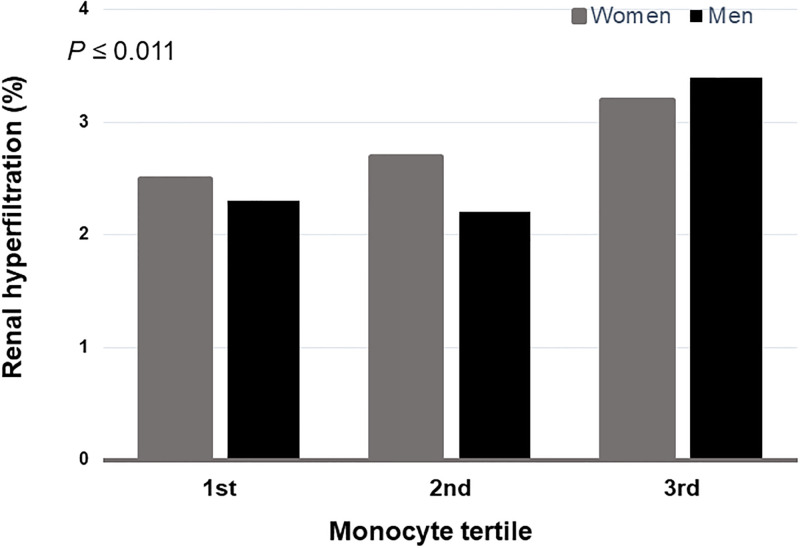
The prevalence of RHF was the highest in participants with the highest tertile of monocyte both in men and women (P ≤ 0.011) (Fig 3). We examined the risk of incident proteinuria according to monocyte tertiles and RHF. In participants without RHF, the 2nd tertile and the 3rd tertile of monocyte count showed significantly increased risks of developing new onset proteinuria (RR: 1.188; 95% CI: 1.009–1.400; RR: 1.352; 95% CI: 1.150–1.588, respectively). Compared to subjects with the lowest tertile of monocyte and no RHF, those having the 3rd tertile of monocyte count in the RHF group were found to have a 3.314-fold higher risk of developing incident proteinuria (95% CI: 1.893–5.802) (Table 3).

Fig 3. The prevalence of renal hyperfiltration (RHF) stratified by monocyte count tertiles.

Fig 3

The highest monocyte tertile was most frequent in RHF both men and women (P≤0.011).

Table 3. Risks for the development of proteinuria by monocyte tertiles and renal hyperfiltration (RHF).

Monocyte tertiles No RHF RHF
HR (95% CI) HR (95% CI)
1st 1.00 1.036 (0.386–2.786)
2nd 1.188 (1.009–1.400) 1.118 (0.416–3.005)
3rd 1.352 (1.150–1.588) 3.314 (1.893–5.802)

Risks are adjusted by age, sex, systolic blood pressure, body mass index, estimated glomerular filtration rate, hemoglobin, aspartate transaminase, aspartate aminotransferase, total cholesterol, diabetes, hypertension, malignancy, and coronary artery disease.

Monocyte count can predict the decline of eGFR in RHF

There was no association between monocyte count and subsequent decline of 30% eGFR in participants without RHF. However, monocyte count could predict the development of 30% decline of eGFR in participants with RHF. The 2nd tertile and the 3rd tertile of monocyte in the RHF group showed 3.420-fold and 3.822-fold increased risk of 30% eGFR decline (RR: 3.420; 95% CI; 1.188–9.847 in the 2nd tertile; RR: 3.822; 95% CI: 1.327–11.006 in the 3rd tertile) (Table 4).

Table 4. Risks for the development of 30% decline in eGFR by monocyte tertiles and renal hyperfiltration (RHF).

Monocyte tertiles No RHF RHF
HR (95% CI) HR (95% CI)
1st 1.00 2.438 (0.824–7.218)
2nd 0.988 (0.521–1.874) 3.420 (1.188–9.847)
3rd 1.645 (0.922–2.935) 3.822 (1.327–11.006)

Risk are adjusted by age, sex, systolic blood pressure, body mass index, estimated glomerular filtration rate, fasting glucose, hemoglobin, aspartate transaminase, aspartate aminotransferase, alkaline phosphatase, total cholesterol, diabetes, hypertension, coronary artery disease, and malignancy.

Discussion

In this retrospective study based on health checkup data of 55,992 apparently healthy subjects, we found that 1,511 (2.7%) showed age and sex adjusted RHF. These subjects had significantly increased risk of developing incident proteinuria and having a decline of eGFR. We also found that higher monocyte count could predict the development of proteinuria and the decline of eGFR in participants with RHF.

RHF is well-known to precede albuminuria and progressive CKD in type 1 diabetes mellitus. Many cross-sectional studies have demonstrated that RHF is also associated with prediabetes, obesity, sickle cell anemia, and smoking [17]. Recently, RHF has been demonstrated to be an independent risk factor for all-cause mortality or cardiovascular mortality [8, 9]. However, transient RHF occurring during pregnancy or after consumption of protein rich diet is not associated with increased filtration fraction. Thus, it is not considered to be pathological [1]. However, whether RHF observed in an apparently healthy population represents an early stage CKD that precedes the onset of proteinuria and progressive decline of GFR remains uncertain. Markers that can differentiate pathological vs. normal physiologic RHF are unknown either. Given the irreversible nature of CKD, identification of patients in their very early stage of CKD is of paramount importance in the prevention of progressive CKD.

In our study, we first identified 1,511 subjects who belonged to age and sex adjusted 97.5 percentile of eGFR. They were younger. They were more likely to have BMI ≥ 30 kg/m2 or < 20 kg/m2. creatinine based eGFR could lead to overestimation of eGFR in participants with lower muscle mass such as BMI <20 kg/m2, resulting in falsely categorized as RHF. So, we compared the percent lean body mass between RHF vs no RHF group in a given range of BMI.

We noticed that percent lean body mass was not different between RHF group and no RHF group in participants with BMI <20 kg/m2 (S1 Table). These data can support that reduced muscle mass does not account for elevated eGFR in these groups.

Interestingly, we observed that in participants with BMI≥20 kg/m2, percent body fat body fat was significantly higher (P≤0.032), while percent lean body mass was significantly lower (P≤0.034) in RHF group compared to no RHF group (S1 Table). These data can suggest that RHF and further decline of eGFR in these participants might be related to obesity or obesity related inflammation.

RHF group had higher prevalence of diabetes mellitus (5.9% in the no RHF group vs. 10.9% in the RHF group), and lower HDL-cholesterol level. Prevalence of dipstick positive proteinuria, serum fasting glucose level, and liver enzymes were also significantly elevated in the RHF group. This indicates that RHF is closely associated with components of metabolic syndrome. Similar findings have already been demonstrated previously [1214]. The higher prevalence of proteinuria at baseline (5.8% in the no RHF group vs. 7.6% in the RHF group, p = 0.003) might be related to the higher prevalence of diabetes in the RHF group. However, decline of 30% eGFR was significantly higher in RHF group than no RHF group (0.3% vs. 8.9%, P<0.001) excluding diabetes mellitus, high SBP, and proteinuria. In multivariate analysis, RHF group showed 4.913-fold increase for the decline in 30% eGFR (95% CI, 1.959–12.320) compared to no RHF group. In addition, monocyte count could predict the development of 30% decline of eGFR in participants with RHF excluding HTN, DM, and proteinuria (S2 Table). These data strongly suggest that RHF per se represent a very early stage of CKD and decline of eGFR is not caused by underlying diabetes, hypertension or underlying CKD.

To determine whether RHF might be a precursor of CKD, we compared the risk of incident proteinuria only in subjects without proteinuria at baseline. Cox regression analysis after adjusting for possible confounding variables including age, sex, BMI, diabetes, hypertension, and so on showed that the relative risk of incident proteinuria during a median follow-up period of 46 months was 1.571-fold higher in the RHF group compared with that in the no RHF group. This is comparable with a recent report also showing an increased risk of incident proteinuria in an analysis of Korean Nationwide health screening data over 11,559,520 adults [15]. Interestingly, this association was only observed in male subjects. We also performed a separate analysis according to gender and found that female subjects with RHF had no increased risk of incident proteinuria (RR, 1.039; 95% 0.462–2.337). However, underlying mechanisms leading to different effects of gender on the development of proteinuria remain uncertain.

In addition to the development of incident proteinuria, we also compared the risk of eGFR decline ≥ 30% and ≥ 40% of baseline. Significantly higher percentage of subjects with initial RHF developed eGFR decline of more than 30% or 40% during a median follow-up of 46 months compared with those without RHF. Multivariate analysis after adjusting for age, sex, and other factors showed that the relative risk of those with decline of eGFR ≥ 30% of baseline in RHF compared to those without RHF was 8.299 (95% CI: 4.003–17.205). Despite a relatively low event rate, this is a the first study demonstrating that RHF precedes the GFR decline in an apparently healthy population. However, not all subjects with RHF progressed to CKD. To identify factors that could predict CKD in RHF, we determined monocyte counts. Monocyte is a unique cell type of bone marrow origin with substantial plasticity. It plays a critical role in many different animal models of kidney diseases such as CKD, diabetic kidney disease, and other chronic inflammatory diseases [16, 17]. Although the role of monocytes in human kidney disease is still lacking, recent epidemiologic studies have shown a possible link between monocytes and CKD. For example, Ganda et al. have shown reduced eGFR in the highest quartile of monocytes. Bowe et al. have demonstrated a significant association between increased monocyte count and the risk of incident or progressive CKD [10, 18]. However, in the latter study, the majority of participants were Caucasians (82%) and most males (95%) had higher prevalence of other comorbid conditions, making it difficult to generalize this finding to relatively healthy subjects with different ethnic backgrounds and gender. The association between proinflammatory CD14+ CD16+ monocytes and vascular stiffness in CKD patients has also been demonstrated [19]. We observed a reverse J shaped curve between eGFR and monocyte count. Monocyte count and other inflammatory markers were significantly elevated not only in subjects who had eGFR < 60 ml/min/1.73m2, but also in those with higher eGFR (105–119 or ≥ 120 ml/min/1.73m2). And these were independent on fasting blood glucose levels. These data suggest that those with both low and high eGFR levels are likely to be in the state of chronic inflammation. We also found that subjects with RHF who belonged to the 3rd tertile of monocyte count had 3.314-fold and 3.822-fold increased risks of developing incident proteinuria and eGFR decline ≥ 30% of baseline compared to those without RHF who belonged to the 1st tertile of monocyte count. These data suggest the possibility that elevated monocyte count might be useful for predicting whether RHF would progress to CKD or not.

Despite several novel findings, this study has some limitations. First, proteinuria was defined as dipstick 1+ or higher instead of using more accurate albumin/creatinine ratio. However, according to several previous cohort studies [20, 21], using dipstick urine test is thought to be comparable with urine albumin excretion in predicting outcomes. Second, using creatinine based eGFR in defining RHF might have a possibility of overestimating GFR in underweight, malnourished subjects [22]. Despite BMI was significantly higher in the RHF group, the proportion of subjects with BMI < 18 was slightly higher in the RHF group (2.3% in the no RHF group vs. 3.9% in the RHF group, p < 0.001) in our study, suggesting that glomerular filtration in these subjects were overestimated.

In conclusion, this retrospective, longitudinal study based on health checkup data of 16,946 showed that apparently healthy subjects with RHF had significantly increased risk of developing incident proteinuria and having decline of eGFR. These data suggest that RHF in the general population might also represent an early stage of CKD regardless of known risk factors including diabetes, obesity, and so on. This study also showed that higher monocyte count in these subjects might be used as a predictor of CKD. Prospective studies are needed in the future to confirm our findings.

Supporting information

S1 Fig. Distribution of cut-off value of renal hyperfiltration (RHF) by sex and age.

The 97.5th percentiles are shown in 10-year age groups. RHF was defined as an estimated glomerular filtration rate over the age- and sex-specific 97.5th percentile.

(TIF)

S1 Table. The comparison of body composition between RHF and no RHF group according to body mass index (BMI).

(DOCX)

S2 Table. Risks for the development of 30% decline in eGFR by monocyte tertiles and renal hyperfiltration (RHF) in participants without diabetes, high SBP, and proteinuria.

(DOCX)

Data Availability

Data cannot be shared publicly because of ethical restrictions on sharing the de-identified data set imposed by the Institutional Review Board (IRB) of Korea University Anam Hospital Clinical Trial Center. Data are available from the IRB (contact via tel: 82-2-920-6566) for researchers who meet the criteria for access to confidential data.

Funding Statement

The author(s) received no specific funding for this work.

References

  • 1.Helal I, Fick-Brosnahan GM, Reed-Gitomer B, Schrier RW. Glomerular hyperfiltration: definitions, mechanisms and clinical implications. Nat Rev Nephrol. 2012; 8(5):293–300. 10.1038/nrneph.2012.19 [DOI] [PubMed] [Google Scholar]
  • 2.Keller CK, Bergis KH, Fliser D, Ritz E. Renal findings in patients with short-term type 2 diabetes. J Am Soc Nephrol. 1996; 7(12):2627–35. [DOI] [PubMed] [Google Scholar]
  • 3.Ficociello LH, Perkins BA, Roshan B, Weinberg JM, Aschengrau A, Warram JH, et al. Renal hyperfiltration and the development of microalbuminuria in type 1 diabetes. Diabetes Care. 2009; 32(5):889–93. 10.2337/dc08-1560 [DOI] [PMC free article] [PubMed] [Google Scholar]
  • 4.Chagnac A, Herman M, Zingerman B, Erman A, Rozen-Zvi B, Hirsh J, et al. Obesity-induced glomerular hyperfiltration: its involvement in the pathogenesis of tubular sodium reabsorption. Nephrol Dial Transplant. 2008; 23(12):3946–52. 10.1093/ndt/gfn379 [DOI] [PubMed] [Google Scholar]
  • 5.Palatini P, Dorigatti F, Saladini F, Benetti E, Mos L, Mazzer A, et al. Factors associated with glomerular hyperfiltration in the early stage of hypertension. Am J Hypertens. 2012; 25(9):1011–6. 10.1038/ajh.2012.73 [DOI] [PubMed] [Google Scholar]
  • 6.Maeda I, Hayashi T, Sato KK, Koh H, Harita N, Nakamura Y, et al. Cigarette smoking and the association with glomerular hyperfiltration and proteinuria in healthy middle-aged men. Clin J Am Soc Nephrol. 2011; 6(10):2462–9. 10.2215/CJN.00700111 [DOI] [PMC free article] [PubMed] [Google Scholar]
  • 7.Palatini P. Glomerular hyperfiltration: a marker of early renal damage inpre-diabetes and pre-hypertension. Nephrol Dial Transplant. 2012; 27(5):1708–14. 10.1093/ndt/gfs037 [DOI] [PubMed] [Google Scholar]
  • 8.Reboldi G, Verdecchia P, Fiorucci G, Beilin LJ, Eguchi K, Imai Y, et al. Glomerular hyperfiltration is a predictor of adverse cardiovascular outcomes. Kidney Int. 2018; 93(1):195–203. 10.1016/j.kint.2017.07.013 [DOI] [PubMed] [Google Scholar]
  • 9.Park M, Yoon E, Lim YH, Kim H, Choi J, Yoon HJ. Renal hyperfiltration as a novel marker of all-cause mortality. J Am Soc Nephrol. 2015; 26(6):1426–33. 10.1681/ASN.2014010115 [DOI] [PMC free article] [PubMed] [Google Scholar]
  • 10.Bowe B, Xie Y, Xian H, Li T, Al-Aly Z. Association between Monocyte Count and Risk of Incident CKD and Progression to ESRD. Clin J Am Soc Nephrol. 2017; 12(4):603–13. 10.2215/CJN.09710916 [DOI] [PMC free article] [PubMed] [Google Scholar]
  • 11.Oh SW, Han KH, Han SY. Associations between renal hyperfiltration and serum alkaline phosphatase. PLoS One. 2015; 10(4):e0122921 10.1371/journal.pone.0122921 [DOI] [PMC free article] [PubMed] [Google Scholar]
  • 12.Tomaszewski M, Charchar FJ, Maric C, McClure J, Crawford L, Grzeszczak W, et al. Glomerular hyperfiltration: a new marker of metabolic risk. Kidney Int. 2007; 71(8):816–21. 10.1038/sj.ki.5002160 [DOI] [PubMed] [Google Scholar]
  • 13.Har R, Scholey JW, Daneman D, Mahmud FH, Dekker R, Lai V, et al. The effect of renal hyperfiltration on urinary inflammatory cytokines/chemokines in patients with uncomplicated type 1 diabetes mellitus. Diabetologia. 2013; 56(5):1166–73. 10.1007/s00125-013-2857-5 [DOI] [PubMed] [Google Scholar]
  • 14.Sasson AN, Cherney DZ. Renal hyperfiltration related to diabetes mellitus and obesity in human disease. World J Diabetes. 2012; 3(1):1–6. 10.4239/wjd.v3.i1.1 [DOI] [PMC free article] [PubMed] [Google Scholar]
  • 15.Lee SM, Park JY, Park MS, Park JH, Park M, Yoon HJ. Association of renal hyperfiltration with incident proteinuria—A nationwide registry study. PLoS One. 2018; 13(4):e0195784 10.1371/journal.pone.0195784 [DOI] [PMC free article] [PubMed] [Google Scholar]
  • 16.Heine GH, Ortiz A, Massy ZA, Lindholm B, Wiecek A, Martínez-Castelao A, et al. ; European Renal and Cardiovascular Medicine (EURECA-m) working group of the European Renal Association-European Dialysis and Transplant Association(ERA-EDTA). Monocyte subpopulations and cardiovascular risk in chronic kidney disease. Nat Rev Nephrol. 2012; 8(6):362–9. 10.1038/nrneph.2012.41 [DOI] [PubMed] [Google Scholar]
  • 17.Gregg LP, Tio MC, Li X, Adams-Huet B, de Lemos JA, Hedayati SS. Association of Monocyte Chemoattractant Protein-1 with Death and Atherosclerotic Events in Chronic Kidney Disease. Am J Nephrol. 2018; 47(6):395–405. 10.1159/000488806 [DOI] [PMC free article] [PubMed] [Google Scholar]
  • 18.Ganda A, Magnusson M, Yvan-Charvet L, Hedblad B, Engström G, Ai D, et al. Mild renal dysfunction and metabolites tied to low HDL cholesterol are associated with monocytosis and atherosclerosis. Circulation. 2013; 127(9):988–96. 10.1161/CIRCULATIONAHA.112.000682 [DOI] [PMC free article] [PubMed] [Google Scholar]
  • 19.Rogacev KS, Seiler S, Zawada AM, Reichart B, Herath E, Roth D, et al. CD14++CD16+ monocytes and cardiovascular outcome in patients with chronic kidney disease. Eur Heart J. 2011; 32(1):84–92. 10.1093/eurheartj/ehq371 [DOI] [PubMed] [Google Scholar]
  • 20.Wen CP, Yang YC, Tsai MK, Wen SF. Urine dipstick to detect trace proteinuria: an underused tool for an underappreciated risk marker. Am J Kidney Dis. 2011; 58(1):1–3. 10.1053/j.ajkd.2011.05.007 [DOI] [PubMed] [Google Scholar]
  • 21.Zeller A, Haehner T, Battegay E, Martina B. Diagnostic significance of transferrinuria and albumin-specific dipstick testing in primary care patients with elevated office blood pressure. J Hum Hypertens. 2005; 19(3):205–9. 10.1038/sj.jhh.1001803 [DOI] [PubMed] [Google Scholar]
  • 22.Delanaye P, Cavalier E, Pottel H. Serum Creatinine: Not So Simple! Nephron. 2017; 136(4):302–8. 10.1159/000469669 [DOI] [PubMed] [Google Scholar]

Decision Letter 0

Tatsuo Shimosawa

1 Jun 2020

PONE-D-20-14418

Renal hyperfiltration as a risk factor for chronic kidney disease

PLOS ONE

Dear Dr. Jo,

Thank you for submitting your manuscript to PLOS ONE. After careful consideration, we feel that it has merit but does not fully meet PLOS ONE’s publication criteria as it currently stands. Therefore, we invite you to submit a revised version of the manuscript that addresses the points raised during the review process.

Please submit your revised manuscript by Jul 16 2020 11:59PM. If you will need more time than this to complete your revisions, please reply to this message or contact the journal office at plosone@plos.org. When you're ready to submit your revision, log on to https://www.editorialmanager.com/pone/ and select the 'Submissions Needing Revision' folder to locate your manuscript file.

Please include the following items when submitting your revised manuscript:

  • A rebuttal letter that responds to each point raised by the academic editor and reviewer(s). You should upload this letter as a separate file labeled 'Response to Reviewers'.

  • A marked-up copy of your manuscript that highlights changes made to the original version. You should upload this as a separate file labeled 'Revised Manuscript with Track Changes'.

  • An unmarked version of your revised paper without tracked changes. You should upload this as a separate file labeled 'Manuscript'.

If you would like to make changes to your financial disclosure, please include your updated statement in your cover letter. Guidelines for resubmitting your figure files are available below the reviewer comments at the end of this letter.

If applicable, we recommend that you deposit your laboratory protocols in protocols.io to enhance the reproducibility of your results. Protocols.io assigns your protocol its own identifier (DOI) so that it can be cited independently in the future. For instructions see: http://journals.plos.org/plosone/s/submission-guidelines#loc-laboratory-protocols

We look forward to receiving your revised manuscript.

Kind regards,

Tatsuo Shimosawa, M.D., Ph.D.

Academic Editor

PLOS ONE

Journal Requirements:

When submitting your revision, we need you to address these additional requirements.

1. Please ensure that your manuscript meets PLOS ONE's style requirements, including those for file naming. The PLOS ONE style templates can be found at https://journals.plos.org/plosone/s/file?id=wjVg/PLOSOne_formatting_sample_main_body.pdf and https://journals.plos.org/plosone/s/file?id=ba62/PLOSOne_formatting_sample_title_authors_affiliations.pdf

2. Please address the following:

- Please refer to any post-hoc corrections to correct for multiple comparisons during your statistical analyses. If these were not performed please justify the reasons. Please refer to our statistical reporting guidelines for assistance (https://journals.plos.org/plosone/s/submission-guidelines.#loc-statistical-reporting).

- Please provide the dates upon which the patient data was accessed.

- Please provide a participant flowchart.

3. We note that you have indicated that data from this study are available upon request. PLOS only allows data to be available upon request if there are legal or ethical restrictions on sharing data publicly. For information on unacceptable data access restrictions, please see http://journals.plos.org/plosone/s/data-availability#loc-unacceptable-data-access-restrictions.

In your revised cover letter, please address the following prompts:

a) If there are ethical or legal restrictions on sharing a de-identified data set, please explain them in detail (e.g., data contain potentially identifying or sensitive patient information) and who has imposed them (e.g., an ethics committee). Please also provide contact information for a data access committee, ethics committee, or other institutional body to which data requests may be sent.

b) If there are no restrictions, please upload the minimal anonymized data set necessary to replicate your study findings as either Supporting Information files or to a stable, public repository and provide us with the relevant URLs, DOIs, or accession numbers. Please see http://www.bmj.com/content/340/bmj.c181.long for guidelines on how to de-identify and prepare clinical data for publication. For a list of acceptable repositories, please see http://journals.plos.org/plosone/s/data-availability#loc-recommended-repositories.

We will update your Data Availability statement on your behalf to reflect the information you provide.

4. We note that you have included the phrase “data not shown” in your manuscript. Unfortunately, this does not meet our data sharing requirements. PLOS does not permit references to inaccessible data. We require that authors provide all relevant data within the paper, Supporting Information files, or in an acceptable, public repository. Please add a citation to support this phrase or upload the data that corresponds with these findings to a stable repository (such as Figshare or Dryad) and provide and URLs, DOIs, or accession numbers that may be used to access these data. Or, if the data are not a core part of the research being presented in your study, we ask that you remove the phrase that refers to these data.

5. Please amend either the title on the online submission form (via Edit Submission) or the title in the manuscript so that they are identical.

6. PLOS requires an ORCID iD for the corresponding author in Editorial Manager on papers submitted after December 6th, 2016. Please ensure that you have an ORCID iD and that it is validated in Editorial Manager. To do this, go to ‘Update my Information’ (in the upper left-hand corner of the main menu), and click on the Fetch/Validate link next to the ORCID field. This will take you to the ORCID site and allow you to create a new iD or authenticate a pre-existing iD in Editorial Manager. Please see the following video for instructions on linking an ORCID iD to your Editorial Manager account: https://www.youtube.com/watch?v=_xcclfuvtxQ

Additional Editor Comments (if provided):

Two reviewers and I have concern on muscle mass differences and diverse clinical background in cohort. The authors should analyze confounding factors that affect eGFR more in details.

[Note: HTML markup is below. Please do not edit.]

Reviewers' comments:

Reviewer's Responses to Questions

Comments to the Author

1. Is the manuscript technically sound, and do the data support the conclusions?

The manuscript must describe a technically sound piece of scientific research with data that supports the conclusions. Experiments must have been conducted rigorously, with appropriate controls, replication, and sample sizes. The conclusions must be drawn appropriately based on the data presented.

Reviewer #1: Yes

Reviewer #2: Partly

**********

2. Has the statistical analysis been performed appropriately and rigorously?

Reviewer #1: Yes

Reviewer #2: N/A

**********

3. Have the authors made all data underlying the findings in their manuscript fully available?

The PLOS Data policy requires authors to make all data underlying the findings described in their manuscript fully available without restriction, with rare exception (please refer to the Data Availability Statement in the manuscript PDF file). The data should be provided as part of the manuscript or its supporting information, or deposited to a public repository. For example, in addition to summary statistics, the data points behind means, medians and variance measures should be available. If there are restrictions on publicly sharing data—e.g. participant privacy or use of data from a third party—those must be specified.

Reviewer #1: Yes

Reviewer #2: Yes

**********

4. Is the manuscript presented in an intelligible fashion and written in standard English?

PLOS ONE does not copyedit accepted manuscripts, so the language in submitted articles must be clear, correct, and unambiguous. Any typographical or grammatical errors should be corrected at revision, so please note any specific errors here.

Reviewer #1: Yes

Reviewer #2: Yes

**********

5. Review Comments to the Author

Please use the space provided to explain your answers to the questions above. You may also include additional comments for the author, including concerns about dual publication, research ethics, or publication ethics. (Please upload your review as an attachment if it exceeds 20,000 characters)

Reviewer #1: This paper presents a retrospective analysis of health examination data from healthy adults and shows that RHF is a risk factor for proteinuria and decline of eGFR, and suggests that increased monocyte count may be a predictor of CKD progression. In CKD, which often follows an irreversible progression, it is "paramount" to recognize patients at a very early stage as preliminary CKD patients, as described by the authors, and the results of this study will make a significant contribution to future CKD treatment.

The points I noticed were as follows;

(1) Was there any difference in the data between the RHF group with a BMI>30 and the RHF group with a BMI<20?

(2) It has been shown in Table 2 that RHF seems to be involved in the progression of proteinuria and reduction of eGFR after adjusting for risk such as diabetes, but if the authors analyzed the RHF population excluding diabetes, what would be the differences in characteristics between the no RHF population?

(3) In the first paragraph of the introduction, line 11, the word "Monocyte" seemed to appear out of the blue. Wouldn't it be better to break a new line or put a preposition of some kind?

(4) On page 13, line 3, RFH should be corrected to RHF.

(5) On page 13, line 4, “this is a first study” → “this is the first study”?

Reviewer #2: Overall impression; The subject is interesting, but the authors should show the conclusive and convincing evidence demonstrating that RHF per se is a risk factor for CKD.

Major criticisms;

1) The RHF group has statistically significantly more eGFR and less BUN. This suggests that the muscle mass in the RHF group might be less. This is supported by the statistically different distributions of BMI in the two groups, i.e. there are more patients with BMI less than 20 in the RHF group. If this is correct, the high eGFR doesn't simply indicate renal hyperfiltration.

2) There are statistically significantly more patients who have hypertension (>130 mmHg), proteinuria and DM in the RHF group than those in the non-RHF group. More patients with CKD risk factors in the RHF group can explain the higher susceptibility of CKD progression.

3) The higher high-sensitivity C-reactive protein was already shown to be the risk factor of CKD progression. (https://www.ncbi.nlm.nih.gov/pmc/articles/PMC5090815/)(https://www.ncbi.nlm.nih.gov/pmc/articles/PMC6133564/)

(https://www.sciencedirect.com/science/article/abs/pii/S0002870319301723)

The higher monocyte count in peripheral blood was already shown to be the risk factor of CKD progression.

So what is novel in this manuscript is the higher susceptibility of CKD progression with higher monocyte count and higher CRP levels in the RHF group, but we cannot tell if the higher susceptibility of CKD progression in the RHF group is explained by the RHF per se or higher prevalence of hypertension and DM etc in the RHF group.

**********

6. PLOS authors have the option to publish the peer review history of their article (what does this mean?). If published, this will include your full peer review and any attached files.

If you choose “no”, your identity will remain anonymous but your review may still be made public.

Do you want your identity to be public for this peer review? For information about this choice, including consent withdrawal, please see our Privacy Policy.

Reviewer #1: No

Reviewer #2: No

[NOTE: If reviewer comments were submitted as an attachment file, they will be attached to this email and accessible via the submission site. Please log into your account, locate the manuscript record, and check for the action link "View Attachments". If this link does not appear, there are no attachment files.]

While revising your submission, please upload your figure files to the Preflight Analysis and Conversion Engine (PACE) digital diagnostic tool, https://pacev2.apexcovantage.com/. PACE helps ensure that figures meet PLOS requirements. To use PACE, you must first register as a user. Registration is free. Then, login and navigate to the UPLOAD tab, where you will find detailed instructions on how to use the tool. If you encounter any issues or have any questions when using PACE, please email PLOS at figures@plos.org. Please note that Supporting Information files do not need this step.

PLoS One. 2020 Sep 3;15(9):e0238177. doi: 10.1371/journal.pone.0238177.r002

Author response to Decision Letter 0


19 Jul 2020

Dear reviewers

Thank you for your thoughtful comments regarding our manuscript. We took your comments seriously and revised our manuscript.

Review Comments to the Author

Reviewer #1: This paper presents a retrospective analysis of health examination data from healthy adults and shows that RHF is a risk factor for proteinuria and decline of eGFR, and suggests that increased monocyte count may be a predictor of CKD progression. In CKD, which often follows an irreversible progression, it is "paramount" to recognize patients at a very early stage as preliminary CKD patients, as described by the authors, and the results of this study will make a significant contribution to future CKD treatment.

The points I noticed were as follows;

Was there any difference in the data between the RHF group with a BMI>30 and the RHF group with a BMI<20?

Thank you for your comment. We re-analyzed the data about the renal function decline and RHF group stratified by BMI. The incidence of 30% eGFR decline was significantly higher in RHF group with a BMI <20 kg/m2 (10.0% vs. 0.1%, P<0.001) and ≥30 kg/m2 (6.2% vs. 0.4%, P=0.003). In multivariate analysis, RHF was an independent risk factor for 30% eGFR decline in participants with both BMI <20 kg/m2 and ≥30 kg/m2 (P≤0.024).

We agree with your concern that creatinine based eGFR could lead to overestimation of eGFR in participants with lower muscle mass such as BMI <20 kg/m2, resulting in falsely categorized as RHF. So, we compared the percent lean body mass between RHF vs no RHF group in a given range of BMI.

We noticed that percent lean body mass was not different between RHF group and no RHF group in participants with BMI <20 kg/m2 (supplement table 1). These data can support that reduced muscle mass does not account for elevated eGFR in these groups.

Interestingly, we observed that percent body fat body fat was significantly higher (P≤0.032), while percent lean body mass was significantly lower (P≤0.034) in RHF group compared to no RHF group (supplement table 1). These data can suggest that RHF and further decline of eGFR in these participants might be related to obesity or obesity related inflammation. We added these data as supplemental table 1 and also in discussion section.

It has been shown in Table 2 that RHF seems to be involved in the progression of proteinuria and reduction of eGFR after adjusting for risk such as diabetes, but if the authors analyzed the RHF population excluding diabetes, what would be the differences in characteristics between the no RHF population?

Thank you for indicating the very important aspect of our data. According to your suggestion, we reanalyzed the data excluding patients with diabetes and found that even after excluding diabetes, eGFR decline was also significantly higher in RHF group than no RHF group (0.4 % vs. 8.4%, P<0.001). After adjusting risk factors, RHF was found to be an independent risk factor for the progression of eGFR 30% decline (RR, 7.123, 95% CI, 3.314-15.310) in non diabetic participants.

However, incident proteinuria was not associated with RHF if we exclude patients with diabetes. Currently, we do not know exactly why there is a discrepancy. The possible explanation might be related to definition of proteinuria by dipstick test that is less accurate than quantitative measurement of albumin or protein.

Another possibility is that proteinuria by dipstick test detects only albumin and CKD progression from non diabetic etiologies, characterized by interstitial inflammation and fibrosis might not be related to the development or progression of albuminuria.

We added the new data in the results section and also possible explanation in the discussion section.

In the first paragraph of the introduction, line 11, the word "Monocyte" seemed to appear out of the blue. Wouldn't it be better to break a new line or put a preposition of some kind?

I added following sentences before the word “monocyte”: Inflammation plays an important role in the loss of renal function in CKD.

On page 13, line 3, RFH should be corrected to RHF.

Thank you for your correction.

(5) On page 13, line 4, “this is a first study” → “this is the first study”?

Thank you for your correction.

Reviewer #2: Overall impression; The subject is interesting, but the authors should show the conclusive and convincing evidence demonstrating that RHF per se is a risk factor for CKD.

Major criticisms;

The RHF group has statistically significantly more eGFR and less BUN. This suggests that the muscle mass in the RHF group might be less. This is supported by the statistically different distributions of BMI in the two groups, i.e. there are more patients with BMI less than 20 in the RHF group. If this is correct, the high eGFR doesn't simply indicate renal hyperfiltration.

Thank you for your comment

We agree with your concern that creatinine based eGFR could lead to overestimation of eGFR in participants with lower muscle mass such as BMI <20 kg/m2, resulting in falsely categorized as RHF. So, we compared the percent lean body mass between RHF vs no RHF group in a given range of BMI.

We noticed that percent lean body mass was not different between RHF group and no RHF group in participants with BMI <20 kg/m2 (supplement table 1). These data can support that reduced muscle mass does not account for elevated eGFR in these groups.

Interestingly, we observed in participants with BMI≥20 kg/m2, that percent body fat body fat was significantly higher (P≤0.032), while percent lean body mass was significantly lower (P≤0.034) in RHF group compared to no RHF group (supplement table 1). These data can suggest that RHF and further decline of eGFR in these participants might be related to obesity or obesity related inflammation. We added these data as supplemental table 1 and also in discussion section

There are statistically significantly more patients who have hypertension (>130 mmHg), proteinuria and DM in the RHF group than those in the non-RHF group. More patients with CKD risk factors in the RHF group can explain the higher susceptibility of CKD progression.

Thank you for your comments and we re-evaluated the eGFR decline in RHF group after excluding the participants with SBP >130 mmHg, proteinuria at baseline, or diabetes (N=39,781). Decline of 30% eGFR was significantly higher in RHF group than no RHF group (0.3% vs. 8.9%, P<0.001). In multivariate analysis, RHF was found to have 4.913-fold increase for the decline in 30% eGFR (95% CI, 1.959-12.320) compared to no RHF group. These data strongly suggest that RHF per se represent a very early stage of CKD and decline of eGFR is not caused by underlying diabetes, hypertension or underlying CKD. We added these data as supplemental table 2 and also in the discussion section.

3) The higher high-sensitivity C-reactive protein was already shown to be the risk factor of CKD progression. (https://www.ncbi.nlm.nih.gov/pmc/articles/PMC5090815/)(https://www.ncbi.nlm.nih.gov/pmc/articles/PMC6133564/)

(https://www.sciencedirect.com/science/article/abs/pii/S0002870319301723)

The higher monocyte count in peripheral blood was already shown to be the risk factor of CKD progression.

So what is novel in this manuscript is the higher susceptibility of CKD progression with higher monocyte count and higher CRP levels in the RHF group, but we cannot tell if the higher susceptibility of CKD progression in the RHF group is explained by the RHF per se or higher prevalence of hypertension and DM etc in the RHF group.

Thank you for your critiques

As you pointed out, higher monocyte count has been demonstrated to be a risk factor of CKD progression by Bowe et.al. in CJASN 2017 Apr 3; 12(4): 603–613. The population in that study consisted of predominantly white male (while 83%, male 97.9%) with significantly higher prevalence of diabetes (28.6%), hypertension (68.2%) with mean eGFR of 76.3. In contrast, we analyzed apparently healthy population undergoing regular health check ups with significantly lower prevalence of diabetes or hypertension, significantly higher eGFR (86.5±13.7, 126.5±14.1; no RHF, RHF).

And according to your comments, we excluded patients with diabetes, hypertension and baseline proteinuria, and still found that RHF had 4.913-fold increase for the decline in 30%eGFR (95% CI, 1.959-12.320) compared to no RHF group, indicating that RHF per se is a risk factor of CKD.

We also observed that monocyte count could predict the development of 30% decline of eGFR in participants with RHF even after excluding HTN, DM, and proteinuria. The 2nd tertile and the 3rd tertile of monocyte in the RHF group showed 9.110-fold and 5.336-fold increased risk of 30% eGFR decline (95% CI; 2.918-28.447 in the 2nd tertile;; 95% CI: 1.551-18.358in the 3rd tertile) (supplement table 2).

So, we suggest that 1) RHF per se represent a very early stage of CKD and 2) increased monocyte count can be a marker of progressive CKD in healthy population with RHF. We added this points in the discussion section.

Thank you for your consideration.

Sincerely,

Sang Kyung Jo, MD, PhD

Professor

Division of Nephrology, Department of Internal Medicine, Korea University Medical College

Address : Korea University Anam Hospital, Koreadae-Ro 73, Sungbuk-Gu

Seoul, Republic of Korea 02841

e-mail : sang-kyung@korea.ac.kr

Attachment

Submitted filename: respose_reviewers_0715.docx

Decision Letter 1

Tatsuo Shimosawa

31 Jul 2020

PONE-D-20-14418R1

Renal hyperfiltration as a risk factor for chronic kidney disease: A health checkup cohort study

PLOS ONE

Dear Dr. Jo,

Thank you for submitting your manuscript to PLOS ONE. After careful consideration, we feel that it has merit but does not fully meet PLOS ONE’s publication criteria as it currently stands. Therefore, we invite you to submit a revised version of the manuscript that addresses the points raised during the review process.

Please clarify if Figure 2 is mislabeled.

Please submit your revised manuscript by Sep 14 2020 11:59PM. If you will need more time than this to complete your revisions, please reply to this message or contact the journal office at plosone@plos.org. When you're ready to submit your revision, log on to https://www.editorialmanager.com/pone/ and select the 'Submissions Needing Revision' folder to locate your manuscript file.

Please include the following items when submitting your revised manuscript:

  • A rebuttal letter that responds to each point raised by the academic editor and reviewer(s). You should upload this letter as a separate file labeled 'Response to Reviewers'.

  • A marked-up copy of your manuscript that highlights changes made to the original version. You should upload this as a separate file labeled 'Revised Manuscript with Track Changes'.

  • An unmarked version of your revised paper without tracked changes. You should upload this as a separate file labeled 'Manuscript'.

If you would like to make changes to your financial disclosure, please include your updated statement in your cover letter. Guidelines for resubmitting your figure files are available below the reviewer comments at the end of this letter.

If applicable, we recommend that you deposit your laboratory protocols in protocols.io to enhance the reproducibility of your results. Protocols.io assigns your protocol its own identifier (DOI) so that it can be cited independently in the future. For instructions see: http://journals.plos.org/plosone/s/submission-guidelines#loc-laboratory-protocols

We look forward to receiving your revised manuscript.

Kind regards,

Tatsuo Shimosawa, M.D., Ph.D.

Academic Editor

PLOS ONE

[Note: HTML markup is below. Please do not edit.]

Reviewers' comments:

Reviewer's Responses to Questions

Comments to the Author

1. If the authors have adequately addressed your comments raised in a previous round of review and you feel that this manuscript is now acceptable for publication, you may indicate that here to bypass the “Comments to the Author” section, enter your conflict of interest statement in the “Confidential to Editor” section, and submit your "Accept" recommendation.

Reviewer #1: (No Response)

Reviewer #2: All comments have been addressed

**********

2. Is the manuscript technically sound, and do the data support the conclusions?

The manuscript must describe a technically sound piece of scientific research with data that supports the conclusions. Experiments must have been conducted rigorously, with appropriate controls, replication, and sample sizes. The conclusions must be drawn appropriately based on the data presented.

Reviewer #1: Yes

Reviewer #2: Yes

**********

3. Has the statistical analysis been performed appropriately and rigorously?

Reviewer #1: Yes

Reviewer #2: Yes

**********

4. Have the authors made all data underlying the findings in their manuscript fully available?

The PLOS Data policy requires authors to make all data underlying the findings described in their manuscript fully available without restriction, with rare exception (please refer to the Data Availability Statement in the manuscript PDF file). The data should be provided as part of the manuscript or its supporting information, or deposited to a public repository. For example, in addition to summary statistics, the data points behind means, medians and variance measures should be available. If there are restrictions on publicly sharing data—e.g. participant privacy or use of data from a third party—those must be specified.

Reviewer #1: Yes

Reviewer #2: Yes

**********

5. Is the manuscript presented in an intelligible fashion and written in standard English?

PLOS ONE does not copyedit accepted manuscripts, so the language in submitted articles must be clear, correct, and unambiguous. Any typographical or grammatical errors should be corrected at revision, so please note any specific errors here.

Reviewer #1: Yes

Reviewer #2: Yes

**********

6. Review Comments to the Author

Please use the space provided to explain your answers to the questions above. You may also include additional comments for the author, including concerns about dual publication, research ethics, or publication ethics. (Please upload your review as an attachment if it exceeds 20,000 characters)

Reviewer #1: The previous reviewer's comments have been adequately addressed and improvements in content have been recognized.

(1) Figure 1 attached to the manuscript, which is divided into Figure 1 A-D, is described in the text as Figure 2 A-D, and there is no Figure 1 appended to the "Materials and Methods, Participants" section.

(2) Similarly, Figure 2, which is attached to the manuscript, is shown as Figure 3 in the text (page 10, line 7).

(3) Figure legends is similarly described as Figure 1 and 2, which is not consistent with Figure 1-3 in the manuscript.

From the text, it looks like a new Figure 1 has been added, and if so, I'd like to check that Figure.

Reviewer #2: You answered all the questions asked at the first submission and corrected the manuscript with proper supplemental information.

**********

7. PLOS authors have the option to publish the peer review history of their article (what does this mean?). If published, this will include your full peer review and any attached files.

If you choose “no”, your identity will remain anonymous but your review may still be made public.

Do you want your identity to be public for this peer review? For information about this choice, including consent withdrawal, please see our Privacy Policy.

Reviewer #1: No

Reviewer #2: No

[NOTE: If reviewer comments were submitted as an attachment file, they will be attached to this email and accessible via the submission site. Please log into your account, locate the manuscript record, and check for the action link "View Attachments". If this link does not appear, there are no attachment files.]

While revising your submission, please upload your figure files to the Preflight Analysis and Conversion Engine (PACE) digital diagnostic tool, https://pacev2.apexcovantage.com/. PACE helps ensure that figures meet PLOS requirements. To use PACE, you must first register as a user. Registration is free. Then, login and navigate to the UPLOAD tab, where you will find detailed instructions on how to use the tool. If you encounter any issues or have any questions when using PACE, please email PLOS at figures@plos.org. Please note that Supporting Information files do not need this step.

PLoS One. 2020 Sep 3;15(9):e0238177. doi: 10.1371/journal.pone.0238177.r004

Author response to Decision Letter 1


9 Aug 2020

Dear reviewers

Thank you for your thoughtful comments regarding our manuscript. We took your comments seriously and revised our manuscript again.

Reviewer #1: The previous reviewer's comments have been adequately addressed and improvements in content have been recognized.

(1) Figure 1 attached to the manuscript, which is divided into Figure 1 A-D, is described in the text as Figure 2 A-D, and there is no Figure 1 appended to the "Materials and Methods, Participants" section.

Thank you for your correction.

(2) Similarly, Figure 2, which is attached to the manuscript, is shown as Figure 3 in the text (page 10, line 7).

Thank you for your correction.

(3) Figure legends is similarly described as Figure 1 and 2, which is not consistent with Figure 1-3 in the manuscript.

Thank you for your correction.

From the text, it looks like a new Figure 1 has been added, and if so, I'd like to check that Figure.

Reviewer #2: You answered all the questions asked at the first submission and corrected the manuscript with proper supplemental information.

Thank you for your consideration.

Sincerely,

Sang Kyung Jo, MD, PhD

Professor

Division of Nephrology, Department of Internal Medicine, Korea University Medical College

Address : Korea University Anam Hospital, Koreadae-Ro 73, Sungbuk-Gu

Seoul, Republic of Korea 02841

e-mail : sang-kyung@korea.ac.kr

Attachment

Submitted filename: response_reveiwers_0809.docx

Decision Letter 2

Tatsuo Shimosawa

12 Aug 2020

Renal hyperfiltration as a risk factor for chronic kidney disease: A health checkup cohort study

PONE-D-20-14418R2

Dear Dr. Jo,

We’re pleased to inform you that your manuscript has been judged scientifically suitable for publication and will be formally accepted for publication once it meets all outstanding technical requirements.

Within one week, you’ll receive an e-mail detailing the required amendments. When these have been addressed, you’ll receive a formal acceptance letter and your manuscript will be scheduled for publication.

An invoice for payment will follow shortly after the formal acceptance. To ensure an efficient process, please log into Editorial Manager at http://www.editorialmanager.com/pone/, click the 'Update My Information' link at the top of the page, and double check that your user information is up-to-date. If you have any billing related questions, please contact our Author Billing department directly at authorbilling@plos.org.

If your institution or institutions have a press office, please notify them about your upcoming paper to help maximize its impact. If they’ll be preparing press materials, please inform our press team as soon as possible -- no later than 48 hours after receiving the formal acceptance. Your manuscript will remain under strict press embargo until 2 pm Eastern Time on the date of publication. For more information, please contact onepress@plos.org.

Kind regards,

Tatsuo Shimosawa, M.D., Ph.D.

Academic Editor

PLOS ONE

Additional Editor Comments (optional):

Reviewers' comments:

Acceptance letter

Tatsuo Shimosawa

26 Aug 2020

PONE-D-20-14418R2

Renal hyperfiltration as a risk factor for chronic kidney disease: A health checkup cohort study

Dear Dr. Jo:

I'm pleased to inform you that your manuscript has been deemed suitable for publication in PLOS ONE. Congratulations! Your manuscript is now with our production department.

If your institution or institutions have a press office, please let them know about your upcoming paper now to help maximize its impact. If they'll be preparing press materials, please inform our press team within the next 48 hours. Your manuscript will remain under strict press embargo until 2 pm Eastern Time on the date of publication. For more information please contact onepress@plos.org.

If we can help with anything else, please email us at plosone@plos.org.

Thank you for submitting your work to PLOS ONE and supporting open access.

Kind regards,

PLOS ONE Editorial Office Staff

on behalf of

Prof. Tatsuo Shimosawa

Academic Editor

PLOS ONE

Associated Data

    This section collects any data citations, data availability statements, or supplementary materials included in this article.

    Supplementary Materials

    S1 Fig. Distribution of cut-off value of renal hyperfiltration (RHF) by sex and age.

    The 97.5th percentiles are shown in 10-year age groups. RHF was defined as an estimated glomerular filtration rate over the age- and sex-specific 97.5th percentile.

    (TIF)

    S1 Table. The comparison of body composition between RHF and no RHF group according to body mass index (BMI).

    (DOCX)

    S2 Table. Risks for the development of 30% decline in eGFR by monocyte tertiles and renal hyperfiltration (RHF) in participants without diabetes, high SBP, and proteinuria.

    (DOCX)

    Attachment

    Submitted filename: respose_reviewers_0715.docx

    Attachment

    Submitted filename: response_reveiwers_0809.docx

    Data Availability Statement

    Data cannot be shared publicly because of ethical restrictions on sharing the de-identified data set imposed by the Institutional Review Board (IRB) of Korea University Anam Hospital Clinical Trial Center. Data are available from the IRB (contact via tel: 82-2-920-6566) for researchers who meet the criteria for access to confidential data.


    Articles from PLoS ONE are provided here courtesy of PLOS

    RESOURCES