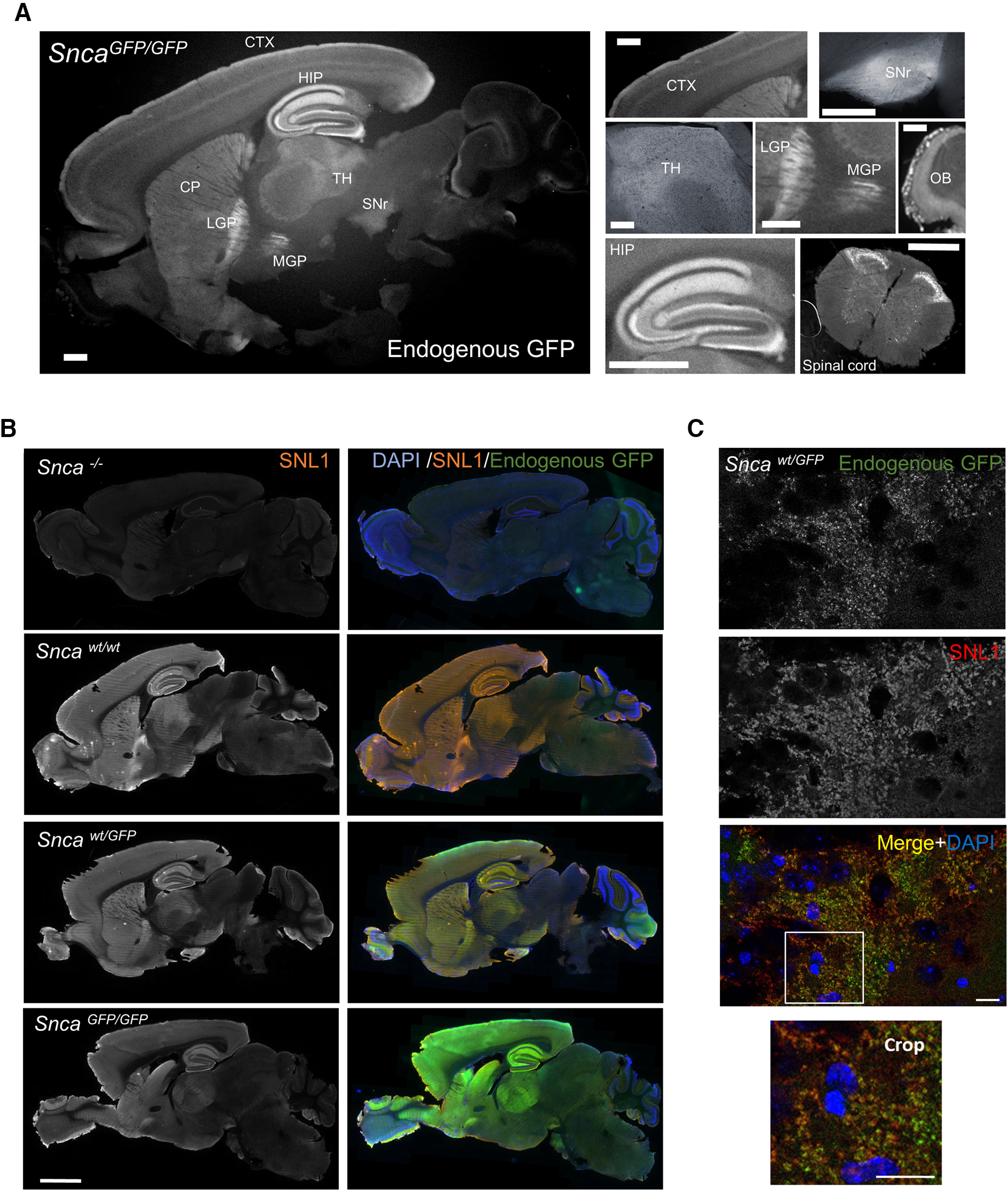
Figure 2.

Distribution of aSyn-GFP in brain and spinal cord. A, Expression pattern of aSyn-GFP in the brain from a homozygous (SncaGFP/GFP) animal. Endogenous GFP fluorescence is shown. Individual brain areas are shown on the right. B, Immunofluorescence showing aSyn expression patterns in wt, Sncawt/GFP, SncaGFP/GFP, and Snca−/− mice using a pan-aSyn antibody (SNL1). C, Co-localization of aSyn-GFP and endogenous aSyn in the hippocampus (CA3) of a Sncawt/GFP mouse labeled with SNL1. CTX = cortex; HIP = hippocampus; CP = caudate putamen; LGP = lateral globus pallidus; MGP = medial globus pallidus; TH = thalamus; SNr = substantia nigra pars reticulata; OB = olfactory bulb; SI = substantia innominata. Scale bars: 1 mm (A), 2 mm (B), 20 µm (C).
