Symbiotic microorganisms are essential for the lives of many multicellular eukaryotes (1). In insects, decades of symbiosis and—more recently—microbiome research have shown that microbial symbionts can supplement limiting nutrients, aid in digestion or detoxification, and defend their host against antagonists, thereby expanding the ecological and evolutionary potential of their hosts (2). In order to ensure that their offspring are endowed with the beneficial symbionts, insects have evolved a range of transmission routes to pass the symbionts vertically from parents to offspring or acquire them horizontally from unrelated host individuals or from the environment (3, 4). However, while, at first glance, the transmission from one host individual to another might seem like the most intricate problem for a symbiotic partnership, maintaining the symbiosis throughout the host’s development may be no less of a challenge (5).
In particular, holometabolous insects like beetles, butterflies and moths, flies, ants, bees, and wasps experience a complete restructuring of the body during metamorphosis from the larva to the adult individual. While gut microbes can be maintained throughout the reorganization of the gut (6), and other extracellular symbionts can persist outside of the host’s body (7), how intracellular mutualists located in special organs (bacteriomes) are maintained and sometimes even translocated during metamorphosis remained poorly understood (but see ref. 8). Maire et al., in PNAS (9), now elucidate the complex journey of the symbiont-bearing organs and their inhabitants in the rice weevil Sitophilus oryzae (Fig. 1), an organism with a long history of study of its symbiotic interaction with the Gammaproteobacteria Sodalis pierantonius (10). These symbionts were previously found to supply limiting nutrients including aromatic amino acids to their host and thereby support cuticle sclerotization and melanization (11).
Fig. 1.
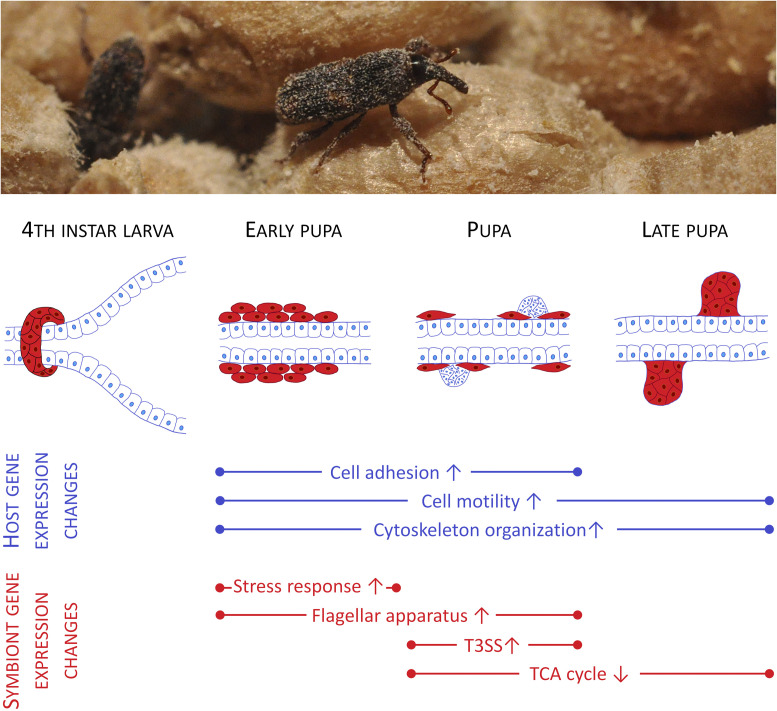
(Top) An adult rice weevil (S. oryzae). (Bottom) Schematic representation of the symbionts’ journey during the weevil’s metamorphosis and some of the associated gene expression changes in host and symbiont. The gut-associated bacteriome dissociates into individual bacteriocytes (red) in the early pupa, which later become spindle shaped and migrate along the gut, accumulating at epithelial cell clusters. Upon symbiont invasion, these putative stem cells differentiate into the mesenteric caeca serving as adult bacteriomes.
In weevil larvae, the symbionts inhabit a bacteriome localized at the foregut to midgut junction, whereas the adult beetles harbor the bacteria in numerous caeca along the midgut. How these organs form and how the symbionts are translocated during metamorphosis remained unknown. Following up on early histological investigations of the symbiosis, Maire et al. (9) traced the fate of the bacteriome cells, the so-called bacteriocytes, by fluorescence microscopy. The bacteriome first elongates in the early pupa before disintegrating into individual bacteriocytes that—astonishingly—change their morphology to a spindle shape and migrate distally along the gut, carrying their symbiont cargo within them (Fig. 1). In the midstage pupa, these bacteriocytes accumulate at sites with clusters of small epithelial cells that the authors interpret to be stem cells based on the high density of nuclei. It is from these cells that the new bacteriomes form, and the symbionts appear to spread into all of the cells of the developing mesenteric caeca (9). Previous studies had already shown that these caeca are of transient nature, being recycled when cuticle development is finalized and the symbionts are no longer needed (11). Only the females retain a symbiont population in ovary-associated bacteriomes that are formed early on in larval development and serve for symbiont transmission to the offspring.
In order to elucidate the molecular genetic basis underlying the translocation of bacteriocytes and the symbiont infection of the mesenteric caeca, Maire et al. (9) performed host and symbiont transcriptome sequencing in a late larval stage as well as throughout pupation. Coinciding with the movement of bacteriocytes and their change in morphology, RNA sequencing revealed the up-regulation of host genes involved in cell adhesion, cell motility, cellular shape, and cytoskeletal reorganization (Fig. 1). Importantly, RNA interference-mediated knockdown of a key gene involved in cell adhesion and motility, that is, galectin, impaired the migration of bacteriocytes along the gut, supporting its role in delivering the symbiont-bearing cells to the target location at the base of the newly forming caeca.
Maire et al., in PNAS, now elucidate the complex journey of the symbiont-bearing organs and their inhabitants in the rice weevil Sitophilus oryzae, an organism with a long history of study of its symbiotic interaction with the Gammaproteobacteria Sodalis pierantonius.
The symbionts also show distinct transcriptional changes throughout metamorphosis (9), which is insofar surprising as many intracellular symbionts encode a highly reduced set of transcriptional regulators in their eroded genomes and, thus, show strongly streamlined gene expression profiles (12). The rice weevil’s Sodalis symbiont, however, retains a comparatively large genome with many transcriptional and posttranscriptional regulators, which is likely due to the recent origin of the symbiotic association (13). During the early and midpupal stages, symbiont genes involved in stress response, motility (flagellum), and infection (type III secretion system [T3SS]) were found to be up-regulated (9), indicating that the colonization of the newly forming caeca is an active process involving the expression of bacterial genes that are known virulence factors in pathogens. Interestingly, a similar up-regulation of T3SS and flagellar genes was previously observed in the Sodalis symbiont of the closely related weevil Sitophilus zeamais (14), and a functional T3SS was found to be essential for successful colonization of host cells in Sodalis glossinidius, a symbiont of tsetse flies (15). Concordantly, the authors speculate that the T3SS and the flagellar apparatus of the rice weevil’s symbiont are instrumental for infecting the clusters of epithelial stem cells that subsequently differentiate into bacteriocytes (9).
While investigating the infection process of the newly forming symbiotic midgut caeca by transmission electron microscopy, Maire et al. (9) made a surprising discovery: The symbionts were not only localized in the cytosol of the host cells, but some even entered the nuclei. As the authors point out, this observation allows for fascinating speculations on the host−symbiont interaction during infection, all the more since an intranuclear localization has recently also been described for Sodalis-related symbionts of psyllids (16). Do the intranuclear symbionts interfere with host gene expression, for example, through the manipulation of epigenetic marks? Could they even be involved in host cell reprogramming, including polyploidization and differentiation into bacteriocytes? These questions open up another level of host−symbiont cross-talk that will be an exciting direction for future symbiosis research.
The study by Maire et al. (9) reveals the complex journey of an endosymbiont during metamorphosis of its host by a combination of bacteriocyte-mediated translocation along the gut and an active infection of epithelial stem cells by the symbiont. This process differs considerably from the well-studied transport of Buchnera symbionts from bacteriomes to the ovaries in aphids through a coordinated series of exocytosis and endocytosis events (17) and the early descriptions of symbiont translocations from midgut-associated organs into modified Malpighian tubules throughout the life stages of some chrysomelid beetles (18). However, movement of symbionts by the transport of entire bacteriocytes has been recently discovered as a means of transgenerational transmission in the whitefly Bemisia tabaci (19). Thus, the mechanisms of symbiont translocation during development and from somatic tissues into the germline differ widely across insect taxa (5). Increasing the number of symbiotic systems for which detailed knowledge is available will be crucial to understand general principles underlying the developmental origin of symbiotic organs and the molecular interplay of host and symbiont during translocation events.
The age of a symbiotic association has a strong impact on the nature of host−symbiont interactions (20). For this reason, the comparatively recent association between rice weevils and Sodalis provides an important complement to the well-studied and ancient symbioses in many sap-sucking Hemiptera. This study reveals that the rice weevil’s symbiont is capable of major gene regulatory changes during host development and expresses virulence factors for host cell infection. In addition, previous research by the same group uncovered that the host keeps the symbiont in check by antimicrobial peptides (21) and mounts a full-fledged immune response if exposed to the symbiont’s peptidoglycan fragments outside of the bacteriome (22). Thus, despite the mutualistic nature of this symbiosis, it also features aspects that are reminiscent of host−pathogen interactions, revealing the potential for conflict in host−symbiont interactions. Considering that Sodalis likely replaced the widespread weevil symbiont Nardonella (23), an interesting open question is whether the mechanism for symbiont translocation evolved in the symbiosis with Nardonella and was coopted by Sodalis, or, alternatively, whether the molecular host−symbiont interplay and developmental fate of the bacteriocytes drastically changed upon symbiont replacement. Considering that Nardonella, like most other ancient endosymbionts, is lacking an infection machinery including T3SS (24), the latter seems more likely. In conclusion, comparing the host−symbiont molecular interplay across insect symbioses of different evolutionary age not only embraces the astonishing diversity of symbiotic associations in nature but also provides a glimpse into the evolutionary sequence of events that can culminate in the tightly controlled and intimate mutualisms observed in many ancient insect−bacteria symbioses (20). The study by Maire et al. (9) makes an important contribution in this direction by pioneering research on intracellular symbiont translocation events during metamorphosis in a holometabolous insect at molecular detail.
Acknowledgments
I thank Laura Flórez, Tobias Engl, and Aurélien Vigneron for valuable comments on the manuscript. Funding by the European Research Council (ERC Consolidator Grant 819585 “SYMBeetle”) and the German Research Foundation (Grants KA2846/2-2, KA2846/5-1, and KA2846/6-1) is gratefully acknowledged.
Footnotes
The author declares no competing interest.
See companion article, “Spatial and morphological reorganization of endosymbiosis during metamorphosis accommodates adult metabolic requirements in a weevil,” 10.1073/pnas.2007151117.
References
- 1.McFall-Ngai M., et al. , Animals in a bacterial world, a new imperative for the life sciences. Proc. Natl. Acad. Sci. U.S.A. 110, 3229–3236 (2013). [DOI] [PMC free article] [PubMed] [Google Scholar]
- 2.Feldhaar H., Bacterial symbionts as mediators of ecologically important traits of insect hosts. Ecol. Entomol. 36, 533–543 (2011). [Google Scholar]
- 3.Bright M., Bulgheresi S., A complex journey: Transmission of microbial symbionts. Nat. Rev. Microbiol. 8, 218–230 (2010). [DOI] [PMC free article] [PubMed] [Google Scholar]
- 4.Salem H., Flórez L., Gerardo N., Kaltenpoth M., An out-of-body experience: The extracellular dimension for the transmission of mutualistic bacteria in insects. Proc. Biol. Sci. 282, 20142957 (2015). [DOI] [PMC free article] [PubMed] [Google Scholar]
- 5.Hammer T. J., Moran N. A., Links between metamorphosis and symbiosis in holometabolous insects. Philos. Trans. R. Soc. Lond. B Biol. Sci. 374, 20190068 (2019). [DOI] [PMC free article] [PubMed] [Google Scholar]
- 6.Johnston P. R., Rolff J., Host and symbiont jointly control gut microbiota during complete metamorphosis. PLoS Pathog. 11, e1005246 (2015). [DOI] [PMC free article] [PubMed] [Google Scholar]
- 7.Kaltenpoth M., Goettler W., Koehler S., Strohm E., Life cycle and population dynamics of a protective insect symbiont reveal severe bottlenecks during vertical transmission. Evol. Ecol. 24, 463–477 (2010). [Google Scholar]
- 8.Stoll S., Feldhaar H., Fraunholz M. J., Gross R., Bacteriocyte dynamics during development of a holometabolous insect, the carpenter ant Camponotus floridanus. BMC Microbiol. 10, 308 (2010). [DOI] [PMC free article] [PubMed] [Google Scholar]
- 9.Maire J., et al. , Spatial and morphological reorganization of endosymbiosis during metamorphosis accommodates adult metabolic requirements in a weevil. Proc. Natl. Acad. Sci. U.S.A. 117, 19347–19358 (2020). [DOI] [PMC free article] [PubMed] [Google Scholar]
- 10.Heddi A., Grenier A. M., Khatchadourian C., Charles H., Nardon P., Four intracellular genomes direct weevil biology: Nuclear, mitochondrial, principal endosymbiont, and Wolbachia. Proc. Natl. Acad. Sci. U.S.A. 96, 6814–6819 (1999). [DOI] [PMC free article] [PubMed] [Google Scholar]
- 11.Vigneron A., et al. , Insects recycle endosymbionts when the benefit is over. Curr. Biol. 24, 2267–2273 (2014). [DOI] [PubMed] [Google Scholar]
- 12.Moran N. A., Dunbar H. E., Wilcox J. L., Regulation of transcription in a reduced bacterial genome: Nutrient-provisioning genes of the obligate symbiont Buchnera aphidicola. J. Bacteriol. 187, 4229–4237 (2005). [DOI] [PMC free article] [PubMed] [Google Scholar]
- 13.Oakeson K. F., et al. , Genome degeneration and adaptation in a nascent stage of symbiosis. Genome Biol. Evol. 6, 76–93 (2014). [DOI] [PMC free article] [PubMed] [Google Scholar]
- 14.Dale C., Plague G. R., Wang B., Ochman H., Moran N. A., Type III secretion systems and the evolution of mutualistic endosymbiosis. Proc. Natl. Acad. Sci. U.S.A. 99, 12397–12402 (2002). [DOI] [PMC free article] [PubMed] [Google Scholar]
- 15.Dale C., Young S. A., Haydon D. T., Welburn S. C., The insect endosymbiont Sodalis glossinidius utilizes a type III secretion system for cell invasion. Proc. Natl. Acad. Sci. U.S.A. 98, 1883–1888 (2001). [DOI] [PMC free article] [PubMed] [Google Scholar]
- 16.Ghosh S., et al. , An intranuclear Sodalis-like symbiont and Spiroplasma coinfect the carrot psyllid, Bactericera trigonica (Hemiptera, Psylloidea). Microorganisms 8, 692 (2020). [DOI] [PMC free article] [PubMed] [Google Scholar]
- 17.Koga R., Meng X.-Y., Tsuchida T., Fukatsu T., Cellular mechanism for selective vertical transmission of an obligate insect symbiont at the bacteriocyte-embryo interface. Proc. Natl. Acad. Sci. U.S.A. 109, E1230–E1237 (2012). [DOI] [PMC free article] [PubMed] [Google Scholar]
- 18.Stammer H. J., Studien an Symbiosen zwischen Käfern und Mikroorganismen I. Die Symbiose der Donaciinen (Coleopt. Chrysomel.). Z. Morphol. Oekol. Tiere 29, 585–608 (1935). [Google Scholar]
- 19.Luan J., Sun X., Fei Z., Douglas A. E., Maternal inheritance of a single somatic animal cell displayed by the bacteriocyte in the whitefly Bemisia tabaci. Curr. Biol. 28, 459–465.e3 (2018). [DOI] [PMC free article] [PubMed] [Google Scholar]
- 20.McCutcheon J. P., Boyd B. M., Dale C., The life of an insect endosymbiont from the cradle to the grave. Curr. Biol. 29, R485–R495 (2019). [DOI] [PubMed] [Google Scholar]
- 21.Login F. H., et al. , Antimicrobial peptides keep insect endosymbionts under control. Science 334, 362–365 (2011). [DOI] [PubMed] [Google Scholar]
- 22.Maire J., et al. , Weevil pgrp-lb prevents endosymbiont TCT dissemination and chronic host systemic immune activation. Proc. Natl. Acad. Sci. U.S.A. 116, 5623–5632 (2019). [DOI] [PMC free article] [PubMed] [Google Scholar]
- 23.Lefèvre C., et al. , Endosymbiont phylogenesis in the dryophthoridae weevils: Evidence for bacterial replacement. Mol. Biol. Evol. 21, 965–973 (2004). [DOI] [PubMed] [Google Scholar]
- 24.Anbutsu H., et al. , Small genome symbiont underlies cuticle hardness in beetles. Proc. Natl. Acad. Sci. U.S.A. 114, E8382–E8391 (2017). [DOI] [PMC free article] [PubMed] [Google Scholar]


