

Abstract
Scaly‐tailed squirrels, the most poorly known group of gliding mammals, hold the record for variety of remarkable integument peculiarities. One of the most striking of these features is the scales on the tail, which apparently allow them to reduce energy costs when positioning themselves on a tree trunk. No less interesting is a peculiar spur that supports the flying membrane: the unciform element (‘spur’). Despite the peculiarity of such elements, their nature has not yet been studied. Using anatomical, histological methods and scanning electron microscopy we studied the structure of the skin and its derivatives in five of the six species from both genera of extant gliding scaly‐tailed squirrels (Anomaluridae, Rodentia): Idiurus macrotis, Idiurus zenkeri, Anomalurus beecrofti, Anomalurus pusillus and Anomalurus derbianus. In addition to the common mammalian skin structures, such as hair, vibrissae, sebaceous glands, meibomian glands of eyelids and eccrine sweat glands of the palmar and plantar pads, these animals have unique species‐specific skin derivatives (the tail scaly organ and its specific glands, vibrissae of the withers, patagium and its hair brush) that play a significant role in their adaptation to gliding and to their environment in general. The structure of the elbow spur is also described and hypotheses on its evolutionary origin from the tendon of the triceps muscle are presented.
Keywords: hair, patagium, scaly organ, scaly‐tailed squirrels, skin, unciform element, vibrissae
The scaly‐tailed squirrels are characterized by a unique set of the integument features. Long hairs on the edge of the membrane improve its properties as an airfoil, and specific sensitive hairs on the nape allow animals feel the space behind their back. The unique collagen‐cartilaginous spur that grows from the elbow helps to maintain the membrane in flight. These and other peculiarities are associated with specific habits of anomalures which combines gliding and roosting in hollow tree trunks.

1. INTRODUCTION
The scaly‐tailed squirrels of the African rodent superfamily Anomaluroidea include seven species of three genera. Previously, the fourth genus, Anomalurops, was erected in some studies (e.g. Rosevear, 1969; Jackson, 2012, 2012; Jackson and Thorington, 2012), but following the most up‐to‐date classifications (Jackson, 2016; Fabre et al. 2018) we consider it to be part of Anomalurus. Recent molecular studies (Heritage et al. 2016; Fabre et al. 2018) showed that the arboreal Zenkerella, as a sister taxon of the Idiurus–Anomalurus clade, should be placed in its own family Zenkerellidae; paleontological data suggest a similar conclusion (Marivaux et al. 2017). All other living anomalures capable of gliding are now considered closely related and constitute another family Anomaluridae. They have a set of specific skin derivatives that markedly distinguish them from other arboreal rodents (Johnson‐Murray, 1987; Jackson, 2012, 2012; Kingdon, 2015). Anomalurus and Idiurus genera are characterized by well‐developed large gliding membranes. A large, spine‐like cartilaginous elbow spur (the unciform element) projects laterally when gliding like an extra limb segment and supports the anterior part of extended patagium. It resembles the similar styliform cartilage of the flying squirrels (Pteromyini) but has a different origin. Another feature for which these animals were given the name ‘scaly‐tailed’ is a unique keratinous (horn) structure, the scaly organ (plate) located on the ventral surface of the tail root. According to frequently cited opinion, this scaly organ helps anomalures in climbing tree trunks or even facilitates landing after the glide (which is inappropriate for Zenkerella), but the most plausible hypothesis is that the scales on the tail provide support for the body while resting by clinging to the trunk (e.g.Rosevear, 1969; Heritage et al. 2016). However, assumptions regarding the role of the tail organ mostly appear in general reference books (e.g. Nowak, 1999; Jackson, 2016), while publications summarizing observations in nature (Adams, 1894; Jones, 1971) do not provide data on the use of scales.
The color of the scaly‐tailed squirrel's hair is diverse, and varies in different species from silver‐gray and brownish to orange and bright yellow. The animals have large eyes, as they are nocturnal (Nowak, 1999). They live in the forests of Central and West Africa, both rain forests and seasonally dry deciduous forests (Kingdon, 2013). They feed predominantly on tree bark along with a variety of plant food such as fruits, flowers, leaves and nuts; insects are also occasionally consumed (Rosevear, 1969; Kingdon, 2015). It is known that anomalures live in pairs or form colonies of dozens of individuals. Anomalurids represent one of several independent developments of gliding ability in mammals along with the flying squirrels of Eurasia and North America (Pteromyini, Rodentia), Southeast Asian flying lemurs (Dermoptera), and three groups of marsupial gliding possums from Australia (Phalangeriformes, Diprotodontia).
Studies on anomalurid anatomy are extremely scarce in the literature (Winton, 1898; Parsons, 1899; Potapova, 2018) since these secretive animals are hard to obtain in the wild. The morphology of the unciform elements, the skin and its derivatives in the anomalures remains almost unexplored. The aim of the present comparative study, therefore, was to reveal the morphological features of the integument structures of the scaly‐tailed squirrels and to identify their adaptations to the environment and habits.
2. MATERIALS AND METHODS
Five specimens, preserved in alcohol, of the adult representatives of the family Anomaluridae (Anomaluroidea, Anomaluromorpha, Rodentia) loaned from the Royal Museum for Central Africa, Tervuren, Belgium (Table 1) were studied. They are attributed to five species of two genera: the long‐eared flying mouse (Idiurus macrotis Miller, 1898), the Pygmy scaly‐tailed flying squirrel (Idiurus zenkeri Matschie, 1894), Beecroft's scaly‐tailed squirrel (Anomalurus beecrofti Fraser, 1853), Lord Derby's scaly‐tailed squirrel (Anomalurus derbianus Gray, 1842), and the Dwarf scaly‐tailed squirrel (Anomalurus pusillus Thomas, 1887).
Table 1.
Measurements of the studied specimens of Anomaluridae
| Species | Sex | Weight, g | Length of, mm | S1 a range, n = 5, mm2 | ||||
|---|---|---|---|---|---|---|---|---|
| Body | Tail | Feet without claws/with claws | Ear | Tail scaly organ | ||||
| Idiurus macrotis |

|
> 22 b | 82 | 118 | 18/20 | 17 | ~12 c | 0.13–0.16 |
| Idiurus zenkeri |

|
> 15 b | 71 | 102 | 15/16.5 | 13 | ~13 c | 0.15–0.20 |
| Anomalurus beecrofti |

|
228 | 210 | 145 | 38/42 | 27 | 40 | 14.5–15.2 |
| Anomalurus derbianus |

|
470 | 260 | 228 | 52/56 | 34 | 64 | 14.1–31.7 |
| Anomalurus pusillus |

|
40 | 124 | 127 | 31/34 | 22.5 | 34 | 6.8 –11.1 |
S1, area of individual scale of the tail scaly organ; n, number of measurements.
Weight without viscera.
Posterior border of the scaly organ is not clear.
Since the material obtained for the study is extremely rare, we were not able to conduct a comprehensive exploration of characters in every species, but we tried to describe the most typical features of the skin, skin derivatives, and associated structures using particular species as an example without causing significant damage to the samples. Total preparations of hairs of the withers, chest, flying membrane, tail and the largest vibrissae were examined with an Amplival light microscope (VEB Carl Zeiss, Jena) and a Leica DM‐R with a JVC 3CCCD digital video camera (Leica, Germany) using a 10 × eyepiece and a 10 × lens. The hair types were determined with a binocular magnifier to divide them into categories and orders, and the thickness of hair shaft and medulla (if present) were measured under a light microscope. The tail scaly organ and its scales were photographed using a Keyence Digital Microscope VHX‐1000 microscope (Keyence Corp., Japan).
Electronic images of the hair and vibrissae were obtained using conventional methods for examining objects in scanning electron microscopy, under a JSM 840A scanning electron microscope (Jeol, Japan). Before that, hair and vibrissae were cleaned and degreased in shampoo, then passed through alcohol solutions with increasing concentration, pasted with colorless varnish on the table, and covered with gold using Edwards S‐150A (Edwards, UK). The resulting images were arranged in a specific order, indicated in the figure captions: cross‐sections of different areas of the shaft (usually the base and ‘granna’, the widest part of a shaft); longitudinal sections of the base and granna of the hair; and surface ornamentation of the cuticle from the base to the most expanded part of the shaft.
The general membrane structure was examined in the skins of five specimens fixed in ethanol. The morphology of the spur was also described in five specimens according to the results of dissection using a Micromed MC‐4‐ZOOM LED stereo microscope.
Unfortunately, since the material was initially fixed in alcohol, the quality of histological preparations was not high. Using histological methods, we studied the structure of the unciform element and skin on the withers, upper eyelid and metatarsal pad. Samples were degreased in 70° alcohol, processed by alcohols with increasing concentration, and embedded in paraffin. Paraffin blocks were made using semi‐automatic equipment (Medite, Germany). Transverse sections of unciform element 10 μm thick underwent Mallory's staining. Longitudinal and transverse (cross‐) sections of skin 6–8 μm thick were stained with Ehrlich hematoxylin and eosin and enclosed in LEICA CV MOUNT liquid glass. The preparations were studied under a Keyence Biorevo BZ‐9000 motorized microscope (Keyence Corp.) and stereoscopic zoom microscope Lomo MSP‐2 (Lomo, Russia).
The microscopic and electronic images were scaled and edited in the computer program Adobe Photoshop Elements 11 (Adobe Systems, Inc., USA), but the changes concerned only their size, brightness and contrast. Measurements of thin structures were carried out using the TESCAN ATLAS program (TESCAN, Czech Republic). We measured (a) the thickness of the hair; (b) the thickness of the medulla (relative to the thickness of the hair in longitudinal and cross‐sections, in percentage); (c) the medulla area (relative to the area of the cross‐section of the shaft, in percentage); (d) the thickness of medullary inner septa; and (e) the Cuticle Index, calculated as a ratio of the height of the scales to the thickness of the shaft at its location. The number of measurements of each parameter was five (n = 5).
The study was conducted using the equipment of the Joint Usage Center ‘Instrumental methods in ecology’ at the Institute of Ecology and Evolution Russian Academy of Sciences, Moscow.
3. RESULTS
3.1. General structure of the gliding membrane
In all five species, the gliding membrane (patagium) is formed by the same set of parts (Figure 1A), but the attachment of these parts to the body is somewhat different within anomalures. The anterior part (propatagium) extends from the shoulder joint along the anterior side of the forelimb, and when unfolded by elbow extension it forms a triangle connecting the arm and forearm. In A. pusillus, the propatagium terminates proximally on the forearm, approximately at its middle. In all other species, by contrast, the propatagium spreads far distally along the forearm, almost reaching the antebrachiocarpal joint.
Figure 1.

Gliding apparatus of the scaly‐tailed squirrels. (A) General view (as per Idiurus macrotis): ventral view, left; dorsal view, right. (B) Scheme of the gliding membrane attachment to the pes in Anomalurus; line of attachment is dashed. (C) Scheme of the scaly organ and uropatagium attachment to the tail in Anomalurus. From left to right: Anomalurus beecrofti, Anomalurus derbianus, Anomalurus pusillus. (D) Scheme of the spur attachment. Arrows indicate the ligament fixing the spur. Lateral view, above; medial view, below
The middle part (plagiopatagium) is attached in front along the posterior side of the forelimb. It descends distally along the antebrachium to about the same extent as the propatagium on the anterior side (i.e. in A. pusillus, the termination of its attachment is shifted most proximally). On the anterior side of the hind limb of Idiurus, the plagiopatagium reaches the tarsus. In Anomalurus, it descends further distally up to the proximal phalanx of the digit I, attaching along the dorsal surface of the pes, close to its preaxial edge.
The caudal part (uropatagium) is attached to the posterior side of the hind limb. In Idiurus, the distal termination of the attachment is at the heel (Figure 1A); here, the membrane attachment curves onto the dorsal side of the ankle, so the heel is entirely below it. In Anomalurus, the attachment of the uropatagium descends more distally (Figure 1B), up to the proximal phalanx of the fifth digit; here, the attachment line is shifted to the dorsal surface of the pes and considerably spaced from its postaxial edge (Figure 1B, dashed line). The attachment of the uropatagium on the tail is restricted to its proximal part. In Idiurus, the membrane attaches to approximately one‐tenth of the tail length, not reaching the scaly organ (Figure 1A). In Anomalurus, by contrast, the membrane reaches the scaly organ: in A.pusillus it terminates at its base, in A. derbianus it comes up to its middle, whereas in A. beecrofti it descends to the distal end of the scaly organ (Figure 1C). In the latter case, the membrane occupies about half the length of the tail.
On the whole, the membrane is attached in such a manner that when it is spread, the forelimbs protrude approximately equally above and below its plane, the hind limbs are disposed mostly below the membrane (the femur to a lesser extent, the shank to a greater extent, the pes entirely below the plane of the membrane), and the tail is completely below the membrane. When gliding, the trunk is almost entirely below the plane of the membrane since the attachment of the plagiopatagium extends from above the shoulder joint to the hip joint approximately along the vertebral column; therefore, above the membrane is only the epaxial part of the trunk, whose height is quite small compared to the hypaxial part, which forms a voluminous abdomen. The manus in all studied species is not included in the membrane, the pes is free of the membrane only in Idiurus, and in Anomalurus the entire proximal part of the pes is enclosed in the membrane (only digits are free).
The spur extending outward from the elbow joint penetrates the membrane to the very edge, so that the thin bent tip of the spur forms a small protrusion of the smooth edge of the membrane. When limbs are most spread apart, and, accordingly, the membrane is maximally stretched, the spur is directed almost perpendicular to the body. Moreover, because the tip of the spur is bent forward and the membrane reaches its very tip posteriorly, an extension of the wing span is formed behind the spur. Thus, the distal end of the spur marks the widest span of the membrane.
3.2. Unciform element
3.2.1. Morphological structure of the spur
The unciform element is a flexible rod thinning towards the distal end. Its diameter at the base is comparable to that of the ulna in its proximal part. In I. macrotis, there is an additional slight thickening at the tip. The proximal end of the spur is tightly attached to the caudal edge of the olecranon (Figure 1D). The strongest ligament fixing the spur to the ulna originates on the dorsal surface of the olecranon, and inserts on the dorsal apex of the proximal end of the spur (Figure 1D, arrows). Actually, this ligament can be considered as a morphological extension of the distal triceps tendon. This tendon accretes to the olecranon apex and then spreads on the base of the spur. The spur is attached to the lateral surfaces of the olecranon by a small amount of collagen filaments to varying degrees expressed in representatives of different species.
No skeletal muscle attaches the base of the spur; however, here many long muscle fibers originate, penetrating into the lateral membrane behind the spur and stretching toward the hind limb.
3.2.2. Histological structure of the spur
The spur is semicircular in transverse section (Figure 2), its flat side faces the ulna. Histology shows that the spur itself is formed by fibrous cartilage wherein collagen fibers occupy a little more space than chondrocytes. Perichondrium collagen fibers run along the perimeter, whereas in the core area the fibers extending transversely from the flat side to the convex predominate. The spur is coated with a layer of striated musculature, thicker on the dorsal and ventral sides, of medium thickness on the side facing caudally, and thinnest on the side facing the ulna. Morphological analysis shows that this striated muscle is formed by basal parts of the fibers stretching from the spur into the plagiopatagium. Over this musculature on the flat side, i.e. between the spur and the ulna, there is a mass of loose connective tissue, profusely penetrated by vessels (arteries and veins).
Figure 2.

Histological structure of the unciform element (spur). Transverse section, Mallory's staining. (A) Whole view. (B, C, D) Magnified areas. Photomicrographs. Scale bars 0.1 mm. 1, chondrocytes; 2, striated musculature; 3, collagen fibers; 4, perichondrial collagenic fibers; 5, blood vessels; 6, loose connective tissue
3.3. Microstructure of hairs of different location
In the studied species, the differentiation and topographic variability of the size and structure of the hair is quite weak, but there are differences. The fur is without pile. The hairs of the studied species are dense, silky and mostly short, but longer in A. derbianus. The coat is represented mainly by guard hairs of a different order (there are up to four categories in length and thickness). Downy hairs are practically absent (with the exception of the ventral surface of the patagium in I. macrotis, see below).
3.3.1. Hair of the trunk
Hairs are distributed in uneven rows by one or in groups of three. The sebaceous glands are poorly developed and located sparsely (not at every hair follicle). There are thickened areas of skin with growing hair. The configuration of the hair shaft varies from a regular cylindrical one with a narrowed base (Idiurus) to an irregular cylindrical configuration (Anomalurus). The degree of pigmentation of the hair is not high, the hair generally has different shades of gray, and there are hairs with a light tip.
The hairs of I. macrotis and I. zenkeri are similar. Their length is 10–12 mm on the withers and chest. The hair shaft has a narrowing in its upper third. Guard hairs of several dimensional orders (thickness is 23; 17; 11 μm) are flattened in the granna. The cortex (cortex pili) is uniform and dense. The hairs have well‐developed pigmented or colorless medulla (medulla; occupying 50%–70% of the thickness of the shaft or 28%–34% of the cross‐sectional area), which is absent at the base, neck, and the top of the hair (Figures 3, 4,3, 4,3, 4). The medulla belongs to the uniserial ladder type (Figure 3B,D)3. Its inner septa (‘discs’) separating the air cavities are thick, 5.6 ± 1.0 μm (Figure 4E), and do not contain pigment granules.
Figure 3.

Microstructure of the hairs on withers in anomalures. (A, B) Idiurus macrotis. (C, D) Idiurus zenkeri. (E, F) Anomalurus beecrofti. (G, H) Anomalurus pusillus. (I, J) Anomalurus derbianus. (A, C, E, G, I) Cross‐sections of the shaft. (B, D, F, H, J) Longitudinal sections of the shaft. Photomicrographs. Scale bars 10 μm. 1, cortical layer; 2, medulla; 3, disc
Figure 4.

Microstructure of the hairs on withers in Anomalurus derbianus (A, B, C) and Idiurus macrotis (D, E, F). (A, D) Cross‐sections of the shaft. (B, E) Longitudinal sections of the shaft. (C, F) Ornamentation of cuticle from base to top of the shaft (left to right). Scanning electron microscope images. Scale bars 10 μm. Numbering definitions as in Figure 3, additionally: 4, air cavity; 5, cuticle
The cuticle ornamentation (squama cuticulae) does not differ along the shaft (Figure 4D). The scales completely or half‐wrap the shaft (annular‐ or semi‐circular‐shaped, respectively). Their configuration is more specific in the lowest parts of the shaft. The free apical edges of the scales are wavy but without nicks. The height of the scales varies from 10.3 ± 1.9 μm at the hair base to 5.5 ± 0.6 μm at the hair top, and is 1.1 and 0.7 (cuticle index), respectively, relative to the thickness of the shaft.
The hair of the studied species of the genus Anomalurus is similar (Figure 3). It is weakly differentiated. Lead hairs (overhair) are missing. The shafts of hairs are straight and have a non‐regular cylindrical configuration judging by the cross‐sections (Figure 3).
Anomalurus beecrofti has dark hairs on the withers (up to 12 mm long) and the dorsal part of the tail (7 mm), and lighter hairs on the chest (9 mm long), straight, with only one narrowing above the long non‐pigmented base of the shaft, with a long, dark pointed tip. The guard hairs are represented by three orders of magnitude, 56, 45 and 23 μm thick, respectively. The medulla is a single‐ or double‐rowed ladder, pigmented to varying degrees (Figure 3E,F). It occupies up to 71% of the width of the shaft (Figure 3F), or 44% of the cross‐sectional area (Figure 3E). The width of discs is 6.3 ± 1.4 μm.
In A. pusillus, the hairs of the withers and chest are short (25–30 mm and 17–18 mm) and very thin (Figure 3G,H). The hairs of the chest have a long non‐pigmented top. They are very weekly differentiated (32, 17, 11 μm thick), with a well‐developed medulla (62%–65% of the shaft thickness or 25.1% of the cross‐sectional area).
In A. derbianus, the hair is longer (up to 24 mm on the withers and up to 15 mm on the chest); there is no narrowing of the shaft (Figure 3A–C). Hairs are more differentiated (56, 45, 34, 23, 17 μm width), with a well‐developed medulla (70% of the shaft thickness or 67% of the cross‐sectional area). The structure of these hairs is similar to those of I. macrotis with the exception of the cuticle ornament at the base of the shaft (Figure 4C, first three images from left to right). The height of the scales varies from 19.5 ± 2.4 μm at the hair base and 6.4 ± 0.6 μm at the granna to 3.0 ± 0.6 μm at the top. Along the shaft the cuticle indices are 1.4 > 0.8 > 0.7 > 0.4 > 0.3, respectively.
On the whole, the medulla is moderately developed and is present in the hair of all categories with the exception of the base and the awl‐shaped apex of the hair. In small species (Idiurus), it is a uniserial ladder, with thick cortical partitions (septum) in the form of ordered ‘discs’, and in the larger ones (Anomalurus), it has the same structure at the base of the hair, but in the expanded part of the shaft (granna), the double‐rowed ladder medulla has alternating discs. Such a medulla is generally typical for hair in small rodents (Sokolov, 1982; Chernova and Tselikova, 2004).
The ornamentation of a simple semi‐ or ring‐shaped tall cuticle is similar in the two species of Idiurus, but quite specific in A. derbianus because of the strong flattening of cuticular scales along the shaft (cuticle index varies from 1.4 to 0.3, against 1.1 up to 0.7 in Idiurus) and the presence of a specific cuticle at the base of the stem.
3.3.2. Hair and the hair ‘brush’ of the patagium
The pelage of the membrane is distinct from that of the body and this difference is obvious especially in the anterior part of the membrane. The hair and glands on the ventral side of the patagium are much less developed than on the dorsal side.
In I. macrotis, the hairs of the patagium dorsal side are the same as on the withers, but on the ventral side they are colorless, only 6–11 μm thick, entirely without medulla, and as such they can be classified as downy.
On the dorsal surface of the tail there are long, strongly distended, yellowish hairs 34 mm long; short hairs (7 mm) grow on the sides of the tail. Long hairs are straight, without narrowing, thick (56 μm), with well‐developed (60%–80%) poorly pigmented medulla. Short hairs are thinner (34, 17, 11 μm).
All the studied species had specialized hairs on the dorsal surface of the patagium, at the area of its attachment to the spur, especially well‐pronounced in the larger species. Here, the straight and hard, highly pigmented, thick (45–94 μm) and long (8–17 mm) hairs are oriented across the long axis of the body, and form a stiff brush (Figure 5A). They differ strikingly from the thinner and softer hairs of the upper and ventral surface of the membrane, bordering this area. In A. derbianus, these hairs have a very long and thick root lying deeply in the skin, and a significant thickness of the shaft, which gives the brush good strength properties. The medulla of these hairs is well developed, occupying up to 70% of the shaft thickness and up to 36% of the cross‐sectional area (Figure 5B,D). Its structure differs from that of the thin hairs of the trunk as it is alveolar (at the cross‐section; Figure 5D) and double‐rowed ladder‐shaped with its alternating and partially overlapping membranous partitions (at the longitudinal section; Figure 5E). The thickness of these walls varies greatly, from 6.7 to 11.7 μm. The ornamentation of the semi‐circular or annular cuticle is somewhat ordinary, similar to that of the vibrissae (see below, Figure 6C,E). The scales are flattened and wavy with a smooth free edge (Figure 5F). The height of scale in the granna is only 4.6 0.4 ± 0.7 μm, and the cuticle index is 0.08. The cuticle of the top of the hair is severely damaged (Figure 5F), which suggests mechanical load.
Figure 5.

Topography (A), microstructure (B, C) and architectonics (D, E, F) of the specialized hairs of the ‘brush’ (shown by arrow) in the patagia of Anomalurus derbianus (A) and Anomalurus beecrofti (B, C, D, E, F). (B, D) Cross‐sections of the shaft; (C) total shaft; (E) longitudinal section of the shaft. (F) Ornamentation of the cuticle of the granna and top of the shaft (left to right). (B, C) Photomicrographs; (D, E, F) Scanning electron microcopy. Scale bars 10 mm (A), 10 μm (B–F). Numbering definitions as in Figure 3, additionally: 5, cuticle; 6, specialized hairs; 7, spur; 8, septum
Figure 6.

Microstructure of the whiskers of Idiurus macrotis (A, B, C) and Anomalurus beecrofti (D, E). (A, D) Cross‐sections from base to top of the shaft (A, left to right; D, bottom upwards). (B) Medulla on longitudinal section of the base and granna of the shaft (left to right). (C) Ornamentation of the cuticle from the granna to top of the shaft (left to right). (E) Ornamentation of the cuticle from the granna to base of the shaft (bottom upwards). Scanning electron microscopy. Scale bars 100 μm. Numeration as in Figure 3, additionally: 5, cuticle; 8, septum
We did not find the arrector pili muscles (musculus arrector pilli) in this hair. When the membrane is stretched, the hairs of the brush are passively oriented parallel to the unciform element, and perpendicular to the longitudinal axis of the body.
3.3.3. Vibrissae
In the studied species, the vibrissae apparatus is highly developed but there are species‐specific features of the topography and the structure of the vibrissae. In I. macrotis and I. zenkeri, the numerous light‐colored whiskers (vibrissae mystaciales) are straight, rigid and long (up to 38–42 mm), forming a bundle of ~ 10 vibrissae. We did not find any supraorbital (vibrissae supraorbitalis) or buccal (vibrissae genales) vibrissae in these species. In I. macrotis and A. beecrofti, in addition to the vibrissae on the face, we found sparse vibrissae in the skin of the withers, which cannot be distinguished by external observation, but histological sections showed a typical set of vibrissae structures in these hairs (see below: histology of the skin).
All species of Anomalurus in addition to long, elastic, rigid and dark whiskers, have bunches of the supraorbital and buccal vibrissae. In A.beecrofti, the length of the whiskers reaches 68–70mm, and the length of two supraorbital vibrissae and three buccal tufts is about 12mm. In A.derbianus, the whiskers are up to 78–81mm long, the supraorbital tuft of four vibrissae are up to 33 mm long, and five buccal vibrissae are up to 25 mm long. The supraorbital tuft is located above the front third of the upper eyelid. In A.pusillus, the maximum length of the whiskers reaches 42mm. The supraorbital and buccal vibrissae are ~ 22 mm long and grouped in bundles by three.
In the small Idiurus, the length of the whiskers is only two times shorter than the body length, while in the medium‐sized A. pusillus and the slightly bigger A. beecrofti it is three times shorter, and in the largest of the studied anomalures, A. derbianus, it is 3.2 times shorter.
Large whiskers have cylindrical (Figure6A) or uneven (Figure6D) shafts. The configuration of the vibrissa shaft is different in the two studied species. The maximum core thickness in the small I.macrotis is 130μm in the granna, which is 10 times greater than at the base of the hair (Figure6A–C). The much larger A.beecrofti has very large vibrissae (Figure6D,E). Its thickness reaches 327μm, which exceeds the thickness of the base only 1.2 times. The medulla occupies a middle position of the shaft and is not well developed;it constitutes15% of shaft area in I.macrotis and only 8% in A.beecrofti in the cross‐sections. The structure of the medulla differs from that of usual hairs (compare Figures4, 5 and 6). In I.macrotis, the medulla structure is chaotic, its walls are scarious branching formations elongated along the hair, but the air cavities are well developed (Figure 6B). At the base of the vibrissae, the medulla structure is denser than in the granna. The presence of cuticle ornament is quite common for thick hairs, ring‐shaped and flattened (Figure 6C,E). The cuticle is badly broken off at the tip of the vibrissa (Figure 6C, second image from left to right).
3.4. Tail scaly organ
In all studied species, on the ventral surface of the tail (closer to its root), there is a scaly organ developed to varying degrees, with different ornamentation, and consisting of horny scales, whose configuration, sizes and thorns are species‐specific (Figure 7; Table 1).
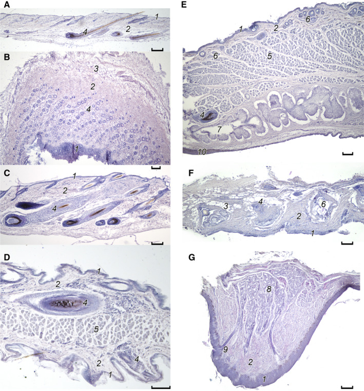
Figure 7.

Topography and structure of the tail scaly organ in anomalures. (A, B) Idiurus macrotis (C, D, E) Idiurus zenkeri. (F, G) Anomalurus beecrofti. (H, I, J) Anomalurus derbianus. (K, L) Anomalurus pusillus. (A, D, G, I, J, L) Close up view of scales. (B) Distal part of tail, lateral view. (C) General view of the ventral surface of the tail. (E) Ventral surface of the tail distal to the scaly organ. (F) Proximal part of tail with the scaly organ, ventral view. (H, K) Fragments of the tail skin with the scaly organ. Scale bars 10 mm (B, C, F, H, K), 1 mm (A, D, E, G, I, J, L)
The scaly organ in both species of Idiurus consists of small scales, without spikes. Scales are arranged in numerous transverse rows. Between the scales, scattered single hairs grow. In the distal part of the organ, the scales become sparser, many missed ‘sites’ appear; the exact position of the border between the scaly organ itself and skin folds lying distally on the ventral surface of the tail is very difficult to determine.
In I.macrotis, the length of the organ is ~ 12 mm, which makes up the seventh part of the tail length. The organ consists of a set of rows of light yellow or whitish scales (Figure 7A), which are closely spaced and arranged in rows across the long axis of the tail (five scales in each row). Thorns or spikes on scales or their bases were not found. The scales are heavily worn, drop‐shaped, as their anterior section is slightly pointed and the posterior section is broadened and rounded. The size of scales varies from 633 × 372 to 646 × 310 μm; their length is ~ 1.7–2.1 times their width. The area of scales varies from 0.13 to 0.16 mm2. Three thin (up to 1 mm long, up to 35 μm thick), colorless for the most part, broken‐off hairs grow from under the rear edge in the central part of each scale. These hairs have enlarged sebaceous glands (see below: histology of the skin). On the dorsal side of the tail, fluffed, long (up to 51 mm), bright, straight hairs (up to 56 µm thick) with a well‐developed medulla grow, while the lateral sides of the organ plate are bordered by thicker short (8–9 mm) yellowish hairs (up to 4–5 mm; Figure 7A,B).
In I.zenkeri, the organ length reaches ~ 13 mm, which is slightly less than one‐eighth of the tail length (Figure 7C). The scales are arranged in transverse rows (Figure 7D), each consisting of four large spikeless scales, sometimes supplemented with one or two small lateral scales, but the rows are not straight and scales are not as ordered as in I.macrotis. There are areas of the organ with fallen scales. The configuration of the scales is generally similar to that of I. macrotis, but they are more oval (Figure 7D), their size varies from 412 × 294 to 760 × 405 μm, and the area from 0.15 to 0.20 mm2. Two thin (up to 0.7 mm long, up to 35 μm thick), colorless for the most part, broken‐off hairs grow from under the rear edge in the central or lateral part of each scale (Figure 7D). On the tail posterior to the organ, there is a longitudinal row of thin but rather long hairs dividing the ventral surface of the tail into two halves (Figure 7E). Along both sides of the organ plate and the ventral surface of the tail posterior to the plate, there are thick strips of short (5–6 mm), slightly thickened (23–34 μm), yellowish hairs (Figure 7C–E). On the dorsal side of the tail, the hair size is up to 25–32 mm long and no more than 23 μm thick.
The scaly organ in species of Anomalurus consists of large scales with sharp spikes atop. Along the tail scales form two longitudinal rows shifted with respect to each other, so that the scales of the left and right rows alternate in a checkerboard pattern. Scales are few, they are easy to count, in all studied Anomalurus their number is 14 (Figure1C). The proximal eight to 10 scales are most prominent, the more distal are smaller in size, and the last two to three scales are quite small. The scales are located close to each other, and the scaly organ has a clear contour. In contrast to Idiurus, the hairs do not form a bristle fringe by the sides of the scaly organ.
In A. beecrofti, the length of the organ is 40 mm, which is a quarter of the length of the tail (Table 1). It consists of 16 closely spaced nail‐shaped scales, which form a wheat‐ear ornament (Figure 7F,G). No more than two scales, alternating, lie across the tail. Between the scales there are skin zones, and the hair is missing here. Light‐brown scales have a long granular surface. In the middle of the caudal edge of the scale, there is a nail‐like rounded small outgrowth (probably a broken thorn) visually separated from the main plate by a dark rim 27–29 µm wide (Figure 7G, arrow). The size of major scales varies from 6.0 × 3.1 to 6.9 × 3.1 mm; their length is almost two times the width. The area of scales ranges from 14.5 to 15.2 mm2. The largest scales lie closer to the root of the tail.
In A. derbianus, the organ length is 64 mm, which is approximately one‐quarter of the tail length. The hair on the scaly plate does not grow (Figure 7H). The organ consists of 14 light‐brown, relatively smooth triangular scales, lying in two longitudinal rows and arranged in a mosaic pattern (alternating left and right scales) (Figure 7H–J). The proximal scales, except the one closest to the tail root, are very large, they gradually decrease distally, and the last two scales (like the most proximal one) are very small. The size of major scales varies from 10.7 × 6.0 to 9.7 × 4.9 mm. The area of scales varies from 14.1 to 31.7 mm2, and decreases along the tail. At the distal protruding triangular edge of each scale, one oval base (size 1.0 × 0.7 – 2.3 × 0.1 mm) of a broken thorn (Figure 7J, arrow) is observed. Judging by the bases, the spikes were larger on the scales of the proximal part of the organ. One short (0.8 mm) and dark thorn (Figure 7I, arrow) persisted. Between the scales, the areas of bare skin can be seen.
Since it is known that A. derbianus has a well‐defined bundle of long hair on the tail, we investigated these hairs, but could not find any specific features. These hairs are not specialized, quite similar to those of the trunk, except for the length (hair of the tail is 25 mm long, hair of the tail brush is 30 mm).
In A. pusillus, the organ length is 34 mm, which is ~ 0.3 of the tail length (Table 1). The organ consists of 14 elongated scales of irregular configuration (Figure 7K,L). The scales lie in pairs across the tail and are only slightly shifted relative to each other. The surface of the scales is smooth. The size of major scales varies from 3.4 × 2.0 to 4.7 × 2.6 mm, and the area from 6.8 to 11.1 mm2. One non‐grated, short (939 μm) thorn is located in the middle back of the scale and directed caudally. A bunch of four small (2.1 mm long, 23 μm thick) whitish hairs protrude from under the middle of the rear edge of each scale.
3.4.1. Sebaceous glands of the tail scaly organ
In I.macrotis, these multi‐lobed glands lie deeply in the skin under the horny scales (Figure 9F). They are large (123.4 ± 4.8 – 96.8 ± 1.4 μm on cross‐sections) and non‐pigmented. Their wide ducts open into the follicles of hair of the tail scaly organ.
Figure 9.
Microstructure of the skin and specific skin glands in some anomalures. (A, B) Withers of Idiurus macrotis. (C) Withers of Anomalurus derbianus. (D) Patagium of Anomalurus beecrofti. (E) Upper eyelid of A.derbianus. (F) Sebaceous glands of the tail scaly organ of I. macrotis. (G) Eccrine glands in a plantar pad (thenar) of Anomalurus pusillus. (A, C, F) Cross‐section (longitudinal); (B) oblique tangential section; (D, G) transverse section; (E) Cross‐section (vertical, transverse). Photomicrographs, hematoxylin and eosin staining. Scale bars 100μm. 1, epidermis; 2, derma; 3, fat tissue; 4, hair follicle; 5, muscles; 6, sebaceous gland; 7, meibomian gland; 8, eccrine glands; 9, excretory duct of eccrine gland; 10, eyelid mucosa
3.5. Palmar and plantar pads
3.5.1. Notes on nomenclature
Many groups of mammals are characterized by the development of swellings of bare skin on the palmar and plantar surfaces of the autopodium, commonly called volar pads [torior pulvini]. In most publications, it is customary to distinguish three rows of volar pads (e.g. Whipple, 1904; Brown and Yalden, 1973; Krättli, 2001), however, their nomenclature varies considerably in different sources (e.g. Pocock, 1922; Greene, 1935; Brown and Yalden, 1973; Ari et al. 2018). The Nomina Anatomica Veterinaria (ICVGAN, 2012) does not help clarify this issue as it provides names only for the pads of the distal row; therefore, there is a need to discuss the nomenclature, adopt a compilation version and introduce some Latin names based on the topography of pads.
The most unambiguous naming is given to the pads of the distal row located at the tips of the digits. In literature, they are commonly referred to as digital pads (e.g. Pocock, 1922; Greene, 1935; Miller et al. 1964; Heaney, 1985; Haffner, 1998) or apical pads (Marquart, 2014), the Latin name 'tori digitales' also applies (Schummer et al. 1981; Kumar, 2015).
The middle row pads lying at the base of the digits, by contrast, have the most controversial nomenclature. In zoological studies, they are usually called the interdigital pads (e.g. Brown and Yalden, 1973; Heaney, 1985; Haffner, 1998; Marquart, 2014). The term metacarpal/metatarsal pads also occurs (Pocock, 1922; Ari et al. 2018), moreover, this option is well established in veterinary anatomy, since in carnivorans, the interdigital pads fuse together and form one large cushion lying at the level of the distal joints of metapodium (Miller et al. 1964; Schummer et al. 1981; Kumar, 2015). However, this nomenclature causes serious confusion, as most zoologists apply the name metacarpal/metatarsal to the third, most proximal row of pads (e.g. Brown and Yalden, 1973; Heaney, 1985; Haffner, 1998). In terms of topography and unambiguous interpretation, it would be optimal to refer to the swellings of the middle row as interdigital pads (tori interdigitales), if not for their shape in carnivorans (due to their fusion, the term interdigital is poorly applicable in this case). Therefore, since the Latin designations 'tori metacarpei' and 'tori metatarsei' are already fixed in veterinary anatomy (Schummer et al. 1981; Kumar, 2015), we propose to equate the terms interdigital pads and metacarpal/metatarsal pads and henceforth use them interchangeably only for the middle row pads.
The most proximal row of pads is usually represented by two formations, the thenar and hypothenar. These names are generally accepted and almost unambiguously used in the biological literature. However, the row itself, to which these pads belong, is called metacarpal/metatarsal by some authors, as already mentioned, but the term carpal/tarsal is also used frequently (Pocock, 1922; Schummer et al. 1981; Marquart, 2014; Kumar, 2015; Ari et al. 2018). From the topographic point of view, either pair of terms is appropriate because the well‐developed thenar and hypothenar extend both in the basipodium region and in the metapodium one, but since the first variant is already used for the middle row, we propose to abandon it in order to avoid confusion in favor of the latter one, the carpal/tarsal row or tori carpei/tori tarsei.
3.5.2. External morphology
A substantial part of the sole surface of the manus and pes in anomalures is bare, not covered with hair. These surfaces carry well‐developed palmar and plantar pads, whose topography and configuration are very specific (Figure 8A). For the first time, the pattern of volar pads in anomalurids was described by E.R. Alston (1875).
Figure 8.

Topography of the palmar and plantar pads of anomalures, volar view. (A) Scheme of the foot. (B, C) Anomalurus pusillus. (D, E) Idiurus zenkeri. (F, G) Anomalurus beecrofti, (H, I) Callosciurus flavimanus. (B, D, H) Left hand. (A, C, E, G, I) Left foot. (F) Multiple pores of eccrine glands on the hypothenar (above) and central pad (below) of the hand. Scale bars 5 mm (B–E), 1 mm (F)
A distinctive feature of all scaly‐tailed squirrels is the poor development of pads at the tips of digits (tori digitales). They are represented only by slight elevations at the base of the claws, characterized by distinctly smooth skin, completely devoid of hair. A special case is the pad of the digit I (torus digitalis pollicis). The first toe is shortened in rodents (in anomalurids, the pollex is entirely subsumed within the palm). Therefore, the digital pad of pollex is aligned with the row of interdigital pads and has a similar appearance. Interdigital pads (tori metacarpei and tori metatarsei) at the base of the fingers are very well developed. There are four of them on the manus, and six on the pes. In addition, all species have large proximal palmar (carpal) and plantar (tarsal) pads, the main of which are the thenar and hypothenar.
Homologization of pads is complicated by the fact that their configuration differs from that of the studied mammalian species. The research describing the development of plantar and palmar pads has been carried out mainly in rats and humans (Kimura et al. 1994, 2002). Without developmental data, it is not possible to distinguish with certainty some of the components of pads formed by the thenar and hypothenar from those formed by the interdigital pads (Kimura et al. 1994). Therefore, in the present study, in naming the pads, we focused on the maximum similarity in position in accordance with the simplified classical topography of pads developed by Wilder (in Whipple, 1904).
At the finger bases in anomalurids there are five pads. The lateral four of these are metacarpal pads, and the medial (radial) one is the proximally shifted digital pad of the pollex. The latter pad lies on the edge of the manus. It is approximately equal in length to the adjacent metacarpal pads, but wider and of nearly rounded shape. Three main metacarpal pads of approximately equal size are located in between the digits II–V. They are oval and elongated along the longitudinal axis of the manus. The most lateral (ulnar) pad of this row lies on the lateral edge of the manus at the base of the fifth digit; it is the smallest, about half as long as the others, and almost round. Metacarpal pads are located quite far apart in A. pusillus, A. derbianus and I. macrotis, while in I.zenkeri they are swollen and tightly pressed against each other. A. becrofti demonstrates an intermediate state.
In all studied species, except A. pusillus, there are three carpal pads; in A. pusillus there are four. The largest pad lies on the radial side of the manus (thenar). In large Anomalurus, it is elongated and oval; in Idiurus, it is nearly round and occupies a significant part of the radial half of the wrist ventral surface; A. pusillus is characterized by an intermediate state. The hypothenar lies on the ulnar side of the wrist, it is similar in shape to the thenar, varies in form depending on the species: oval in Anomalurus, or round in Idiurus. The hypothenar on the manus is slightly shifted proximal to the thenar. Between the distal ends of the thenar and hypothenar, in all species lies a small and round central pad. In fact, this pad is located in the intermediate row between the carpal and the metacarpal ones, where, as is known from developmental data (Kimura et al. 1994), both distal components of the thenar and hypothenar, as well as proximal components of metacarpal pads can lie. It is impossible to establish its origin without observations on ontogenesis, but judging by the topography, it is more likely carpal than metacarpal. Only A. pusillus has, besides this, a separate distal component of hypothenar (hypothenar distalis).
On one hand, the pads occupy the largest area in I.zenkeri; all together they cover ~ 60% of the hairless palmar surface. On the other hand, A. pusillus has the most gracile pads; the area occupied by all of them is ~ 30% of the hairless palmar surface.
The overall pattern of plantar pads on the pes differs from the palmar version only in the number of interdigital pads (six instead of four). Interdigital pads are swollen and brought close together, even in species that have pads on the manus placed apart. The four central interdigital pads lying at the base of the interdigital spaces are large and oval. The two outer ones are smaller and more rounded, and lie on the very edges of the pes. Unlike the manus, the medial pad is no larger than the lateral one, in fact they are approximately equal. The hypothenar is subdivided into the proximal and distal components in all Anomalurus; in Idiurus it is small, there is only the part of it that we consider to be the proximal component. Contrary to the manus, the hypothenar is displaced distally relative to the beginning of the thenar. In all the studied species, the thenar is very large, stretching along the entire metatarsus up to the interdigital pads, sometimes abutting them. In Anomalurus, it ends opposite to the interdigital pad I; in Idiurus it ends opposite the gap between interdigital pads I–II. The central pad is more or less developed in all species, it is small and rounded as on a manus. In Anomalurus, it is located approximately in the middle of the bare plantar skin, between the distal end of the thenar and the distal hypothenar. In Idiurus, it is displaced proximally, and is practically pressed into the gap between the thenar and hypothenar diverging at an angle.
On the foot, the pads are more swollen and closely located than on the hand. The plantar pads are placed most closely to each other in A. beecrofti and I. zenkeri, and most sparsely in A. pusillus. The plantar pads occupy the largest area in I.zenkeri, all together they cover ~ 50% of the hairless plantar surface. In A. pusillus, the area occupied by all of them is only ~ 40% of the hairless plantar surface.
3.5.3. Histology of the skin and eccrine glands of the volar pads
The firm skin of the pads is distinguished by a thickened epidermis, especially in the apical part. For instance, at a total height of the palmar pad in A. pusillus of 1005 ± 14 μm, its vertex is covered by the epidermis with a thickness of 85.4 ± 5.3 μm, while on the lateral sides the epidermis is twice as thin (47.5 ± 7.8 μm; Figure 9G). The stratum corneum of the epidermis is quite thin and peelable. Beneath is an intermittent layer of granular cells (stratum granulosum), and the base of the epidermis is formed by a multi‐row layer of spiny cells (stratum spinosum), which is underlain by the basal layer (stratum germinativum). The scaly‐tailed squirrels have a poorly developed papillary joint of the epidermis and dermis on the pads: dermal papillae are sparse, short and wide, more pronounced on the apex of the pad. The dermis is dense, without fat cells, and it consists of randomly interwoven thick bundles of collagen fibers. In the middle part of the dermis and in its deepest layers, there are the secretory tubules of eccrine glands and the blood vessels. The subcutaneous tissue (tela subcutanea) is poorly developed; it contains only a few small fat cells. The corpus of the pad is underlain by the fibers of the striated muscle, namely m. palmaris brevis, connecting the lateral and medial edges of the pad. This muscle has its portions in the thenar and hypothenar, the rest pads are devoid of striated musculature.
In anomalurids, all the surface of the skin of pads is pitted with numerous rather large pores of eccrine glands (Figure 8F). For instance, in the palm of A. pussilus these glands form a dense glandular field in the depth of the derma (Figure 9G), which covers ~ 20% of the cross‐section of the pad. Secretory tubes (their diameter is 23.5 ± 4.3 μm) lie at a depth of 384 ± 77.5 μm. They are very convoluted and curled into balls of 565 ± 45 × 333 ± 56 μm. Judging by the tall (8–10 μm) and loose secretory epithelium, the glands actively secrete. The tube cavity is filled with basophilic contents. At the mid‐height of the pad, the secretory tubes sharply pass into narrow ducts, stretching not to the sides of the pad but vertically to its apex, where the epidermis is most thick (82.0 ± 6.4 μm). The excretory ducts expand twice in the superficial derma and enter the epidermis through dermal papillae between epidermal ridges.
3.6. Histology of the skin and specific skin glands
The hairy skin has the typical mammalian structure (Montagna and Parakkal, 1974). It is unpigmented, with a thin, relatively smooth epidermis. The border between the papillary and the reticular layers of dermis is not prominent. The papillary layer contains thin bundles of randomly arranged collagen fibers. In the reticular layer, there are clusters of fat cells associated with deep‐lying follicles of growing hair. Subcutaneous fatty tissue is developed only locally.
3.6.1. Skin of the withers
In I.macrotis, the skin of the withers is non‐pigmented and thin (165.5 ± 7.2 μm), with a thin (11–15 μm) weakly folded epidermis (Figure 9A). Thickened (up to 249 μm) regions of skin with large growing hairs are found. The corneous layer (stratum corneum) is also very thin (no more than 6 μm), consists of the basal (stratum germinativum) and spinous (stratum spinosum) layers, but the granular layer (stratum granulosum) is absent. The papillary dermis occupies approximately one‐third of the thickness of the dermis, but in case of growing hairs two‐thirds. It does not clearly differ from the reticular layer in thickness and configuration of bundles of thin collagen fibers. These collagen bundles are strongly crimped and intertwined in different directions. The hair grows singly, in irregular rows, the diameter of their follicles is 23–45 μm, but in growing hair it reaches 68 μm (Figure 9B). They have very thin muscles (musculus arrector pilli), and some of them have one small (34 × 34–79 μm) sac‐shaped sebaceous gland (glandulae sebaceous pili). Bulbs of growing hairs are surrounded by clusters of fat cells measuring 11 × 34–45 μm. Sweat glands (glandulae suborifera) are absent. The subcutaneous fatty tissue (tela subcutanea) is developed. Follicles of three found vibrissae are large (131.1 × 89.1; 100.0 × 195.1; 113.0 × 195.1 μm), with a very thick connective tissue capsule, a well‐developed annular blood sinus and large sebaceous glands (Figure 10A–C).
Figure 10.

Vibrissae in the skin of withers in Idiurus macrotis (A, B, C) and Anomalurus beecrofti (D, E). Photomicrographs, hematoxylin and eosin staining, oblique tangential section. Scale bars 100μm. Number definitions as in Figure 9, additionally: 11, vibrissae
In A. beecrofti, the thickness of the folded non‐pigmented skin is 344 ± 33.0 μm in areas with growing hairs (Figure 9C). The epidermis is thin with a subtle corneous layer and without a granular layer. The papillary layer occupies two‐thirds of the thickness of the dermis. Small hairs (23–34 μm in diameter) grow singly or in groups of three. Large growing hairs have follicles with a diameter of up to 56 μm, which lie to a depth of subcutaneous tissue. Sack‐shaped small (34–45 × 34) sebaceous glands are found not in every hair. The arrector pilli muscles are small or absent. Fat cells surround the bulbs of large hairs. Two large vibrissae were found (per 1 mm of the longitudinal section; Figure 10D,E). They have a prominent collagen capsule, small blood sinuses, and are equipped with two large sebaceous glands.
In A. derbianus, the skin is non‐pigmented, folded, up to 226 μm thick in areas with non‐growing hairs, and up to 445 μm thick in places of intensive hair growth (Figure 9C). The papillary layer occupies half the thickness of the skin. The papillary and reticular layers differentiate poorly. Most bundles of collagen wrinkle weakly, stretching parallel to the surface of the skin. Clusters of large (68 × 68 – 79 × 79 μm) fat cells are located along the bulbs of large hairs. Subcutaneous fatty tissue is poorly developed. Hairs grow singly or three grow out of one skin funnel. Their follicles range in size from 34 × 34 to 56 × 56 μm. Some large hairs have two sac‐shaped small (45 × 34; 56 × 34 μm) sebaceous glands.
3.6.2. Skin of the patagium
The skin of the patagium is very specific. The space between the dorsal and the ventral skin is very narrow. The deep skin folds in the dead specimens indicate that the skin is elastic and can stretch and contract (Figure 9D). Further details are considered in A. beecrofti. The structure of the strongly folded skin differs on the dorsal and ventral sides of the patagium (Figures 9D, 11). In the area of the hair ‘brush’ the skin is thick (489 ± 58.1 μm). The core of the patagium between the dorsal and the ventral skin layers is filled with the powerful striated musculature. The thickness of the core muscle layer is 37.5% of the total patagium thickness, 40.8% falls on the dorsal layer of the membrane skin, and 21.7% on its ventral layer. The epidermis on the dorsal and ventral sides is thin, does not contain pigment and granular cells, but is covered with a weak intensely exfoliating horny layer. The dermis is homogeneous. It contains thin collagen and elastin fibers that are strongly crimped and stretching along the skin surface. The dermis is plentifully vascularized, has numerous foci of inflammation and encapsulated parasites. Both sides differ in the size of the hair follicles. On the dorsal side, the follicles of large hairs of the ‘brush’ reach 404 × 151 μm, and in small hairs of the ventral skin, they are 56 × 68 μm. Bulbs of large hairs reach the muscle layer, and bulbs of small hairs occupy two‐thirds of ventral skin thickness. Follicles of both types have one baggy sebaceous gland on the front wall, the size of which in large hairs (113 × 68 μm) is twice that in small ones (56 × 34 μm). Subcutaneous fat cells are absent.
Figure 11.

Microstructure of the patagium of Anomalurus beecrofti on a cross‐section. Dorsal side of the patagium, above; ventral side, below. Photomicrographs, Mallory's staining. Scale bars 0.1 mm. 1, epidermis; 2, hair follicle; 3, collagen fibers of derma; 4, blood vessel; 5, muscles
The degree of the sebaceous glands development (their number, size, etc.) in the hairs of the patagium is low. Probably because of the absence of such lubrication, the epidermis of the patagium peels off very actively; the corneous layer is 32–49 μm thick in the patagium, while it is only 11–17 μm in the withers.
3.6.3. Meibomian skin glands
The meibomian glands (glandulae tarsales) of the eyelids have a location and structure typical for mammals (for review see Sokolov and Chernova, 2001). They lie in a row along the edges of the eyelids. Meibomian alveolar glands of the upper eyelid of A. derbianus have typical location and structure of multi‐lobed sebaceous glands with a system of wide branching ducts (Figure 9E). Their size is 567.1 ± 28.9 – 243.9 ± 49.1 μm (on cross‐sections). These glands have no pigmentation, and open on the inner surface of the eyelid, without any connection with large eyelashes.
4. DISCUSSION
4.1. Patagium
The scaly‐tailed squirrels have all three sections of the patagium typical of mammal gliders. Although videos of gliding are not available because of the relative rareness of the anomalures and difficulties with regard to field observations, nevertheless, judging by the preserved specimens, it can be confidently stated that the propatagium does not spread so much on the head as in colugos (Dermoptera) and flying squirrels (Sciuridae, Rodentia), but rather stretches from the region of the shoulder joint (base of the neck) as in sugar gliders (Marsupialia). The uropatagium includes only the proximal part of the tail as in all gliders with long bushy tails. The uropatagium is quite well developed, more like in the giant flying squirrel Petaurista than in the marsupial gliders. A distinctive feature of the plagiopatagium is that it is additionally supported by the unciform element connected to the elbow joint, which is not found in any other extant or fossil mammals.
4.2. Unciform element
There are essential differences in the position of the spur and its origin between the flying squirrels (pteromyins) and the scaly‐tailed squirrels (anomalurids). The unciform element of anomalures is attached to the ulnar bone in the elbow joint, while the styliform element of flying squirrels inserts on the pisiform bone of the wrist (Thorington and Stafford, 2001). The data accumulated to date indicate that the spur of Pteromyini is derived as a result of an overgrowth of the hypothenar cartilage characteristic of non‐gliding squirrels (Kawashima et al. 2017).
Histologically, the spur of anomalures is generally similar to that of flying squirrels (Oshida et al. 2000), as far as it is possible to evaluate given the rather poor data on the structure of the styliform element. It also has a large number of chondrocytes in the core interspersed with collagen fibers. However, the spur of flying squirrels seemingly has fewer fibers, whereas in the spur of anomalures the cartilage is more fibrous. In adult flying squirrels, in addition to fibrous cartilage, the base of the spur proximal part contains areas of bone tissue (with age the spur ossifies from the proximal end in the distal direction, the ossification area reaches the middle of the spur in old individuals). In scaly‐tailed squirrels, no areas of ossification were found.
The histological structure of the anomalurid spur argues for its emergence from the distal end of the triceps tendon as a result of the chondrification of a ligamentous precursor. The chondrification or even ossification of the triceps tendon in the region of the olecranon (patella ulnaris) occurs in mammals, at least in bats (Haines, 1940; Amador et al. 2018). In our opinion, this is the most important structural and evolutionary difference between the two structures supporting the membrane of the rodent gliders: one is purely muscular in origin (Anomaluridae), and the other is skeletal one (Pteromyini).
In the course of evolution, at the stage when the gliding membrane reached only the elbow, the spur could have begun to form as a small tendon‐cartilaginous support for the membrane, helping to increase the wing span. In various groups of gliding mammals, the maximal wing span is achieved in a different way. In pteromyin flying squirrels, the membrane tip is supported by the styliform element, in the marsupial sugar glider and its congeners by the fifth digit, and in other gliding possums the wing tip is at the elbow. The design of the flying apparatus in the scaly‐tailed squirrels suggests the following role of the unciform element: when the membrane is maximally stretched, the forelimbs are protruded craniolaterally and the spur is sprawled laterally, and thus supports the membrane at the point of its greatest width, i.e. makes the wing tip. Nevertheless, in the lack of images of gliding anomalures, it is difficult to verify this hypothesis. However, manipulations with fluid‐preserved specimens showed that the widest part in the membrane is precisely in the area of the spur, which indirectly supports our view. In addition, it becomes obvious that the membrane becomes fully stretched only in case the forelimbs are protruded not laterally, but craniolaterally.
Of particular interest are the muscle fibers extending from the spur into the membrane. At this stage of research, it is hard to establish their homology, although the most likely candidate for the source of this musculature is musculus cutaneus trunci. However, it can be confidently stated that the connection of this musculature with the spur is secondary. After the formation of the spur in the evolutionary process some fibers of the membrane muscles proliferated to this area and became attached to the spur. The muscles are not involved in the movements of the spur, its abduction occurs due to tension of the plagiopatagial part of the membrane under the air pressure. Nevertheless, contraction of these muscle fibers (while gliding), can regulate the wing camber. The main function of this musculature, in our opinion, is picking up of the membrane so that it does not interfere with non‐aerial locomotion.
4.3. Pelage
4.3.1. Vibrissae apparatus
Vibrissae apparatus is well developed in all studied species. The main feature of the pelage of scaly‐tailed squirrels is, of course, the presence of the vibrissae on the body, especially in the withers area. This feature is unique among mammals. In most terrestrial rodents, the topography of vibrissae is not very diverse and is mainly limited to the area of the muzzle (facial vibrissae or whiskers), but in some species, in particular in specialized subterranean or arboreal forms, the location of vibrissae is more diverse (Sokolov and Kulikov, 1987). For example, in the red squirrel (Sciurus vulgaris), in addition to facial vibrissae, there are carpal and ventral body vibrissae (Bresslau, 1912). The entire hair sinus system of the squirrel is elaborately developed, which indicates its particular functional importance (Hyvärinen et al. 1977). It can be assumed that the vibrissae apparatus plays an important role in the orientation system. Additional areas possessing vibrissae can serve for tactile control of ‘blind zones’. Thus, moles (Talpa spp.) have vibrissae on the tail, whereas in the naked mole rat (Heterocephalus glaber), they are found all over the body. Collectively, in anomalurids, the tips of the widely spread vibrissae cover a significant sensory area in front of the animal's muzzle; the fact they have vibrissae on the withers indicates the importance of the ‘blind zone’ control behind the head. Since the scaly‐tailed squirrels use hollow tree trunks as roosting places, it appears particularly helpful in the complete darkness of the shelter for them to feel the surrounding space not only with the ventral side but also with their back. Vibrissae located on the dorsal surface of the body can serve to do this.
There are topographic differences between the two genera in vibrissae location. For example, in small Idiurus, we did not find supraorbital and buccal vibrissae, which are present in larger Anomalurus. Moreover, larger species have the greater absolute length of the face vibrissae, but their relative length is shorter.
The vibrissae of representatives of two genera have common and different structural features. Common features are: (a) poor development of the medulla, which is typical for hair that needs a strong cortical component; (b) a non‐specialized cuticle usual for robust hairs; (c) a broken cuticle at the ends of the vibrissae through coarse mechanical impact, while in contact with substrate. In the studied species, the structure of the medulla is very different from that of usual hairs of the trunk, as well as from the types of vibrissa medullary structure known in other mammals (Chernova and Kulikov, 2011; Chernova et al. 2012, 2015).
Earlier we identified several types of vibrissae shaft structure (Chernova and Kulikov, 2011; Figure 12). The vibrissa shaft is not different from that of the hair; it has a one‐ or two‐row ladder form (Figure 12A). The medulla contains no large air cavities; it is filled with a row of tightly adjoining, non‐perforated achromous round keratinous polyhedrons or cylinders oriented at an angle to the longitudinal axis of the vibrissa shaft (Figure 12B,C). In the carnivores studied, these cylinders form a one‐row ladder with narrow, air‐filled spaces between them, whereas in rodents, the polyhedrons form a two‐row ladder (Figure 12C). The medulla contains regularly arranged dense pyramidal septa that divide the cavities of the medullary canal into fragments with polymorphic configuration. The septum walls and longitudinal axis of the shaft of vibrissa form an angle of ~ 45°–60°. The body of the pyramidal septum has extremely dense root‐shaped outgrowths attached to the wall of medullary canal (Figure 12D). One more type of the vibrissa shaft structure was found among the studied anomalures. In I.macrotis, air cavities are virtually absent, and the entire medullary canal is filled with longitudinally oriented thin keratin membranes (Figures 6C, 12E). The scarious structure of the medulla walls, similar to that observed in Idiurus, was found in the vibrissae of another African rodent, the gundi (Ctenodactylus gundi, Ctenodactylidae; unpublished data of O.F.C.).
Figure 12.

Mechanical design of the vibrissae medulla in different mammal species. (A) Phodopus roborovskii. (B) Sylvaemus sylvaticus. (C) Mustela nivalis. (D) Hylomys megalotis. (E) Idiurus macrotis. Scanning electron microscopy. Scale bars 10 μm
4.4. Patagium brush
In the pelage of the patagium, the most interesting feature is the presence of the specialized hair brush on the dorsal surface of the patagium, immediately posterior to the unciform element. In shape, size and structure the hairs of the brush differ considerably from the body fur. Considering the peculiar structure of the medulla, they are in some way intermediate between large guard hairs and vibrissae of other mammals. The similarity with vibrissae, despite the absence of blood sinuses, is enhanced by unusual polymorphic and thickened walls of the medullary cavities, which, jointly with the thick collagen sheath of the root and the well‐developed cortex, undoubtedly increase the strength of these hairs. The probable function of the brush is to add thickness to the leading edge of the flying membrane in the region of maximum wingspan. Due to this, the wing acquires better aerodynamic properties. The absence of the arrector pili muscles in these hairs indicates that they passively shape the airfoil profile at the most important part of the wing.
4.5. Volar pads
All studied species of anomalures have well‐developed complexes of pads on the non‐haired volar surface of the hand and foot. The presence of volar pads is characteristic of many mammal species, and it is generally accepted that three series of these are distinguishable: at the tips of the fingers (digital pads), at the bases of the fingers (interdigital pads), and at the proximal pads (the thenar and hypothenar; Whipple, 1904; Brown and Yalden, 1973; Schummer et al. 1981). Different lineages of mammals have a specific set of pads, but the pattern and shape of the pads are much more dependent on the mode of life.
The palm of rodents is characterized by the following features. There are four similar digital pads at the tips of the digits, plus a special pad at the pollex apex, whose position and form depend on the degree of reduction of the digit I. Digital pads can be either underdeveloped, inconspicuous, representing a section of hairless skin in the base of the claws, or markedly swollen, occupying the entire distal phalanx. The latter variant is more characteristic of mammals actively using grasping; among rodents it is found, for example, in the climbing murid Chiropodomys, and it is quite common in primates and arboreal diprotodont marsupials. The greatest development of digital pads is achieved in the marsupial Acrobates, which uses adhesion when moving on smooth vertical substrates (Rosenberg and Rose, 1999). In scaly‐tailed squirrels, these pads are quite poorly developed, similar to S. vulgaris, but are less prominent than in Pteromys volans, and much less than in various semi‐arboreal rats, e.g. Rattus, Niviventer, Maxomys, and especially in scansorial Chiropodomys (our unpublished data). The digit I is entirely submerged in the thickness of the palm, however, it retains two phalanges in addition to the metacarpal element. As a result, the digit I barely reaches the distal ends of the metacarpals of the other digits, so that its digital pad lies in line with the interdigital pads. This pattern is typical, for example, of rats (Kimura et al. 1994), dormice Dryomys (Spitzenberger and Eberl‐Rothe, 1974), Muscardinus (Haffner, 1998), and many others. Another even more proximal location of the digital pad of pollex, at the level of thenar, is common in squirrels (Pocock, 1922; Cartmill, 1974; our unpublished data). In some of them, for example, in Pteromys, Glaucomys and Petaurista, it merges completely into the thenar and becomes indistinguishable from it (Pocock, 1922; our unpublished data). Meanwhile, in Zenkerella, the only non‐gliding representative of the Anomaluridae, the digital pad of pollex takes an intermediate position. The manus of Zenkerella is very elongated and narrow, and the digital pad of the pollex lies on its medial margin, between the metacarpal pad I and the thenar (Heritage et al. 2016). A similar position of the pollex is observed, for example, in Neotamias quadrivittatus (Pocock, 1922).
The number of interdigital pads on the palm of gliding anomalures is four, which exceeds the standard number in rodents, which is three, as well as that in Zenkerella. Four pads are characteristic of some other groups of mammals, for example, tree shrews (e.g. Panyutina et al. 2015). However, we found that squirrels of the subfamily Callosciurinae can have four interdigital pads (Menets berdmorei and Callosciurus prevostii) and even five (Callosciurus flavimanus; Figure 8H). Generally, it can be noted that by location and shape, these pads in scaly‐tailed squirrels are most similar to those of Callosciurus. The pads are elongated into high ridges, whereas usually they are rather rounded, even in the majority of arboreal squirrels (Pocock, 1922).
The proximal row in rodents typically consists of two pads. Both the thenar and hypothenar can be divided into distal and proximal pads. Embryological data show that, in Rattus, both the thenar and hypothenar have distal components, although they are fused to the marginal interdigital pads (Kimura et al.1994). Thus, without developmental observations, it is difficult to distinguish them. However, in some mammals, the distal parts of the thenar and hypothenar are located separately, and then they can be recognized quite confidently. Among our studied species, distal hypothenar and proximal hypothenar on the hand are developed in A. pusillus. The same pattern is typical for Zenkerella, and its distal hypothenar is practically equal in size to the proximal hypothenar (Heritage et al. 2016).
The most striking feature of the set of palmar pads in all members of Anomaluroidea is the presence of a small round pad between the thenar and hypothenar, which we called the central pad. We did not find a structure similar in position in any mammal, either in the studied specimens or in the literature. Based on the palm pattern of Idiurus, it could be assumed that this pad is the distal hypothenar (Figure 8D). However, after study of the palms of A. pusillus (Figure 8B) and Zenkerella (Heritage et al. 2016), which both have two hypothenar components, it becomes obvious that the central pad occupies quite a different position. Judging by the location, this pad appears to be an entirely functional formation, which is needed as a supporting and shock‐absorbing element in the central part of the palm. It is unclear, however, why such an element is not required for arboreal sciurids and especially for the flying squirrels.
The pattern of the pad arrangement on the foot is very similar to that on the hand. The main differences include the ‘normal’ position of the digital pad of hallux because the toe is not reduced. In most rodents (including Zenkerella), there are four interdigital pads (Pocock, 1922; Howell, 1926; Heaney, 1985; Kimura et al.1994; Krättli, 2001; Bezuidenhout and Evans, 2005; Marquart, 2014; Heritage et al. 2016), rarely there are five of them, for example, in Sundasciurus (Heaney, 1985), while Anomalurus and Idiurus have six of these pads. In this, gliding scaly‐tailed squirrels again are most like species of Callosciurus which also have six of them (Figure 8I). The most medial of the interdigital pads in Anomalurus closely adjoins the distal end of the thenar, so the theory that by origin it is in fact the distal thenar cannot be rejected. By contrast, in Idiurus, the thenar abuts the gap between the interdigital pads I and II. It is worth noting that in Callosciurus the thenar contacts the interdigital pad II. The latter is much elongated compared to the other interdigital pads, which suggests that the distal portion of the thenar contributed to its formation. The foot hypothenar in all Anomalurus consists of separate proximal and distal pads, the same is observed in Zenkerella, whereas in Idiurus, only the proximal component is expressed. Like the palm, the plant of gliding anomalures also bears a small round central pad that is not found in other mammals; apparently, Zenkerella does not have it either (Heritage et al. 2016). It looked very pronounced in all adult specimens of Anomalurus and Idiurus, whereas in the young individual of A. pusillus, it was barely distinguishable. A tiny structure similar in position to the central pad was found in several specimens of Nannosciurus melanotis (Heaney, 1985), but the provided description gives no arguments for it reliable identification. In the pes of all scaly‐tails including Zenkerella, both the thenar and hypothenar are very well developed; together they occupy a fairly large area.
The comparison with the family Sciuridae shows that the pattern of tarsal pads in anomalurids is most similar to that of callosciurine squirrels. However, Anomalurus differs from both Idiurus and callosciurines by the subdivided hypothenar. Well‐developed swollen pads resembling those of anomalures are characteristic of the arboreal Callosciurus, whereas the mainly terrestrial Menetes has rather small pads. The tarsal pads are missing in some arboreal squirrels such as P. volans, Glaucomys volans, S. vulgaris, Tamiasciurus hudsonicus (Pocock, 1922; our unpublished data), or presented only by a small thenar in Sciurus carolinensis, S.niger, Myosciurus pumillio, and Exilisciurus spp. (Pocock, 1922; Heaney, 1985). The thenar and hypothenar also completely disappear in the foot of a number of strictly terrestrial and semi‐arboreal squirrels of the subfamily Xerinae (Pocock, 1922; Ognev, 1963).
Based on the general notions of locomotion and the role of the pads, it could be assumed that the small thenar and hypothenar on the hind limb are associated with the digitigrade posture (normally, the proximal part of the foot does not come in contact with the substrate, and the pads on it are therefore needless). In contrast, perfectly developed, elongated thenar and hypothenar of the pes could be regarded as an adaptation to the claw climbing in which foot reversal is used during the descent, and the entire plantar surface is tightly pressed to the substrate. Within this logic, the disappearance of these pads on the foot of terrestrial squirrels is quite understandable (Pocock, 1922). Similarly, the poor state of the proximal plantar pad development in M. berdmorei, somewhat resembling that in rats, corresponds well with the semi‐terrestrial lifestyle of this squirrel (Thorington et al. 2012). Accordingly, the predominantly arboreal Callosciurus has extremely developed and elongated thenar and hypothenar; the same pattern is observed in the arboreal Zenkerella. The flying squirrels P. volans and G. volans, for which the use of foot reversal during descent is not a key specialization (as they can go down by gliding), do not have tarsal pads. Anomalurus and Idiurus also glide, but they spend a lot of time on vertical trunks, which requires static clinging. Available photos show that while positioning on the tree trunk, they press the entire surface of the pes to the substrate. Thus, the differences in shelter preferences could satisfactorily explain the hypertrophy of the proximal plantar pads in the scaly‐tailed squirrels and their absence in the flying squirrels. However, some sciurine squirrels (S. vulgaris and T. hudsonicus) absolutely do not fit into this concept; perhaps this is the most amazing and unexpected finding related to pads. Their feet are completely devoid of the thenar and hypothenar, although these are specialized arboreal animals actively using foot reversal when descending.
Pocock (1922) proposed an elegant explanation for the absence of the thenar and hypothenar in the foot of these squirrels. Having studied the red squirrel (S. vulgaris) in different seasons, he showed that in winter, its plantar surface was noticeably haired, except for the skin of the pads. The zone of the missing thenar and hypothenar was completely covered with hairs. In summer, the plantar skin of these squirrels was almost bare. Based on this, he suggested that the pads could have been eliminated for the sake of increasing the area of the skin that can bear pelage. This can indeed be critically important for small mammals inhabiting northern latitudes. In this context, the disappearance of the thenar and hypothenar in flying squirrels can acquire a different explanation.
Obviously, the role of the volar pads in the locomotion of arboreal rodents is significant, but it is not so far possible to clearly correlate the shape, size and topography of pads with the lifestyle or the locomotion mode. There is probably a wide range of reasons, which determine certain characteristics of a set of pads. This issue seems to us very interesting and promising for further research.
As to the internal structure of the pads, its correlation with the mode of life has been relatively well studied. It is shown that in fossorial forms the surface of pads is flat and the glands are few or absent. Scansorial mammals have broad, soft, cushion‐like pads containing numerous glands, the secret of which supposedly increases the coefficient of friction. In species that actively use the grasping mode (Muscardinus avellanarius and Micromys minutus), pads are abundantly filled with fat cells (Haffner, 1998). In the histologically studied pads of anomalures, fat cells in the dermis are absent, and in the subcutaneous tissue they are rare or also absent. This reduces pad compliance. It can be assumed that compliance is important when grasping, so that the sole fits the substrate well and creates a larger contact surface. During claw‐climbing tree trunks the rigid pads seem to be preferable, as they can act as additional anchors on the roughness of the bark. In this case, instead of adhering to thin branches as do graspers, they wedge into the holes and slits of the substrate.
Interestingly, unlike the pads of many terrestrial mammals (Schaffer, 1940; Montagna and Parakkal, 1974; Petrishchev and Chernova, 1988; Sokolov and Chernova, 2001), scaly‐tailed squirrels have a poorly developed papillary joint of the epidermis and dermis: dermal papillae are short and sparse.
The pads of anomalures contain numerous eccrine glands. Eccrine glands are typical for mammalian volar skin and lubricate it (Schaffer, 1940; Sokolov, 1982; Haffner, 1998). These are tubular glands assembled in a tight ball which lie in the depth of the skin and through narrow, long, non‐convoluted ducts deliver the secret to the skin surface. The ducts are orientated perpendicular to the surface of the skin and expand funnel‐like near the excretory pore. It is interesting to note that the ducts pass the border of the dermis and epidermis through the dermal papillae (cones), as in hyraxes (Hyracoidea), but not through the epidermal protrusions (ridges or cones), as in other rodents (Sokolov and Sale, 1981; Haffner, 1998). In the studied palm of A. pusillus, the ducts of the eccrine glands open at the apex of the pad, rather than on its lateral sides, although this is the longest way to the skin surface contrary to that through the relatively thin epidermis of the lateral walls of the pad. The ducts arranged in this way are not squeezed upon the flattening of the pad when it is in contact with the support. However, most importantly, the ducts opening at the apex of the pad provide its lubrication in the most critical zone in terms of interaction with substrate.
In arboreal rodents, the pads are distinguished by a large number of eccrine glands in combination with the thick stratum corneum (Haffner, 1998). This applies to both grasping and claw‐climbing animals. It can be assumed that a thick stratum corneum in combination with a lubricant serves primarily to minimize the wear of the contacting volar surface. However, increasing friction due to lubrication can also be an essential task, especially for grasping mammals, which control their position while climbing due to the friction that occurs when the hand or foot grips a substrate. It is relevant as well for those claw‐climbing mammals that use vertical supports as resting places, i.e. that are compelled to be fixed on them for a long time. Here, the claws cling to surface roughness and the soles are pressed against the substrate, and due to this pressing the arising frictional force directed vertically can help the pads to partially unload the claws.
In all scaly‐tailed squirrels there are remarkable sickle‐curved claws associated with large bundles of short, but coarse, possibly tactile hairs. The claws on all digits are very long, latero‐medially flattened. Of all the mammals studied by us, the claws of anomalures most resemble in shape those of Cynocephalidae (see Figure 2.9 in Panyutina et al. 2015). They are significantly larger than those of semi‐arboreal and even scansorial rodents. The state of the claws development in Anomaluridae clearly indicates that they use the claw‐climbing mode and do not implement grasping at all.
4.6. Tail scaly organ
The location of the tail scaly organ is similar in all studied anomalures; it occupies the basal part of the ventral surface of the tail. This corneous formation is unique and was the reason behind the common names of these animals. The relative size of the scaly organ in small anomalures is smaller: in Idiurus it is 0.10–0.13 the tail length, while in large Anomaurus it can reach 0.26–0.28 the tail length. The structure of the organ is species‐specific; differences are observed in: (a) number of scale rows running along the tail (five in Idiurus and two in Anomalurus); (b) pattern formed by the scales – irregular transvers rows in Idiurus and alternating left and right scales in Anomalurus; (c) size (including thickness and degree of keratinization); (d) configuration of the scales; (e) coloration; (f) form, size, and degree of wear of their spikes – one at the scale trailing edge in Anomalurus, but their complete absence in Idiurus. In shape, size of scales and their arrangement in two rows, the scaly organ of Zenkerella is similar to that of Anomalurus. We cannot be confident without proper examination, but according to the published photo (Heritage et al. 2016; Figure 1D) Zenkerella also has spikes, like Anomalurus.
Hypertrophic sebaceous glands were found by us in the ventral part of the tail root of I. macrotis (in other species, this region was not histologically investigated). These glands lie in the skin under the scales of the tail scaly organ and excrete sebum through the follicles of hairs growing from under the scales. They certainly provide fatty lubrication of thick horny scales, which helps to preserve their elasticity and protect against mechanical damage and parasitic invasions. We suggest that similar glands are developed in other anomaluroids. We have not yet been able to confirm this assumption because of the technical complexity of the histological processing of this solid scaly skin and the need to maximize the retention of objects.
It is known that some mammals also have specific subcaudal sebaceous glands on the ventral side of the tail base, which play an important role in chemical communication. These are desmans (Desmana moschata and Galemys pyrenaicus), the four‐toed elephant shrew (Petrodromys tetradactylus), the greater white‐toothed shrew (Crocidura russula), pocket mice (Liomys irroratus and Perognathus spp.), Eurasian field mice (Apodemus spp.; for a review, see Sokolov and Chernova, 2001). Many of them mark substrate while landing after jumps. Sometimes the specialization for chemical communication using the subcaudal sebaceous glands even affects the shape of attendant hairs: they acquire a club‐shaped swelling at the tips and turn into peculiar swabs, as was shown in Petrodromus tetradactylus (Sokolov et al. 1980). In anomalures, the hairs of the scaly organ are not specialized, and there is no evidence that the secretion of the scaly organ glands is used for chemical communication. However, such a function of these glands can not be completely rejected as specific chemical substances can easily remain on substrate as a result of the tail contact with a tree trunk.
In conclusion, as a result of morphological study of the skin, its derivatives and associated structures in five species of the African scaly‐tailed squirrels, a number of unique features that might ensure fitness to the environment of these enigmatic mammals were revealed. Peculiarities of their lifestyle leave a significant mark in the integument characters as compared to other rodents:
The design of the membrane supported by the elbow spur indicates that in gliding the scaly‐tailed squirrels keep their forelimbs directed strongly forward, whereas the unciform is abducted laterally and supports the widest point of the membrane;
The structure of the unciform element provides an argument in favour of its origin from the triceps tendon, but not from any element of the skeleton. The musculature, associated with the unciform emerged from the subcutaneous muscle (m. cutaneus trunci) and its attachment to the spur is secondary;
The pattern of the tail scaly organ is species‐specific and largely depends on the size of the animal. Variations in its topography and structure, such as the presence of spikes in large species of Anomalurus, and their absence in small Idiurus, are apparently associated with the difference in the load experienced by the tail organ when clinging to a tree trunk surface. For small and light Idiurus, even a minor roughness is sufficient for creation of the friction necessary for body support, whereas large, heavy Anomalurus require spikes that are close in properties to the claws on the digits;
Excellent development of the vibrissae apparatus in anomalures indicates the high tactile orientation ability. The presence of vibrissae in the withers in representatives of both genera proves the importance of tactile control of the space behind the head. This seems to be important, given that all studied species use hollow tree trunks as roosting places, i.e. actually inhabit tubular‐shaped shelters, where the vibrissa apparatus can not only replace vision but also work as ‘eyes in the back of the head’. The vibrissae of A. derbianus are unique since we have not seen such a medullary structure in any of the numerous mammals that we have previously studied;
The specialized thick and straight hair on the dorsal surface of the patagium, just behind the unciform element, presented in all studied species most likely perform an important function of contributing to the improvement of the airfoil properties. In flight, these hair do not hike up under the oncoming air flow but give additional thickness to the membrane in the region of maximum wingspan, thereby providing a more perfect profile at the leading edge. The lack of muscles that control the hair suggests that they maintain the needed shape passively, which is not surprising, inasmuch as no hair muscles could withstand the air flow;
The glandular apparatus of the skin is poorly developed, as far as can be judged from the study of specific skin glands, which like in most rodents, are represented by the usual sebaceous glands of hair and vibrissae, eyelids (meibomian), and eccrine sweat glands of the soles. Enlarged sebaceous glands of the tail scaly organ were described for the first time. Probably their function is to lubricate the scales to reduce their attrition rate and brittleness avoidance.
To summarize, Anomaluridae are characterized by a unique set of integumentary features. Some of them, but not all, are specific adaptations to gliding. The others are associated with a very specific lifestyle, which combines arboreal locomotion and the use of tube‐shaped shelters.
ACKNOWLEDGEMENTS
This research would not have been possible without the support of Wim Wendelen (Royal Museum for Central Africa, Tervuren, Belgium) who kindly supplied all the scaly‐tailed squirrel specimens for study. The authors are deeply grateful. We also thank T. V. Vassilieva for consulting on histology, as well as A. N. Kuznetsov and A. A. Panteleev for discussion of the manuscript. E. L. Yakhontov helped with the English language of the paper. We are very grateful to Laurent Marivaux for the tremendous work in improving the text. This study was supported by the Russian Foundation for Basic Research (grant 18‐04‐01301).
A.A.P. conceived and designed the study. O.F.C. and A.A.P. acquired the anatomical data, O.F.C. and I.B.S. provided the histological data and measurements. A.A.P. and O.F.C. drafted the manuscript and prepared the figures. Analyses and interpretations were performed by A.A.P. and critically reviewed by O.F.C. and I.B.S. All authors discussed the paper and gave final approval for publication.
Panyutina AA, Chernova OF, Soldatova IB. Morphological peculiarities in the integument of enigmatic anomalurid gliders (Anomaluridae, Rodentia). J. Anat. 2020;237:404–426. 10.1111/joa.13211
Footnotes
The paper discusses the origin of the styliform cartilage from the ulnar sesamoid (according to Gupta, 1966) or the hypothenar cartilage (as in Thorington & Darrow, 2000). This discussion is superfluous, since by analyzing the term's origin, it becomes clear that one and the same structure is meant (Greene, 1935; Holmgren, 1952).
Data Availability Statement
The data that support the findings of this study are available from the corresponding author upon reasonable request.
References
- Adams, W.H. (1894) On the habits of the flying‐squirrels of the genus Anomalurus . Proceedings of the Zoological Society of London, 62, 243–246. [Google Scholar]
- Alston, R.E. (1875) On Anomalurus, its structure and position . Proceedings of the Zoological Society of London, 43 88–97. [Google Scholar]
- Amador, L.I. , Giannini, N.P. , Simmons, N.B. and Abdala, V. (2018) Morphology and evolution of sesamoid elements in bats (Mammalia: Chiroptera). American Museum Novitates, 3905, 1–41. [Google Scholar]
- Ari, H.H. , Kuru, N. , Uslu, S. and Ozdemir, O. (2018) Morphological and histological study on the foot pads of the Anatolian bobcat (Lynx lynx). Anatomical Record, 301, 932–938. [DOI] [PubMed] [Google Scholar]
- Bezuidenhout, A.J. and Evans, H.E. (2005) Anatomy of the woodchuck (Marmota monax). American Society of Mammalogists Special Publication, 13, 1–180. [Google Scholar]
- Bresslau, E. (1912) Die ventralen Tasthaare der Eichhörnchen, ihre Funktion und ihre Verbeitung. Zoologische Jahrbücher, 15(Suppl), 479–492. [Google Scholar]
- Brown, J.C. and Yalden, D.W. (1973) The description of mammals – 2 Limbs and locomotion of terrestrial mammals. Mammal Rev, 3, 107–134. [Google Scholar]
- Cartmill, M. (1974) Primate locomotion In: Jenkins F.A. (ed). Pads and claws in arboreal locomotion. NY: & London: Academic Press, pp. 45–83. [Google Scholar]
- Chernova, O.F. and Kulikov, V.F. (2011) Structural differences between the shafts of mammalian vibrissae and hairs and their causes. Dok Biology Science, 438, 182–185. [DOI] [PubMed] [Google Scholar]
- Chernova, O.F. , Kulikov, V.F. and Abramov, A.V. (2015) The hair structure of the long‐eared gymnure (Otohylomys megalotis). Proceedings of the Zoological Institute RAS, 319, 428–440. [Google Scholar]
- Chernova, O.F. , Kulikov, V.F. , Shchinov, A.V. and Rozhnov, V.V. (2012) Peculiarities of the hair structure of shrew gymnure (Neotetracus sinensis) and short tailed gymnure (Hylomys suillus) (Insectivora, Erinaceomorpha, Erinaceidae). Zoologicheskii Zhurnal, 91, 980–993 (In Russian). [Google Scholar]
- Chernova, O.F. and Tselikova, T.N. (2004) Atlas of mammal hairs. Moscow: КMК Press; (In Russian). [Google Scholar]
- Fabre, P.H. , Tilak, M.K. , Denys, C. et al (2018) Flightless scaly‐tailed squirrels never learned how to fly: A reappraisal of Anomaluridae phylogeny. Zoologica Scripta, 47, 404–417. [Google Scholar]
- Greene, E.C. (1935) Anatomy of the rat. Transactions of the American Philosophical Society, 27, 1–370. [Google Scholar]
- Gupta, B.B. (1966) Notes on the gliding mechanism in the flying squirrel. Occasional papers of the Museum of Zoology, University of Michigan, 645, 1–7. [Google Scholar]
- Haffner, M. (1998) A comparison of the gross morphology and micro‐anatomy of the foot pads in two fossorial and two climbing rodents (Mammalia). Journal of Zoology, 244, 287–294. [Google Scholar]
- Haines, R.W. (1940) Note on the independence of sesamoid and epiphysial centres of ossification. Journal of Anatomy, 75, 101–105. [PMC free article] [PubMed] [Google Scholar]
- Heaney, L.R. (1985) Systematics of oriental pygmy squirrels of the genera Exilisciurus and Nannosciurus (Mammalia: Sciuridae). Occasional papers of the Museum of Zoology, University of Michigan, 170, 1–58. [Google Scholar]
- Heritage, S. , Fernández, D. , Sallam, H.M. et al (2016) Ancient phylogenetic divergence of the enigmatic African rodent Zenkerella and the origin of anomalurid gliding. PeerJ, 4, e2320. [DOI] [PMC free article] [PubMed] [Google Scholar]
- Holmgren, N. (1952) An embryological analysis of the mammalian carpus and its bearing upon the question of the origin of the tetrapod limb. Acta Zoologica, 33, 1–115. [Google Scholar]
- Howell, A.B. (1926) Anatomy of the wood rat; comparative anatomy of the subgenera of the American wood rat (genus Neotoma). Monographs of the American society of mammalogists, 1, 1–225. [Google Scholar]
- Hyvärinen, H. , Kangasperko, H. and Peura, R. (1977) Functional structure of the carpal and ventral vibrissae of the squirrel (Sciurus vulgaris). Journal of Zoology, 182, 457–466. [Google Scholar]
- International Committee on Veterinary Gross Anatomical Nomenclature ( 2012) Nomina anatomica veterinaria, 5th edition. Hannover: ICVGAN Editorial Committee. [Google Scholar]
- Jackson, S. (2012) Gliding mammals of the world. Collingwood: CSIRO Publishing. [Google Scholar]
- Jackson, S. (2016) Family Anomaluridae (Anomalures) In: Wilson D.E., Lacher T.E. and Mittermeier R.A. (Eds.) Handbook of the mammals of the world. 6, Barcelona: Lynx Edicions, pp. 270–279. [Google Scholar]
- Jackson, S.M. and Thorington, R.W. (2012) Gliding mammals: taxonomy of living and extinct species. Smithsonian Contributions to Zoology, 638, 1–117. [Google Scholar]
- Johnson‐Murray, J.L. (1987) The comparative myology of the gliding membranes of Acrobates, Petauroides and Petaurus contrasted with the cutaneous myology of Hemibelideus and Pseudocheirus (Marsupialia: Phalangeridae) and with selected gliding Rodentia (Sciuridae and Anomaluridae). Austr J Zool, 35, 101–113. [Google Scholar]
- Jones, C. (1971) Notes on the anomalurids of Rio Muni and adjacent areas. Journal of Mammalogy, 52, 568–572. [PubMed] [Google Scholar]
- Kawashima, T. , Thorington, R.W. , Bohaska, P.W. and Sato, F. (2017) Evolutionary transformation of the palmaris longus muscle in flying squirrels (Pteromyini: Sciuridae): an anatomical consideration of the origin of the uniquely specialized styliform cartilage. Anatomical Record, 300, 340–352. [DOI] [PubMed] [Google Scholar]
- Kimura, S. , Schaumann, B.A. and Plato, C.C. (1994) Palmar and plantar pads and flexion creases of the rat (Rattus norvegicus). Journal of Morphology, 220, 237–242. [DOI] [PubMed] [Google Scholar]
- Kimura, S. , Schaumann, B.A. and Shiota, K. (2002) Comparative investigations of human and rat dermatoglyphics: palmar, plantar and digital pads and flexion creases. Anatomical Science International, 77, 34–46. [DOI] [PubMed] [Google Scholar]
- Kingdon, J. (2013) Family Anomaluridae: anomalures In: Happold D.C.D. (Ed). Mammals of Africa. Volume III. Rodents, hares and rabbits. London: Bloomsbury Publishing, pp. 602–617. [Google Scholar]
- Kingdon, J. (2015) The Kingdon field guide to African mammals. London: Bloomsbury Publishing. [Google Scholar]
- Krättli, H. (2001) Struktur und Funktion des Extremitäteninteguments bei einheimischen Mäuseartigen (Muridae, Rodentia). PhD Thesis, University of Zurich. [Google Scholar]
- Kumar, M.S.A. (2015) Clinically oriented anatomy of the dog and cat. Ronkonkoma, NY: Linus Learning. [Google Scholar]
- Marivaux, L. , Adnet, S. , Benammi, M. , Tabuce, R. and Benammi, M. (2017) Anomaluroid rodents from the earliest Oligocene of Dakhla, Morocco, reveal the long‐lived and morphologically conservative pattern of the Anomaluridae and Nonanomaluridae during the Tertiary in Africa. Journal of Systematic Palaeontology, 15, 539–569. [Google Scholar]
- Marquart, K. (2014) Habitat use and morphological adaptations of endemic rodents (Muroidea: Nesomyinae) of East Madagascar. PhD Thesis, Fakultät Naturwissenschaften, Universität Hohenheim. [Google Scholar]
- Miller, M.E. , Christensen, G.C. and Evans, H.E. (1964) Anatomy of the dog. Philadelphia & London: W. B. Saunders Company. [Google Scholar]
- Montagna, W. and Parakkal, P.F. (1974) The structure and function of skin. NY &. London: Academic Press. [Google Scholar]
- Nowak, R.M. (1999) Walker's Mammals of the world. Baltimore: Johns Hopkins University Press. [Google Scholar]
- Ognev, S.I. (1963) Mammals of the USSR and adjacent countries. Volume. Rodents. Washington: Smithsonian Institution. [Google Scholar]
- Oshida, T. , Hiraga, H. , Nojima, T. and Yoshida, M.C. (2000) Anatomical and histological notes on the origin of the long accessory styliform cartilage of the Russian flying squirrel, Pteromys volans orii . Mammal Study, 25, 41–48. [Google Scholar]
- Panyutina, A.A. , Korzun, L.P. and Kuznetsov, A.N. (2015) Flight of mammals: from terrestrial limbs to wings. Cham: Springer. [Google Scholar]
- Parsons, F.G. (1899) Position of Anomalurus as indicated by its myology. The Journal of the Linnean Society of London, 27, 317–334. [Google Scholar]
- Petrishchev, B.I. and Chernova, O.F. (1988) Structure of the mammal plantar skin In: Sokolov V.E. and Zhenevskaya R.P. (Eds.) Ecological and functional morphology of the mammal skin. Moscow: Nauka, pp. 81–90 (In Russian). [Google Scholar]
- Pocock, R.I. (1922) On the external characters of the beaver (Castoridae) and of some squirrels (Sciuridae). Proceedings of the Zoological Society of London, 92, 1171–1212. [Google Scholar]
- Potapova, E.G. (2018) The tongue structure in scaly‐tailed squirrels (Rodentia, Anomaluridae). Biological Bulletin, 45, 865–871. [Google Scholar]
- Rosenberg, H.I. and Rose, R. (1999) Volar adhesive pads of the feathertail glider, Acrobates pygmaeus (Marsupialia; Acrobatidae). Canadian Journal of Zoology, 77, 233–248. [Google Scholar]
- Rosevear, D.R. (1969) The rodents of West Africa. London: British Museum; (Natural History). [Google Scholar]
- Schaffer, J. (1940) Die Hautdrüsenorgane der Säugetiere: mit besonderer Berücksichtigung ihres histologischen Aufbaues und Bemerkungen über die Proktodäaldrüsen. Berlin und Vienna: Urban & Schwarzenberg. [Google Scholar]
- Schummer, A. , Wilkens, H. , Volmerhaus, B. and Habermehl, K.H. (1981) The circulatory system, the skin, and the cutaneous organs of the domestic mammals. Berlin: Springer‐Verlag. [Google Scholar]
- Sokolov, V.E. (1982) Mammal skin. Berkeley: University of California Press. [Google Scholar]
- Sokolov, V.E. and Chernova, O.F. (2001) Mammal skin glands. Moscow: GEOS; (In Russian). [Google Scholar]
- Sokolov, V.E. , Dankova, A.A. and Evgen’eva, TP . ( 1980) Morphology of the subcaudal specific skin gland of the four‐toed elephant shrew Petrodromus tetradactylus (Mammalia, Insectivora). Doklady Akademii Nauk SSSR, 250, 746–749 (In Russian). [Google Scholar]
- Sokolov, V.E. and Kulikov, V.F. (1987) The structure and function of the vibrissae apparatus in some rodents. Mammalia, 51, 125–138. [Google Scholar]
- Sokolov, V.E. and Sale, J. (1981) Structure of skin in hyraxes (Mammalia, Hyracoidea). Zoologicheskii Zhurnal, 60, 1849–1860 (In Russian). [Google Scholar]
- Spitzenberger, F. and Eberl‐Rothe, G. (1974) Der Sohlenhaftmechanismus von Dryomys laniger . Annalen des Naturhistorischen Museums in Wien, 78, 485–494. [Google Scholar]
- Thorington, R.W. and Darrow, K. (2000) Anatomy of the squirrel wrist: bones, ligaments and muscles. Journal of Morphology, 246, 85–102. [DOI] [PubMed] [Google Scholar]
- Thorington, R.W. , Koprowski, J.L. , Steele, M.A. and Whatton, J.F. (2012) Squirrels of the world. Baltimore: Johns Hopkins University Press. [Google Scholar]
- Thorington, R.W. and Stafford, B.J. (2001) Homologies of the carpal bones in flying squirrels (Pteromyinae): a review. Mammal Study, 26, 61–68. [Google Scholar]
- Whipple, I.L. (1904) The ventral surface of the mammalian chiridium with special reference to the conditions found in man. Zeitschrift fur Morphologie und Anthropologie, 7, 261–368. [Google Scholar]
- de Winton, W.E. (1898) On a new genus and species of rodents of the family Anomaluridae, from West Africa. Proceedings of the Zoological Society of London, 66, 450–454. [Google Scholar]
Associated Data
This section collects any data citations, data availability statements, or supplementary materials included in this article.
Data Availability Statement
The data that support the findings of this study are available from the corresponding author upon reasonable request.