Abstract
Importance: The nasal bone is one of the most commonly fractured bones of the midface. However, the frequency of coincident fractures of adjacent bones such as the frontal process of the maxillary bone, nasal septum, and medial or inferior orbital walls has not been fully evaluated.
Objective: The purpose of this study was to investigate the incidence of fractures of adjacent structures in the setting of a nasal bone fracture. Second, we propose a new classification system of nasal bone fractures with involvement of adjacent bony structures.
Design, Setting, and Participants: One thousand, one hundred ninety-three patients with midfacial fractures were retrospectively reviewed. The characteristics of fractures of the nasal bone and the incidence of coincident fractures of the frontal process of maxilla, bony nasal septum, medial, or inferior orbital walls were analyzed.
Exposure: All patients included in the study presented with nasal trauma.
Main Outcomes and Measures: The coincident fractures of adjacent midfacial structures were assessed, and a new classification of midfacial fractures based on computed tomography (CT) scan images was proposed.
Results: Among the 1193 cases, bilateral fractures of the nasal bone were most common (69.24%), and coexistent fracture of the frontal process of the maxilla and bony nasal septum was 66.89% and 42.25%, respectively. Coincident fracture of the orbital walls was observed in 16.51% of cases. The major etiology of fracture for the younger and elderly groups was falls, compared with assault as the most common etiology in the adult group. A classification scheme was generated in which fractures of the nasal bone were divided into five types depending on coexisting fractures of adjacent structures.
Conclusions and Relevance: External force applied to the nasal bone can also lead to coexistent fracture of adjacent bony structures including the frontal process of the maxilla, nasal septum, and orbital walls. The proposed classification of nasal fracture based on CT imaging helps to incorporate coincident disruption of adjacent structures.
Key Points
Question: What is the frequency of coincident fractures of adjacent bony structures after nasal bone trauma?
Findings: A new classification scheme was generated in which fractures of the nasal bone were divided into five types depending on coexisting fractures of adjacent structures.
Meaning: External force applied to the nasal bone can also lead to coexistent fractures of adjacent bony structures. The proposed classification of nasal fracture based on computed tomography imaging helps to incorporate coincident disruption of adjacent structures.
Introduction
Nasal bone fracture is one of most common injuries in the otolaryngology emergency department with traffic accidents, assault, and falls as the major contributing factors.1–3 Common signs and symptoms of a nasal fracture include poor cosmetic deformity, epistaxis, and nasal obstruction.4,5 If displaced fractures of the nasal bone or frontal process of the maxilla are found with aesthetic disruption or nasal obstruction, closed or open reduction may be indicated.4,6
The plain radiograph of the nasal bone (Waters view) was classically used to help guide diagnosis, and treatment, however, is only 82% accurate for diagnosis of nasal bone fractures.7 Similarly, plain radiographs had limited views of adjacent bony trauma such as the medial or inferior orbital walls as well as the bony nasal septum, thus limiting its clinical use. As an alternative, the computed tomography (CT) scan can often clearly define fractures of the nasal bone and adjacent structures such as the frontal process of the maxilla, bony nasal septum, and medial or inferior orbital walls.8 Owing to this advantage, a CT scan is, therefore, used more often for diagnosis of midfacial fractures.9–11
The traditional classification system (defined as naso-orbitoethmoidal [NOE] fracture), which was proposed by Markowitz et al., is widely used in the diagnosis and treatment of midfacial fractures.12 An NOE fracture can be divided into three types.
Type I: single-segment central fragment.
Type II: comminuted central fragment with fractures remaining external to the medial canthal tendon insertion.
Type III: comminuted central fragment extending into bone bearing the canthal insertion.
These fractures are often caused by high-impact traffic accidents.13 In clinical practice, however, there are many patients with directed nasal trauma caused by assault or falls with fractures mainly of the nasal bone that may have concomitant fractures of the frontal process of the maxilla, bony nasal septum, and medial or inferior orbital walls. Moreover, the severity of the trauma is much less than an NOE fracture caused by traffic accidents or similar high-impact injury. The criteria of NOE fracture may not be used for classification of the mentioned group of fractures. Furthermore, the coincidence and characteristics of nasal fractures involving adjacent bony structures when external force is applied to the nasal bone have not been fully assessed.
Regarding the classification of nasal bone fractures, Zhao et al. proposed a classification of external nasal fractures based on CT imaging, which also took the nasal septum into consideration.9 However, the sample size was small (n = 60), and adjacent bony structures such as the frontal process of the maxilla and the orbital wall were not discussed in detail. A similar study by Hwang et al. proposed a classification system of nasal fracture based on CT imaging.7 The nasal bone and nasal septum were included for assessment in their study; however, the status of the orbital walls was excluded.14,15
The purpose of this study is to investigate the incidence and fracture characteristics of the nasal bone with concomitant adjacent bony injury. Second, we propose a novel comprehensive classification system of nasal bone fractures based on CT scan imaging.
Materials and Methods
There were 1576 patients with nasal trauma from January 1, 2015, to December 31, 2015, who presented to the otolaryngology emergency department and were retrospectively reviewed. Among these 1576 patients, there were 1125 patients who were found to have fractures of the nasal bone based on CT scan images. There were also 68 patients with comminuted midfacial fractures (Type II or III NOE fracture) caused by a traffic accident or falling from high-level buildings (1193 patients were enrolled for investigation). The other 383 patients without a diagnosis of midfacial fractures were excluded from this study. The diagnosis of nasal bone fracture was made by a radiologist (B.Y.) and an ENT surgeon (L.L.). Demographic data such as gender, age, and trauma etiology were collected from the case registration system, and the study protocol was approved by the ethics committee of Beijing TongRen Hospital.
The incidence of unilateral or bilateral nasal bone fractures, and the percentage of fractures with coexisting fractures of the frontal process of the maxilla, bony nasal septum, and medial or inferior orbital wall were assessed.
Results
Among the 1193 patients, there were 68 patients with coexistent comminuted midfacial fractures and 1125 patients with nasal bone fractures but without comminuted midfacial fractures. The percentage for male and female patients was 83.2% and 16.8%, respectively. The age ranged from 2 to 96 years old, among which the adult group (between 18 and 64 years) accounted for 84.53%. Assault constituted the major cause (60.44%) of nasal fractures in the adult group.
Bilateral fractures of the nasal bone were most common (826 cases, 69.24%), and unilateral right or left side fracture of the nasal bone occurred in 116 (9.72%) and 151 cases (12.66%), respectively. Fracture in the distal portion of the nasal bone (1072 cases, 89.86%) was more frequent than that in the proximal portion (121 cases, 10.14%) of the nasal bone.
Concomitant fractures of the frontal process of the maxilla occurred in 798 cases (66.89%). Among these 798 patients, 228 cases (28.57%) occurred bilaterally, and 249 cases (31.20%) and 320 cases (40.10%) occurred on the right and left sides, respectively.
There were 504 patients (42.25%) with coexistent fractures of the bony nasal septum. Hematoma of the nasal septum occurred in two cases, and immediate drainage was performed without formation of subsequent abscess.
Fractures with involvement of the orbital wall occurred in 197 cases (16.51%), among which the fracture frequency in the medial wall (109 cases on the right side, 102 cases on the left side) was much higher than that in the inferior orbital wall (37 cases on the right side, 17 cases on the left side). Among these 197 cases, there were 19 cases (9.64%) involving the bilateral medial orbital walls, and there were another 23 patients (11.68%) with coexisting ipsilateral fractures of both the medial and inferior orbital walls.
There were 68 patients with coexistent comminuted midfacial fractures of which structures such as the nasal bone, frontal process of maxilla, bony septum, orbital walls, and bones for canthal insertion were involved. These were classified as complex type of midfacial fractures and reconstruction by multidisciplinary team was performed.
Midfacial fractures in the pediatric group (<18 years) and in the elderly group (≥65 years) were also assessed. The incidence of midfacial fractures in the pediatric group and in the elderly group was 10.66% and 4.81%, respectively, and falls were the major cause for these two age groups. Moreover, bony displacement for the elderly group (72.31%) was much higher than that in the adult group (44.35%) and the pediatric group (22.66%).
To better understand midfacial fractures, we propose a new classification system to subdivide nasal bone fractures into five types based on CT imaging:
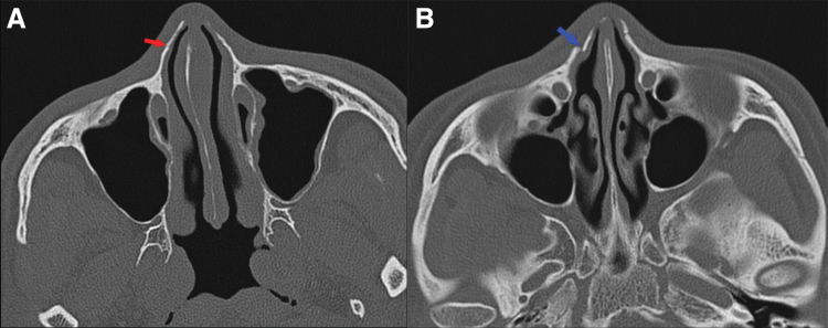
Type 1a: Either unilateral or bilateral fracture of the nasal bone, with no bony displacement (Fig. 1A); Type 1b: Unilateral or bilateral fracture of the nasal bone, with bony displacement (Fig. 1B).
Type 2a: Fracture of both the nasal bone and frontal process of the maxilla without bony displacement (Fig. 2A); Type 2b: Fracture of both the nasal bone and frontal process of the maxilla with bony displacement (Fig. 2B).
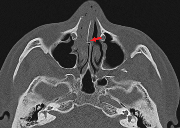
Type 3: Fracture of the nasal bone (with or without fracture of the frontal process of the maxilla), with concomitant fracture of the bony nasal septum (Fig. 3).
Type 4: In addition to fractures of the nasal bone (with or without fracture of the frontal process of the maxilla and/or bony nasal septum), the presence of coexistent fractures of the medial (Fig. 4A) and/or inferior orbital walls (Fig. 4B).
Type 5: Comminuted midfacial fractures (Fig. 4C).
Fig. 1.
(A) Fracture referring to the nasal bone without bony displacement (red arrow); (B) fracture of nasal bone with bony displacement (blue arrow).
Fig. 2.
(A) Coexistent fractures involving both the nasal bone and frontal process of the maxillary bone without bony displacement (red arrow); (B) fractures with involvement of either or both the nasal bone and frontal process of the maxillary bone with bony displacement (blue arrow).
Fig. 3.
Coexistent fracture with involvement of the nasal septum (arrow).
Fig. 4.
(A) Fracture involving the medial orbital wall (arrow); (B) fracture with involvement of the inferior orbital wall (arrow); (C) comminuted midfacial fractures with involvement of the nasal bone (red arrow), frontal process of the maxilla (white arrow), bony nasal septum (green arrow), and orbital wall (yellow arrow).
According to the new classification, the incidence of Type 1a (7.21%), Type 1b (11.90%), Type 2a (9.22%), Type 2b (18.61%), Type 3 (36.55%), Type 4 (10.81%), and Type 5 (5.70%) midfacial fractures is illustrated in Table 1.
Table 1.
The number of patients and the corresponding ratio of the fractures for Types 1 to 5 divided by the new classification system
| Case number | Ratio (%) | |
|---|---|---|
| Type 1a | 86 | 7.21 |
| Type 1b | 142 | 11.90 |
| Type 2a | 110 | 9.22 |
| Type 2b | 222 | 18.61 |
| Type 3 | 436 | 36.55 |
| Type 4 | 129 | 10.81 |
| Type 5 | 68 | 5.70 |
Discussion
Owing to the location of the nasal bone near the frontal process of the maxilla, bony nasal septum, and orbital walls, external mechanical force imposed to the nasal bone has the potential to cause concomitant injuries to these adjacent structures.8,16 The severity of these fractures is much less than that of NOE fractures. However, we found that these types of medium-impact midfacial fractures were far more common in clinical practice than the rare high-impact NOE fractures. Therefore, this study may be a complement to the current classification system of NOE fractures.
The nasal bone protrudes from the midface, which may, therefore, increase its vulnerability to assault.17 In this study, we found that the percentage of bilateral fractures of the nasal bone was much higher than either right- or left-sided unilateral fractures. Moreover, due, in part, to the relative protruding character of the distal nasal bone, fractures occurred at the distal portion of the nasal bone more frequently than the proximal portion. Furthermore, a fracture of the nasal bone was often accompanied with adjacent bony fractures, with an overall coincidence up to 80.89% in our case series.
The frontal process of the maxilla is located close to the nasal bone. When an external force is imposed on the nasal bone, a coexistent fracture of the frontal process of the maxilla was present in 66.89% of cases in this study. In light of the high incidence of fractures of the frontal process of the maxilla, we recommend that the status of the frontal process of maxilla deserves more attention when the diagnosis of nasal bone fracture is made.
Fractures of the nasal septum also often accompany fractures of the nasal bone. Zhao et al. reported that the incidence of nasal septal fracture was 66.7%.9 In the study presented here, the incidence was 42.25%. The continuum of the bony septum and nasal bone might contribute to the higher occurrence of cofracture at the bony portion. Although coexisting fractures of the nasal septum were common, oftentimes they did not require surgical correction, which ultimately depended on the ventilation capacity and sensation of nasal obstruction after swelling dissipated.10,18,19
A septal hematoma caused by septal fracture deserves careful attention.20 Immediate incision and drainage can help to reduce the risk of abscess formation or necrosis of the cartilaginous septum. In this study, there were two cases of septal hematoma, both occurred over the bony septum. Timely incision along the base of the septum was performed, and no subsequent case of abscess, septal perforation, or cosmetic deformity from cartilage necrosis occurred.
Both the inferior and medial orbital walls have the potential to be damaged.16 In this study, we found that the coexisting incidence of fractures involving the medial and inferior orbital walls was 16.51%, and fracture of the medial orbital wall was more frequently observed than that of the inferior orbital wall. There was no entrapment of extraocular muscles or vision disruption in the 1125 cases without comminuted midfacial fracture and, as such, conservative measures were utilized in these 1125 cases.17 For the 68 cases with comminuted multiple midfacial fractures, however, reconstruction by a multidisciplinary team including an otolaryngologist, ophthalmologist, and oral–maxillofacial surgeon was performed to restore aesthetics and function.
In addition to nasal fractures occurring in the adult group, we also investigated the incidence and characteristics of midfacial fractures in the pediatric (<18 years) and elderly patient populations (≥65 years).21,22 By comparison with the adult group, the occurrence of nasal fracture in children and elderly patients was lower, and falls consisted of the primary etiology in our case series.14 Coexisting fractures of the nasal septum and orbital wall in the pediatric group were much lower than that in the adult group. However, the incidence of nasal septum and orbital wall fractures in the elderly group was much higher than that in the adult group. This finding might be correlated with bone fragility in different developmental stages.23,24
The classification system proposed in this study was intimately correlated with the treatment strategy. From Type 1 to Type 4, no comminuted fractures existed, and as such, conservative treatment or closed reduction was indicated. For Type 5 with comminuted fractures, however, open surgery and a multidisciplinary approach were oftentimes needed. For both Type 1a and Type 2a fractures, conservative measures were often indicated when there was no bony displacement. Displaced fracture of the nasal bone (Type 1b) or frontal process of the maxilla (Type 2b) oftentimes required closed reduction in the office immediately or within 7 to 10 days after swelling had decreased.25 Moreover, coexistent fractures of the nasal bone and bony nasal septum were defined as a Type 3 fracture. Surgical intervention may be indicated when nasal obstruction persisted after edema caused by fracture of the nasal septum resolved. When fracture of the orbital walls occurred (Type 4), conservative management was indicated when there was no disorder of ocular mobility, enophthalmos, or eyesight reduction. In the event that any of mentioned symptoms occurred, immediate ophthalmological consultation and appropriate treatment were recommended. Comminuted midfacial fractures (68 cases) were defined as Type 5 in our case series, which is equivalent to traditional NOE fractures. In the setting of comminuted fractures, a multidisciplinary approach was often indicated.
There are also limitations to this study. First, due to the nature of retrospective data that were derived from a single center, the applicability and relevance of the proposed classification at additional institutions still need to be validated. Second, the resultant treatment outcomes for each type of fracture were not included in this study, which needs to be further assessed.
Conclusions
The external force imposed to the nasal bone can also lead to fracture of adjacent bony structures, including the frontal process of the maxilla, nasal septum, and orbital walls. The new classification of nasal bone fracture based on CT imaging may provide a better understanding of the frequency of coincident fractures and aid in ensuring that coincident fractures are appropriately addressed.
Acknowledgment
The authors thank Shuling Li, MD, for help in selection and diagnosis of midfacial fractures.
Author Disclosure Statement
N.R.L. holds stock in Navigen Pharmaceuticals and was a consultant for Cooltech Inc., neither of which are relevant to this study. All other authors declare no relevant conflicts of interest.
Funding Information
The author(s) received no financial support for the research, authorship, and/or publication of this article.
References
- 1. Simmen D. [Nasal fractures—indications for open reposition]. Laryngorhinootologie. 1998;77(7):388–393 [DOI] [PubMed] [Google Scholar]
- 2. Sargent LA, Rogers GF. Nasoethmoid orbital fractures: diagnosis and management. J Craniomaxillofac Trauma. 1999;5(1):19–27 [PubMed] [Google Scholar]
- 3. Motamedi MH. An assessment of maxillofacial fractures: a 5-year study of 237 patients. J Oral Maxillofac Surg. 2003;61(1):61–64 [DOI] [PubMed] [Google Scholar]
- 4. Kelley BP, Downey CR, Stal S. Evaluation and reduction of nasal trauma. Semin Plastic Surg. 2010;24(4):339–347 [DOI] [PMC free article] [PubMed] [Google Scholar]
- 5. Smith HL, Chrischilles E, Janus TJ, et al. Clinical indicators of midface fracture in patients with trauma. Dent Traumatol. 2013;29(4):313–318 [DOI] [PubMed] [Google Scholar]
- 6. Ziccardi VB, Braidy H. Management of nasal fractures. Oral Maxillofac Surg Clin North Am. 2009;21(2):203–208, vi [DOI] [PubMed] [Google Scholar]
- 7. Hwang K, You SH, Kim SG, Lee SI. Analysis of nasal bone fractures; a six-year study of 503 patients. J Craniofac Surg. 2006;17(2):261–264 [DOI] [PubMed] [Google Scholar]
- 8. Carvalho TB, Cancian LR, Marques CG, Piatto VB, Maniglia JV, Molina FD. Six years of facial trauma care: an epidemiological analysis of 355 cases. Braz J Otorhinolaryngol. 2010;76(5):565–574 [DOI] [PMC free article] [PubMed] [Google Scholar]
- 9. Zhao Y, Zhu L, Ma F. [CT analysis of classification of external nasal fracture and the influence of fractured position to nasal septum]. Lin Chung Er Bi Yan Hou Tou Jing Wai Ke Za Zhi. 2014;28(8):527–530 [PubMed] [Google Scholar]
- 10. Mundinger GS, Dorafshar AH, Gilson MM, et al. Analysis of radiographically confirmed blunt-mechanism facial fractures. J Craniofac Surg. 2014;25(1):321–327 [DOI] [PubMed] [Google Scholar]
- 11. Jeon SP, Kang SJ, Kim JW, Kim YH, Sun H. New nasal fracture classification for patients with silicone implants. J Plast Surg Hand Surg. 2013;47(5):363–367 [DOI] [PubMed] [Google Scholar]
- 12. Markowitz BL, Manson PN, Sargent L, et al. Management of the medial canthal tendon in nasoethmoid orbital fractures: the importance of the central fragment in classification and treatment. Plast Reconstr Surg. 1991;87(5):843–853 [DOI] [PubMed] [Google Scholar]
- 13. Erdmann D, Follmar KE, Debruijn M, et al. A retrospective analysis of facial fracture etiologies. Ann Plast Surg. 2008;60(4):398–403 [DOI] [PubMed] [Google Scholar]
- 14. Rajput D, Bariar LM. Study of maxillofacial trauma, its aetiology, distribution, specturm, and management. J Indian Med Assoc. 2013;111(1):18–20 [PubMed] [Google Scholar]
- 15. Han DS, Han YS, Park JH. A new approach to the treatment of nasal bone fracture: radiologic classification of nasal bone fractures and its clinical application. J Oral Maxillofac Surg. 2011;69(11):2841–2847 [DOI] [PubMed] [Google Scholar]
- 16. Kang CM, Han DG. Correlation between operation result and patient satisfaction of nasal bone fracture. Arch Craniofac Surg. 2017;18(1):25–29 [DOI] [PMC free article] [PubMed] [Google Scholar]
- 17. Banerjee R, Basu S, Pachisia S, Sahu S, Mishra M, Ghosh S. Management of nasoorbitoethmoidal fracture: an institutional experience. Indian J Otolaryngol Head Neck Surg. 2019;71(2):225–232 [DOI] [PMC free article] [PubMed] [Google Scholar]
- 18. Wang ZS, Peng MQ, Wei H, Ying CL, Wan L. [The subtle anatomical structures of normal nasal bone in MSCT image and forensic identification]. Fa Yi Xue Za Zhi. 2014;30(3):184–187 [PubMed] [Google Scholar]
- 19. Patel R, Reid RR, Poon CS. Multidetector computed tomography of maxillofacial fractures: the key to high-impact radiological reporting. Semin Ultrasound CT MR. 2012;33(5):410–417 [DOI] [PubMed] [Google Scholar]
- 20. Puricelli MD, Zitsch RP, 3rd. Septal hematoma following nasal trauma. J Emerg Med. 2016;50(1):121–122 [DOI] [PubMed] [Google Scholar]
- 21. Rottgers SA, Decesare G, Chao M, et al. Outcomes in pediatric facial fractures: early follow-up in 177 children and classification scheme. J Craniofac Surg. 2011;22(4):1260–1265 [DOI] [PubMed] [Google Scholar]
- 22. Lopez J, Luck JD, Faateh M, et al. Pediatric nasoorbitoethmoid fractures: cause, classification, and management. Plast Reconstr Surg. 2019;143(1):211–222 [DOI] [PubMed] [Google Scholar]
- 23. Wright RJ, Murakami CS, Ambro BT. Pediatric nasal injuries and management. Facial Plast Surg. 2011;27(5):483–490 [DOI] [PubMed] [Google Scholar]
- 24. Zelken JA, Khalifian S, Mundinger GS, et al. Defining predictable patterns of craniomaxillofacial injury in the elderly: analysis of 1,047 patients. J Oral Maxillofac Surg. 2014;72(2):352–361 [DOI] [PubMed] [Google Scholar]
- 25. Kang CM, Han DG. Objective outcomes of closed reduction according to the type of nasal bone fracture. Arch Craniofac Surg. 2017;18(1):30–36 [DOI] [PMC free article] [PubMed] [Google Scholar]


