PLOS ONE logoLink to PLOS ONE
. 2020 Sep 8;15(9):e0238572. doi: 10.1371/journal.pone.0238572

Growth inhibition associated with disruption of the actin cytoskeleton by Latrunculin A in rhabdomyosarcoma cells

Julia Würtemberger 1, Daria Tchessalova 2, Carla Regina 1, Christoph Bauer 1, Michaela Schneider 1, Amy J Wagers 2,3, Simone Hettmer 1,*
Editor: Sandro Pasquali4
PMCID: PMC7478754  PMID: 32898143

Abstract

Functional genomic screening of KRAS-driven mouse sarcomas was previously employed to identify proliferation-relevant genes. Genes identified included Ubiquitin-conjugating enzyme E2 (Ube2c), Centromere Protein E (Cenpe), Hyaluronan Synthase 2 (Has2), and CAMP Responsive Element Binding Protein 3 Like 2 (Creb3l2). This study examines the expression and chemical inhibition of these candidate genes, identifying variable levels of protein expression and significant contributions to rhabdomyosarcoma (RMS) cell proliferation. Chemical treatment of human and murine RMS cell lines with bortezomib, UA62784, latrunculin A and sorafenib inhibited growth with approximate EC50 concentrations of 15-30nM for bortezomib, 25-80nM for UA62784 and 80-220nM for latrunculin A. The multi-kinase inhibitor sorafenib increased in vitro proliferation of 4 of 6 sarcoma cell lines tested. Latrunculin A was further associated with disruption of the actin cytoskeleton and reduced ERK1/2 phosphorylation. Together, this work advances opportunities for developing therapies to block progression of soft-tissue sarcomas and demonstrates that disruption of the actin cytoskeleton in sarcoma cells by latrunculin A is associated with a reduction in RMS cell growth. (167 words).

Introduction

Rhabdomyosarcoma (RMS) represents the most common soft-tissue sarcoma (STS) subtype within the pediatric age group [1, 2]. RAS pathway genes are frequently mutated in STS [3], including PAX3/7:FOXO1 fusion-negative RMS [4]. We previously reported on a customized shRNA-based proliferation screen [5], which tested the contributions of 141 sarcoma-relevant genes, previously identified by transcriptional profiling of genetically engineered mouse sarcomas driven by KRAS(G12v) and CDKN2A deletion [6]. In this screen, the strongest inhibitory effect on sarcoma growth was produced by silencing of asparagine synthetase (ASNS), which established that adequate asparagine availability was a metabolic vulnerability with potential anti-sarcoma therapeutic value [5]. The screen also identified four other potentially druggable genes/ cellular processes: (1) Ubiquitin-conjugating enzyme E2 (Ube2c), which is essential in cell cycle progression by orchestrating proteolysis of cyclin-dependent kinase and its inhibitors [7]; (2) Centromere Protein E (Cenpe), a kinesin-like protein that localizes to the kinetochore during mitosis and is important for bipolar spindle formation [8]; (3) Hyaluronan Synthase 2 (Has2), responsible for the synthesis of hyaluronan, which serves as a scaffold for the extracellular matrix and critically determines the extracellular micromilieu [9]; and, (4) CAMP Responsive Element Binding Protein 3 Like 2 (Creb3l2), a transcription factor and downstream target of mitogen-activated (MAPK) signalling that promotes tumor cell survival [10]. This study aimed at determining the expression of UBE2C, CENPE, HAS2 and CREB3L2 in human STS and examining the growth-attenuating effects of candidate chemicals bortezomib (aimed at UBE2C; [7]), UA62784 (aimed at CENPE; [8]), sorafenib (aimed at Creb3l2; [10]) and latrunculin A (aimed at HAS2; [9]) on the in vitro growth of human and murine sarcomas. Findings from our experiments highlight that inhibition of actin polymerization by latrunculin A is linked to reduced growth of RMS cells.

Materials and methods

Sarcoma cell lines

Mouse sarcoma cell lines were derived from a Kras;CDKN2Anull mouse sarcoma with myogenic differentiation (RMS) and a Kras;CDKN2Anull undifferentiated, non-myogenic mouse sarcoma (NMS). The human RMS cell line RD (PAX3/7:FOXO1-negative) and the human fibrosarcoma line HT1080 originated from ATCC. Human RMS cell lines Rh3, Rh5, Rh10, Rh28, Rh30, Rh41 (all PAX3:FOXO1-positive) and Rh36 (PAX3/7:FOXO1-negative) were gifts from Dr. Peter Houghton (Greehey Children’s Research Institute, San Antonio, TX, USA). All cell lines were grown in DMEM with 10% FBS and 1% Penicillin-Streptomycin.

Customized shRNA proliferation screen

The shRNA proliferation screen was carried out in two Kras;CDKN2Anull mouse sarcoma cell lines as previously described [5]. The screen and details of the statistical analysis were published previously [5]. In brief, each candidate gene was targeted by 5 individual shRNAs. For each shRNA, relative cell proliferation was determined as the percentage growth of shRNA infected cells compared to the mean growth of cells infected with cntrl-shRNAs. Differences in average proliferation between cells infected with shRNAs against one specific target gene and average proliferation of cntrl-shRNA infected cells were tested for statistical significance using T-tests and the algorithm published by J. W. McNicol and G. Hogan [11]. Receiver operator curve analysis established a false discovery rate less than 30% for relative proliferation of less than 52% or 40% of cntrl-shRNA infected cells for the two lines. The growth-inhibitory effects of shRNA-mediated silencing of individual candidate genes were considered significant if p<0.01 and q<0.05 and 3 shRNAs scored with an FDR<30%.

Immunohistochemistry

Candidate gene expression in primary human sarcoma tissue was evaluated using commercially available sarcoma tissue arrays (US Biomax SO2081). Paraffin was removed by placing slides in a Coplin jar at 58 degrees centigrade in a microwave oven. Slides were then rehydrated by immersing them serially in xylene (3 x 5 minutes), 90% ethanol (1 x 3 minutes) and 80% ethanol (1 x 3 minutes) prior to rinsing them in gently running tap water and placing them in PBS for 30 minutes. Antigen retrieval was performed in 10mM sodium citrate buffer pH6 in a microwave oven operated at high power for 5 minutes, and tissue sections were blocked in PBS, 5% BSA, pH7.4. Tissue was stained for CENPE (1 in 500, HPA042294, Sigma; human testis served as positive and brain as negative control tissue), UBE2C (1 in 200, A-650, Boston Biochem Inc; colon served as positive and brain as negative control tissue), CREB3L2 (1 in 200, HPA015068, Sigma; liver served as positive and colon as negative control tissue) and HAS2 (1 in 600, ab140671, Abcam; dermis served as positive and brain as negative control tissue). Primary antibodies were incubated overnight at 4 degree centigrade. Control tissues were obtained from the National Disease Research Interchange (NDRI) (S1 Fig). Primary antibody binding was detected by labeling with biotinylated secondary antibodies (1 in 800, B8895, Sigma) and Streptavidin-HRP (BD, 51-75477E). Slides were then exposed to DAB substrate (BD, 550880), which reacts with HRP to produce a brown-colored signal. CENPE (nuclear), UBE2C (cytoplasmic), CREB3L2 (cytoplasmic) and HAS2 (nuclear) staining was evaluated by two independent operators. If > 25% of cells per core exhibited a positive signal, antigen expression was considered positive.

RNA isolation and qRT-PCR

RNA was isolated from human RD, HT1080, Rh3, Rh5, Rh10, Rh28, Rh30, Rh41 and Rh36 cells and murine SMPO1 cells by TRIzol extraction followed by DNAse digestion and purification using the RNeasy Plus Micro Kit. The use of human muscle as control tissue was approved by the Institutional Review Board at Joslin Diabetes Center. Human fetal muscle was obtained from 20–23 week gestation fetuses and adult muscle from deceased volunteers. Tissue was homogenized in TRIzol using a tissue homogenizer prior to RNA isolation as described above. RNA was reverse transcribed using Superscript III First-Strand Synthesis System for RT-PCR (Invitrogen). qRT-PCR was performed using an ABI 7900 RT-PCR system (Applied Biosystem) with SYBR-green PCR reagents.

CENPE, HAS2, UBE2C and CREB3L2 in human tissue were detected using the following primer sequences: GATTCTGCCATACAAGGCTACAA (CENPE, fw); TGCCCTGGG-TATAACTCCCAA (CENPE, rev); CTCTTTTGGACTGTATGGTGCC (HAS2, fw), AGGGTAGGTTAGCCTTTTCACA (HAS2, rev); GACCTGAGGTATAAGC-TCTCGC (UBE2C, fw), TTACCCTGGG-TGTCCACGTT (UBE2C, rev); CAGAGAAGAGTGTGTCAATGGAG (CREB3L2, fw), CTGGTGGTAAT-GTGGGTGAAG (CREB3L2, rev).

Cenpe, Has2, Ube2c and Creb3l2 in mouse tissue were detected using the following primer sequences: TCAGGAAAGACACACACGATG (Cenpe, fw); TGCGAGCCATTTCAAAGCCA (Cenpe, rev); TGTGAGAGGTTTCTATGTGTCCT (Has2, fw), ACCGTACAGTCCAAATGAGAAGT (Has2, rev); CTCCGCCTTCCCTGAGTCAGC (Ube2c, fw), GGTGCGTTGTAAGGGTAGCC (Ube2c, rev); CATGTACCACACGCACTTCTC (Creb3l2, fw), CCACCTCCATTGACTCGCT (Creb3l2, rev).

Proliferation assays

Mouse Kras;CDKN2Anull SMP-01 RMS and Sca1-01 NMS, human RD, Rh30 and Rh41 RMS and human HT1080 fibrosarcoma cells were exposed to the following chemicals: sorafenib (0.1-1mM, stock 10mM in DMSO, Cayman Chemicals), bortezomib (50-250nM, stock 10mM in DMSO, Cayman Chemicals), latrunculin A (50-1000nM, stock 237 mM in ethanol, Cayman Chemicals), UA62784 (50-250nM, stock 5.66mM in DMSO, Sigma) and vehicle (DMSO, ethanol). Proliferation assays were performed as previously described [1, 6]. Estimated EC50 concentrations were calculated using GraphPad Prism.

Western blot

Cells were washed with PBS and lysed in lysis buffer (New England Biolabs). Protein concentrations were determined using DC protein assays (Biorad). Membranes were incubated with primary antibodies against p44/42 MAPK (Erk1/2; titer 1:2000; 9102, Cell Signaling Technology), phospho-p44/42 MAPK (phospho-Erk1/2; titer 1:2000; 9101; Cell Signaling Technology) and GAPDH (titer 1:10000; 2118, Cell Signaling Technology) at 4 degrees overnight. Secondary antibodies (titer 1:10000; 170–6515, Biorad) were incubated for 1 hour at room temperature.

Immunocytochemistry

Cells were fixed with 4% paraformaldehyde (PFA), incubated with triton X 0.2% and blocked with 10% goat serum 10%. Human cells were stained with Phalloidin iFluor 488 (titer 1:100, A12379, Thermo Scientific), and mouse cells were stained with Phalloidin iFluor 594 (titer 1:100, ab176757, Abcam) at room temperature for one hour. Nuclei were stained with 1μg/ml 4’-6-diamino-2-phenylindole (DAPI, D9542, Sigma Aldrich). Staining was then evaluated by immunofluorescence microscopy using a Zeiss LSM 710 confocal microscope. RD and Rh30 human RMS cells were cultured and stained in uncoated 96-well-dishes. Kras;CDKN2Anull mouse sarcoma cells were cultured and stained on 10% matrigel (354234, Corning).

Statistics

Differences in cell growth were tested for statistical significance using T-tests (ns p≥0.05, * p<0.05, ** p<0.01, *** p <0.001).

Results

Identification of target genes

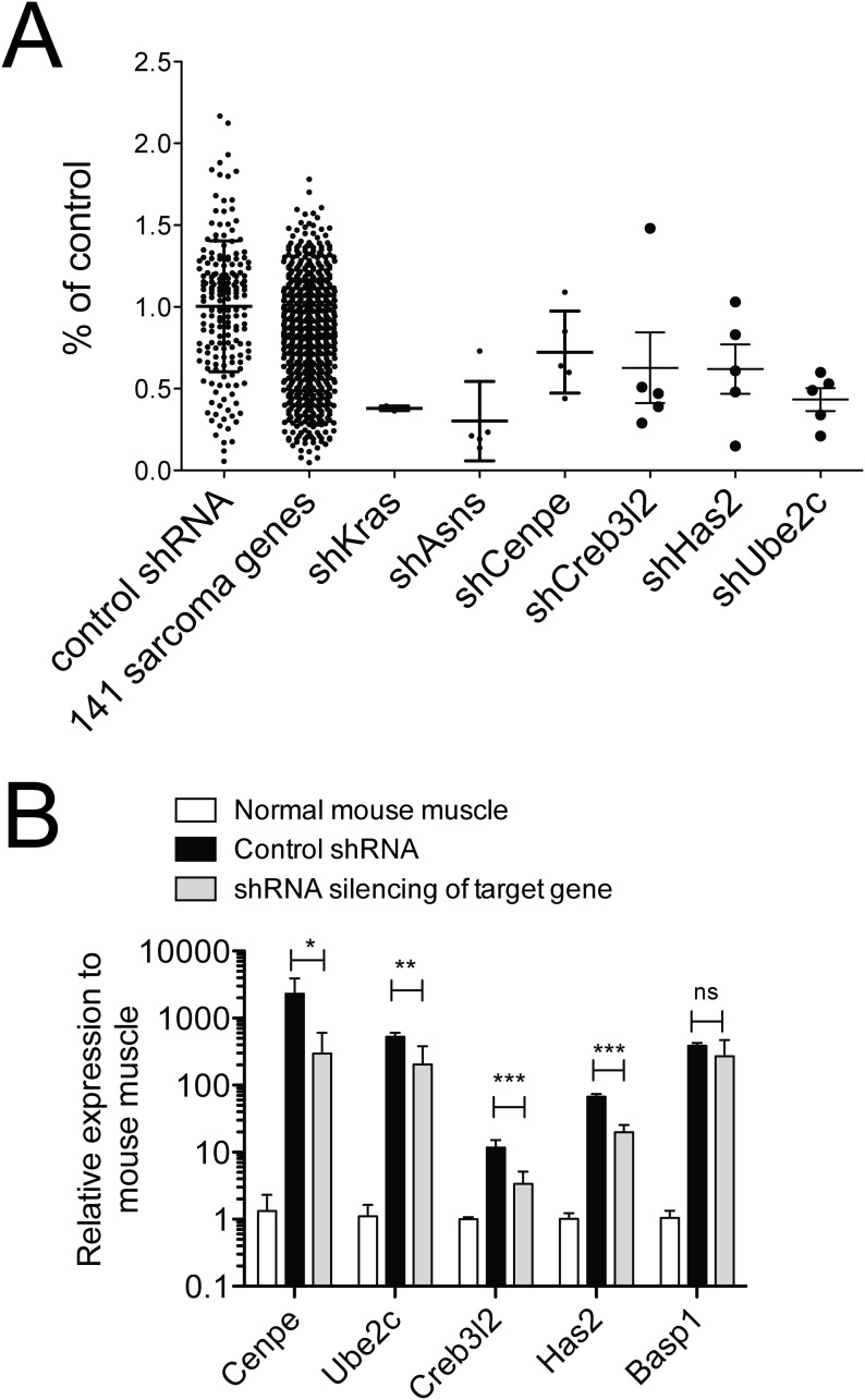
A candidate set of 141 sarcoma-relevant genes (S1 Table), identified by transcriptional profiling of genetically engineered mouse RMS and non-myogenic sarcomas driven by KRAS(G12v) and CDKN2A deletion, was previously published [6]. All 141 candidate genes were tested by shRNS proliferation screening [6], designed to determine the growth-promoting effects of each of these candidates. The screen employed five shRNAs per target gene and two low-passage Kras;CDKN2Anull mouse sarcoma cell lines [5]. Sixteen of 141 candidate genes met significance criteria in one or both cell lines (Table 1; p<0.01, q<0.05, at least 3 shRNAs with FDR<30%; [5]). Published literature was reviewed to identify the genes, within this list of 16, for which chemical modulators had previously been described, because such small molecules could potentially be re-purposed as anti-sarcoma drugs. This review identified five potentially druggable genes/ cellular processes: ASNS, UBE2C, CENPE, HAS2, CREB3L2 (Fig 1A). As prior work already has validated asparagine starvation as an actionable metabolic vulnerability in sarcomas [5, 12], this study concentrates on the potential roles of UBE2C, CENPE, HAS2 and CREB3L2 in STS with a focus on RMS. Effective knockdown, confirmed by PCR (Fig 1B), of each of these targets by the target-specific shRNAs employed in our screen reduced cell proliferation to 40–60% of that seen in control cell cultures (Table 1).

Table 1. Proliferation-relevant sarcoma genes.

Transcriptional profiling of genetically engineered, KRAS-induced mouse rhabdomyosarcomas (RMS) and non-myogenic sarcomas (NMS) identified 141 sarcoma-relevant genes; their function was evaluated using a customized shRBA screen. Sixteen of the 141 sarcoma-relevant genes scored as „hits”in RMS and/ or NMS (p<0,01, q>0,05, at least 3 of 5 shRNAs with FDR< 30%) and are listed here. These 16 hits include 5 potentially druggable targets, i.e. Asns, Cenpe, Crebl2, HAs2 and Ube2c.

Kras; p16p19null RMS Kras; p16p19null NMS
Symbol Cell function Chemical modulation Mean % of ctrl Level p-value q value Mean % of ctrl Level p-value q value
Asns Asparagine synthesis AS5, Asparaginase, Mupirocin 30,16 2 < .0001 0,0058 6,6934 1 < .0001 0,0072
Rbbp8 Cell cycle 56,06 3 0,0022 0,0160 30,7323 2 0,0012 0,0288
Rad54l DNA repair 57,56 3 0,0031 0,0180 40,5093 3 0,0076 0,0421
Ube2c Cell cycle Bortezomib 43,56 3 < .0001 0,0058 38,6873 - 0,0054 0,0389
Cenpe Chromosome stability UA62784, GSK923295A 72,49 - 0,0511 0,0689 39,9054 3 0,0067 0,0402
Has2 Extracellular matrix Latrunculin A 61,76 - 0,0075 0,0335 41,5132 3 0,0088 0,0453
Creb3l2 Transcriptional regulation Sorafenib 62,69 2 0,0090 0,0326 60,2497 - 0,1230 0,1736
Basp1 Transcriptional regulation 45,37 3 0,0002 0,0039 50,8361 - 0,0371 0,0989
Lrrfip1 Transcriptional regulation 54,80 3 0,0017 0,0164 54,4497 - 0,0606 0,1322
Lasp1 Cytoskeletal organization 60,37 3 0,0056 0,0271 58,6252 3 0,1019 0,1706
Pold3 DNA repair 62,96 3 0,0095 0,0290 31,9654 - 0,0015 0,0216
Efhd2 Apoptosis 53,27 - 0,0034 0,0179 46,8886 2 0,0007 0,0252
Myo9b Cytoskeletal organization 67,41 - 0,0445 0,0737 33,5929 2 0,0021 0,0252
Runx1 Transcriptional regulation 64,64 - 0,0455 0,0713 30,6930 3 0,0012 0,0288
Shcbp1 Cell proliferation 71,42 - 0,1118 0,1099 37,7316 3 0,0045 0,0405
Egr2 Transcriptional regulation 74,44 - 0,0689 0,0850 42,9416 3 0,0048 0,0384

Fig 1. Candidate sarcoma targets.

Fig 1

141 sarcoma-relevant genes were identified by transcriptional profiling of genetically engineered Kras-driven mouse sarcomas [6]. Their contributions to sarcoma growth were probed by customized shRNA screening using 5 shRNAs per candidate gene. Control shRNAs were directed against LUC, RFP and LACZ; shKras served as a positive control [5]. (A) Five candidate genes whose targeting by shRNAs in this screen resulted in reduced sarcoma cell proliferation represent potentially druggable genes/ cellular processes: Asns, Ube2c, Cenpe, Has2, Creb3l2. The anti-proliferative effects of the shRNAs directed against these candidate genes in a Kras;CDKN2Anull mouse RMS cell line are shown. (B) Effective knockdown by the target-specific shRNAs in Kras;CDKN2Anull mouse RMS cells was confirmed by qRT-PCR. Target gene expression was determined by gene-specific qRT-PCR (mean +/- SD of 3 technical replicates presented; ns p≥0.05, * p<0.05, ** p<0.01, *** p <0.001, as determined by T-tests compared to cntrl-shRNA infected cells).

Candidate gene expression in sarcomas

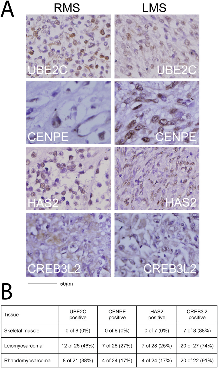
Candidate proteins CENPE, CREB3L2, HAS2 and UBE2C were detected in primary human STS tissue at variable levels (Fig 2A and 2B). In human RMS, UBE2C was detected in 8 of 21 (38%), CENPE and HAS2 in 4 of 24 (17%) and CREB3L2 in 20 of 22 (91%) (Fig 2B). In human leiomyosarcomas (LMS), expression of UBE2C was found in 12 of 26 (46%), of CENPE in 7 of 26 (27%), of HAS2 in 7 of 26 (25%) and of CREB3L2 in 20 of 27 (74%) human leiomyosarcoma cores (Fig 2B). CREB3L2 was also detected in normal human skeletal muscle samples (7 of 8 (88%) samples), but UBE2C, CENPE and HAS2 were not.

Fig 2. Expression of candidate sarcoma targets in human sarcoma tissue.

Fig 2

Immunohistochemical staining of commercially available sarcoma tissue arrays (US Biomax SO2081) was used to confirm candidate expression in human sarcomas. (A) Representative stains in RMS (left column) and high-grade LMS (right column) are shown for UBE2C (top row), CENPE (second row from top), HAS2 (third row from top) and CREB3L2 (bottom row). (B) Candidate proteins were expressed in 17–91% of RMS cores and 27–74% of LMS cores. Please see also S1 and S2 Figs.

Expression levels of candidate genes were further evaluated in nine human sarcoma cell lines by RT-PCR. Increased expression of UBE2C, CENPE and CREB3L2 compared to normal muscle was detected in 9 of 9 human sarcoma cell lines analyzed (S2A–S2C Fig), whereas HAS2 expression was detected in only 5 of 9 human sarcoma lines (S2D Fig). UBE2C, CENPE and CREB3L2 were also detected in normal human skeletal muscle, while HAS2 was detected in fetal muscle only (S2D Fig). Discrepancies between candidate expression levels in sarcoma cell lines and primary tissue could be due to passaging in vitro.

Anti-proliferative effects of chemical targeting of candidate genes

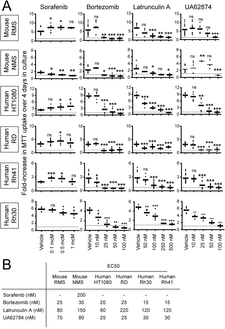
Given that downregulation of Ube2c, Cenpe, Has2 and Creb3l2 by shRNA knockdown diminished mouse sarcoma cell proliferation in vitro (Table 1), we hypothesized that small molecule inhibitors previously reported to impede the cellular functions of these targets could prove useful in inhibiting their growth. We obtained and tested 4 chemical compounds: (1) the proteasome inhibitor bortezomib, (2) the ATPase inhibitor UA62784, (3) the sponge-derived macrolide latrunculin A, and (4) the protein kinase inhibitor sorafenib. Bortezomib has been shown to downregulate UBE2C and mediate accumulation of cyclins A and B1 [7]. UA62784 and GSK923295A were shown to inhibit ATPase activity in the CENPE motor protein [8], although UA62784 has also been implicated with microtubule polymerization and associated with accumulation of mammalian cells in mitosis due to aberrant formation of mitotic spindles [13]. Latrunculin A was previously shown to reduce HAS2 expression in fibroblasts [9], and sorafenib was shown to reduce CREB3L2 expression in glioma cells [10]. Of the four chemicals tested, three inhibited proliferation of mouse Kras;p16p19null RMS and NMS cells, human HT1080 fibrosarcoma cells, human fusion-negative RMS cells (RD) and two different PAX3:FOXO1-positive human RMS cell lines (Rh30, Rh41) (Fig 3A and 3B). Approximate EC50 concentrations were 15-30nM for bortezomib, 25-80nM for UA62784 and 80-220nM for latrunculin A (Fig 3B). Sorafenib had a slight inhibitory effect on NMS proliferation, with an estimated EC50 concentration of 200nM; yet, unexpectedly, sorafenib had no inhibitory effects on the other cell lines tested. In fact, sorafenib increased the growth of mouse RMS and of all but one of the human sarcoma cell lines tested (Fig 3A and 3B).

Fig 3. Chemical inhibition of mouse and human sarcoma cell growth in vitro.

Fig 3

(A) Bortezomib, Latrunculin A, and UA62874, previously reported to modulate cellular functions of UBE2C, HAS2 and CENPE, respectively, reduced proliferation of a Kras;CDKN2Anull mouse sarcoma cell line with myogenic differentiation (mouse RMS), a Kras;CDKN2Anull undifferentiated, non-myogenic mouse sarcoma cell line (mouse NMS), the human fibrosarcoma cell line HT1080, the human embryonal RMS cell line RD (PAX3/7:FOXO1-negative) and the human alveolar RMS cell lines Rh30 and Rh41 (PAX3/7:FOXO1-positive). Sorafenib increased growth of the mouse RMS cell line, HT1080, RD and Rh41 (mean +/- SD of 6 technical replicates obtained in 3 independent experiments are presented; ns p≥0.05, * p<0.05, ** p<0.01, *** p <0.001, as determined by T-tests compared to vehicle-treated cells). (B) Estimated EC50 concentrations ranged between 15–30 nM for Bortezomib, 80–150 nM for Latrunculin A and 25–80 nM for UA62784 (EC50 concentrations were calculated using graphpad). Please see also S3 Fig.

Latrunculin A effects on RMS cells

Latrunculin A inhibited the growth of mouse and human RMS cell lines. Its effects on target gene expression were evaluated by qRT-PCR in mouse and human RMS cell lines. There was no clear effect on HAS2 expression in human RD, human Rh30 and mouse RMS cells (S3 Fig).

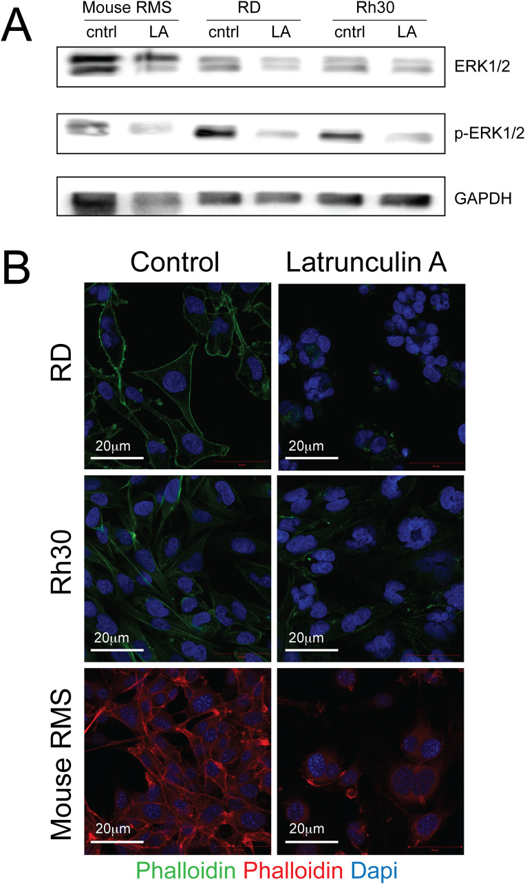
As latrunculin A is known to bind actin monomers and induce depolymerization of the cytoskeleton [14, 15], we evaluated the actin cytoskeleton in Latrunculin-A treated cells by phalloidin staining. Human RD sarcoma cells were exposed to 250nM latrunculin A, human Rh30 RMS cells to 100nM latrunculin A and mouse RMS sarcoma cells to 100nM latrunculin A. Different concentrations were chosen due to differences in latrunculin A sensitivity between sarcoma cell lines. There was profound disruption of the F-actin cytoskeleton after Latrunculin A treatment of all 3 RMS cell lines, including human RD (Fig 4B, to panels) and Rh30 cells (Fig 4B, middle panels) and mouse RMS cells (Fig 4B, bottom panels). Finally, we demonstrated diminished ERK1/2-phosphorylation in human and mouse RMS cells exposed to Latrunculin A (Fig 4A, S1 Raw images).

Fig 4. Protein expression levels of ERK and pERK in human and mouse rhabdomyosarcoma cell lines after treatment with Latrunculin A.

Fig 4

Human RD sarcoma cells were exposed to 250nM latrunculin A, human Rh30 RMS cells to 100nM latrunculin A and mouse RMS sarcoma cells to 100nM latrunculin A. Different concentrations were chosen due to differences in latrunculin A sensitivity between sarcoma cell lines. (A) Evaluation of ERK1/2 (42/ 44kDa), p-ERK1/2 (22/ 44 kDa) and GAPDH (37 kDa) by Western blotting demonstrated reduced ERK1/2 phosphorylation in latrunculin A-treated sarcoma cells. (B) The F-actin cytoskeleton was evaluated using phalloidin staining. Actin organization was profoundly disrupted in latrunculin A-treated (top right panel)) vs. control (top left panel) RD cells, latrunculin A-treated (middle right panel) vs. control (middle left panel) Rh30 cells and latrunculin A-treated (bottom right panel) vs. control (bottom left panel) mouse RMS cells. Please also see S1 Raw images.

Discussion

Radiation and surgery continue to be a mainstay in the treatment of STS, including RMS [16]. The outcome of tumors that have spread regionally and/or systemically is dismal [17]. Unlike many other STS tumors, RMS is relatively sensitive to conventional cytostatic drugs, and currently available multimodal treatment strategies cure approximately 65% of children and adolescents with RMS [18]. Still, more than 70% of those diagnosed with metastatic RMS die from disease [19]. These statistics highlight the need for new drugs to treat STS.

Functional genomic screening of KRAS-driven mouse sarcomas was employed to identify actionable proliferation-relevant genes of potential therapeutic applicability. Proliferation assays in 2 mouse and 4 human sarcoma cell lines, including one mouse and 3 human RMS cell lines, revealed growth inhibition by the proteasome inhibitor bortezomib, the CENPE inhibitor UA62784 and latrunculin A. Yet, interestingly, the multi-kinase inhibitor sorafenib increased in vitro proliferation of 5 of 6 sarcoma cell lines tested, including 3 human RMS cell lines. Bortezomib, CENPE inhibitors and sorafenib were previously evaluated by the Pediatric Preclinical Testing Program for anti-RMS effects as single agents. Bortezomib and sorafenib did not reduce the size of RMS xenografts (n = 6–7, [20, 21]). The CENPE inhibitor GSK923295A only induced an objective response in 2 of 5 xenografts tested [22]. Taken together, these in vivo observations do not support a major role for bortezomib, CENPE inhibitors and sorafenib in RMS treatment.

Latrunculin A is a 2-thiazolidinone macrolide derived from sponges that binds monomeric actin with 1:1 stoichiometry, blocks actin polymerization and results in depolymerization of tumor cell cytoskeleton. Antitumorigenic effects of Latrunculin A have been reported in prostate cancer, hepatocellular carcinoma and gastric cancer [2325]. Latrunculin A was tested in this study, because it was previously shown to reduce HAS2 expression in fibroblasts [9]. Yet, in our experiments, latrunculin A did not decrease HAS2 expression in mouse or human RMS cells. However, we did observe a profound disruption of the actin cytoskeleton and reduced ERK1/2 phosphorylation in latrunculin A-treated sarcoma cells. It was previously shown that disruption of F-actin interrupts growth-factor mediated RAS activation [26]. We speculate that latrunculin A may interfere with RAS pathway activation in RMS by disrupting the actin cytoskeleton. Further studies are needed to fully understand the anti-sarcoma effects of disrupting the actin cytoskeleton by treatment with latrunculin A, its effects on RAS signalling and the anti-RMS efficacy of latrunculin A in vivo.

Taken together, this study supports the anti-sarcoma efficacy of latrunculin A and indicates that the multi-tyrosine kinase inhibitor sorafenib should be viewed with caution in the treatment of RMS.

Supporting information

S1 Raw images. Original gel images (ERK1/2 western blots).

Original uncropped and unadjusted images of the Western blots examining ERK1/2, p-ERK1/2 and GAPDH in RMS cells are provided.

(PDF)

S1 Table. Sarcoma genes.

141 sarcoma-relevant genes were identified by transcriptional profiling of KRAS-driven mouse sarcomas, and their contributions to sarcoma growth were probed by customized shRNA screening. Five candidate genes (marked in bold font) were found to be proliferation-relevant and immediately actionable [5, 6].

(DOCX)

S1 Fig. Validation of immunohistochemical staining of candidate sarcoma targets.

Staining was established using the following control tissues: Positive control tissues were human colon (UBE2C), human testes (CENPE), human skin (HAS2) and human liver (CREB3L2). Negative control tissues were human brain (UBE2C, CENPE, HAS2) and human colon (CREB3L2).

(TIF)

S2 Fig. Expression of candidate sarcoma targets in human sarcoma cell lines.

Expression of Cenpe, Has2, Creb3l2 and Ube2c in human sarcoma cell lines, as well as in human adult and human fetal muscle, was determined by qRT-PCR (Mean +/- SD of 4 technical replicates are presented; ns p≥0.05, * p<0.05, ** p<0.01, *** p <0.001, as determined by T-tests compared to adult muscle).

(TIF)

S3 Fig. Expression of candidate sarcoma targets after latrunculin A treatment in human and murine RMS cell lines.

Cells were incubated with Latrunculin A for 96 hours. Expression of Has2 was determined by qRT-PCR. Latrunculin A treatment did not reduce HAS2 expression (mean +/- SD of 3 technical replicates are presented; ns p≥0.05, * p<0.05, ** p<0.01, *** p <0.001, as determined by T-tests compared to carrier controls).

(TIF)

Acknowledgments

The authors thank Alexandra Fischer for administrative support. The authors declare no competing financial interests.

Data Availability

All relevant data are within the manuscript and its supporting information files.

Funding Statement

This work was funded by a Stand Up To Cancer-American Association for Cancer Research Innovative Research Grant (SU2CAACR- IRG1111; to AJW) and P.A.L.S. Bermuda/St. Baldrick’s (to SH). We did not receive any other external funding to support this study.

References

  • 1.Hettmer S, Li Z, Billin AN, Barr FG, Cornelison DD, Ehrlich AR, et al. Rhabdomyosarcoma: Current Challenges and Their Implications for Developing Therapies. Cold Spring Harb Perspect Med. 2014;4(11). [DOI] [PMC free article] [PubMed] [Google Scholar]
  • 2.Parham DM, Barr FG. Classification of rhabdomyosarcoma and its molecular basis. Adv Anat Pathol. 2013;20(6):387–97. 10.1097/PAP.0b013e3182a92d0d [DOI] [PMC free article] [PubMed] [Google Scholar]
  • 3.Yoo J, Robinson RA, Lee JY. H-ras and K-ras gene mutations in primary human soft tissue sarcoma: concomitant mutations of the ras genes. Mod Pathol. 1999;12(8):775–80. [PubMed] [Google Scholar]
  • 4.Shern JF, Chen L, Chmielecki J, Wei JS, Patidar R, Rosenberg M, et al. Comprehensive genomic analysis of rhabdomyosarcoma reveals a landscape of alterations affecting a common genetic axis in fusion-positive and fusion-negative tumors. Cancer Discov. 2014;4(2):216–31. 10.1158/2159-8290.CD-13-0639 [DOI] [PMC free article] [PubMed] [Google Scholar]
  • 5.Hettmer S, Schinzel AC, Tchessalova D, Schneider M, Parker CL, Bronson RT, et al. Functional genomic screening reveals asparagine dependence as a metabolic vulnerability in sarcoma. Elife. 2015;4. [DOI] [PMC free article] [PubMed] [Google Scholar]
  • 6.Hettmer S, Liu J, Miller CM, Lindsay MC, Sparks CA, Guertin DA, et al. Sarcomas induced in discrete subsets of prospectively isolated skeletal muscle cells. Proc Natl Acad Sci U S A. 2011;108(50):20002–7. 10.1073/pnas.1111733108 [DOI] [PMC free article] [PubMed] [Google Scholar]
  • 7.Bavi P, Uddin S, Ahmed M, Jehan Z, Bu R, Abubaker J, et al. Bortezomib stabilizes mitotic cyclins and prevents cell cycle progression via inhibition of UBE2C in colorectal carcinoma. Am J Pathol. 2011;178(5):2109–20. 10.1016/j.ajpath.2011.01.034 [DOI] [PMC free article] [PubMed] [Google Scholar]
  • 8.Henderson MC, Shaw YJ, Wang H, Han H, Hurley LH, Flynn G, et al. UA62784, a novel inhibitor of centromere protein E kinesin-like protein. Mol Cancer Ther. 2009;8(1):36–44. 10.1158/1535-7163.MCT-08-0789 [DOI] [PMC free article] [PubMed] [Google Scholar]
  • 9.Rock K, Grandoch M, Majora M, Krutmann J, Fischer JW. Collagen fragments inhibit hyaluronan synthesis in skin fibroblasts in response to ultraviolet B (UVB): new insights into mechanisms of matrix remodeling. J Biol Chem. 2011;286(20):18268–76. 10.1074/jbc.M110.201665 [DOI] [PMC free article] [PubMed] [Google Scholar]
  • 10.Sheng Z, Li L, Zhu LJ, Smith TW, Demers A, Ross AH, et al. A genome-wide RNA interference screen reveals an essential CREB3L2-ATF5-MCL1 survival pathway in malignant glioma with therapeutic implications. Nat Med. 2010;16(6):671–7. 10.1038/nm.2158 [DOI] [PMC free article] [PubMed] [Google Scholar]
  • 11.J M. Significance testing after analysis of variance. Annals of Applied Biology 2013;62:288–9. [Google Scholar]
  • 12.Gwinn DM, Lee AG, Briones-Martin-Del-Campo M, Conn CS, Simpson DR, Scott AI, et al. Oncogenic KRAS Regulates Amino Acid Homeostasis and Asparagine Biosynthesis via ATF4 and Alters Sensitivity to L-Asparaginase. Cancer Cell. 2018;33(1):91–107 e6. 10.1016/j.ccell.2017.12.003 [DOI] [PMC free article] [PubMed] [Google Scholar]
  • 13.Tcherniuk S, Deshayes S, Sarli V, Divita G, Abrieu A. UA62784 Is a cytotoxic inhibitor of microtubules, not CENP-E. Chem Biol. 2011;18(5):631–41. 10.1016/j.chembiol.2011.03.006 [DOI] [PubMed] [Google Scholar]
  • 14.Ayscough KR, Stryker J, Pokala N, Sanders M, Crews P, Drubin DG. High rates of actin filament turnover in budding yeast and roles for actin in establishment and maintenance of cell polarity revealed using the actin inhibitor latrunculin-A. J Cell Biol. 1997;137(2):399–416. 10.1083/jcb.137.2.399 [DOI] [PMC free article] [PubMed] [Google Scholar]
  • 15.Fujiwara I, Zweifel ME, Courtemanche N, Pollard TD. Latrunculin A Accelerates Actin Filament Depolymerization in Addition to Sequestering Actin Monomers. Curr Biol. 2018;28(19):3183–92 e2. 10.1016/j.cub.2018.07.082 [DOI] [PMC free article] [PubMed] [Google Scholar]
  • 16.Crago AM, Brennan MF. Principles in Management of Soft Tissue Sarcoma. Adv Surg. 2015;49:107–22. 10.1016/j.yasu.2015.04.002 [DOI] [PMC free article] [PubMed] [Google Scholar]
  • 17.Linch M, Miah AB, Thway K, Judson IR, Benson C. Systemic treatment of soft-tissue sarcoma-gold standard and novel therapies. Nat Rev Clin Oncol. 2014;11(4):187–202. 10.1038/nrclinonc.2014.26 [DOI] [PubMed] [Google Scholar]
  • 18.Hawkins DS, Gupta AA, Rudzinski ER. What is new in the biology and treatment of pediatric rhabdomyosarcoma? Curr Opin Pediatr. 2014;26(1):50–6. 10.1097/MOP.0000000000000041 [DOI] [PMC free article] [PubMed] [Google Scholar]
  • 19.Oberlin O, Rey A, Lyden E, Bisogno G, Stevens MC, Meyer WH, et al. Prognostic factors in metastatic rhabdomyosarcomas: results of a pooled analysis from United States and European cooperative groups. J Clin Oncol. 2008;26(14):2384–9. 10.1200/JCO.2007.14.7207 [DOI] [PMC free article] [PubMed] [Google Scholar]
  • 20.Houghton PJ, Morton CL, Kolb EA, Lock R, Carol H, Reynolds CP, et al. Initial testing (stage 1) of the proteasome inhibitor bortezomib by the pediatric preclinical testing program. Pediatr Blood Cancer. 2008;50(1):37–45. 10.1002/pbc.21214 [DOI] [PubMed] [Google Scholar]
  • 21.Keir ST, Maris JM, Lock R, Kolb EA, Gorlick R, Carol H, et al. Initial testing (stage 1) of the multi-targeted kinase inhibitor sorafenib by the pediatric preclinical testing program. Pediatr Blood Cancer. 2010;55(6):1126–33. 10.1002/pbc.22712 [DOI] [PMC free article] [PubMed] [Google Scholar]
  • 22.Lock RB, Carol H, Morton CL, Keir ST, Reynolds CP, Kang MH, et al. Initial testing of the CENP-E inhibitor GSK923295A by the pediatric preclinical testing program. Pediatr Blood Cancer. 2012;58(6):916–23. 10.1002/pbc.23176 [DOI] [PMC free article] [PubMed] [Google Scholar]
  • 23.Doller A, Badawi A, Schmid T, Brauss T, Pleli T, Zu Heringdorf DM, et al. The cytoskeletal inhibitors latrunculin A and blebbistatin exert antitumorigenic properties in human hepatocellular carcinoma cells by interfering with intracellular HuR trafficking. Exp Cell Res. 2015;330(1):66–80. 10.1016/j.yexcr.2014.09.010 [DOI] [PubMed] [Google Scholar]
  • 24.Konishi H, Kikuchi S, Ochiai T, Ikoma H, Kubota T, Ichikawa D, et al. Latrunculin a has a strong anticancer effect in a peritoneal dissemination model of human gastric cancer in mice. Anticancer Res. 2009;29(6):2091–7. [PubMed] [Google Scholar]
  • 25.Sayed KA, Khanfar MA, Shallal HM, Muralidharan A, Awate B, Youssef DT, et al. Latrunculin A and its C-17-O-carbamates inhibit prostate tumor cell invasion and HIF-1 activation in breast tumor cells. J Nat Prod. 2008;71(3):396–402. 10.1021/np070587w [DOI] [PMC free article] [PubMed] [Google Scholar]
  • 26.Sperka T, Geissler KJ, Merkel U, Scholl I, Rubio I, Herrlich P, et al. Activation of Ras requires the ERM-dependent link of actin to the plasma membrane. PLoS One. 2011;6(11):e27511 10.1371/journal.pone.0027511 [DOI] [PMC free article] [PubMed] [Google Scholar]

Decision Letter 0

Sandro Pasquali

18 Dec 2019

PONE-D-19-29958

Target screening in soft tissue sarcomas.

PLOS ONE

Dear Dr Hettmer,

Thank you for submitting your manuscript to PLOS ONE. After careful consideration, we feel that it has merit but does not fully meet PLOS ONE’s publication criteria as it currently stands. Therefore, we invite you to submit a revised version of the manuscript that addresses the points raised during the review process.

We would appreciate receiving your revised manuscript by Feb 01 2020 11:59PM. When you are ready to submit your revision, log on to https://www.editorialmanager.com/pone/ and select the 'Submissions Needing Revision' folder to locate your manuscript file.

If you would like to make changes to your financial disclosure, please include your updated statement in your cover letter.

To enhance the reproducibility of your results, we recommend that if applicable you deposit your laboratory protocols in protocols.io, where a protocol can be assigned its own identifier (DOI) such that it can be cited independently in the future. For instructions see: http://journals.plos.org/plosone/s/submission-guidelines#loc-laboratory-protocols

Please include the following items when submitting your revised manuscript:

  • A rebuttal letter that responds to each point raised by the academic editor and reviewer(s). This letter should be uploaded as separate file and labeled 'Response to Reviewers'.

  • A marked-up copy of your manuscript that highlights changes made to the original version. This file should be uploaded as separate file and labeled 'Revised Manuscript with Track Changes'.

  • An unmarked version of your revised paper without tracked changes. This file should be uploaded as separate file and labeled 'Manuscript'.

Please note while forming your response, if your article is accepted, you may have the opportunity to make the peer review history publicly available. The record will include editor decision letters (with reviews) and your responses to reviewer comments. If eligible, we will contact you to opt in or out.

We look forward to receiving your revised manuscript.

Kind regards,

Sandro Pasquali, M.D., Ph.D.

Academic Editor

PLOS ONE

Additional Editor Comments (if provided):

Comments from the two reviewers need to be carefully addressed, particularly those from Reviewer #2. Title, introduction, discussion and conclusion should be modified to better reflect the study aim, design and results. The manuscript in the present form is quite misleading, although it can make a contribution to existing literature if properly modified. Please, follow reviewers suggestions.

Journal Requirements:

When submitting your revision, we need you to address these additional requirements.

1. Please ensure that your manuscript meets PLOS ONE's style requirements, including those for file naming. The PLOS ONE style templates can be found at

http://www.journals.plos.org/plosone/s/file?id=wjVg/PLOSOne_formatting_sample_main_body.pdf and http://www.journals.plos.org/plosone/s/file?id=ba62/PLOSOne_formatting_sample_title_authors_affiliations.pdf

2. We noticed you have some minor occurrence(s) of overlapping text with the following previous publication(s), which needs to be addressed:

https://doi.org/10.7554/eLife.09436

In your revision ensure you cite all your sources (including your own works), and quote or rephrase any duplicated text outside the Methods section. Further consideration is dependent on these concerns being addressed.

3. PLOS ONE now requires that authors provide the original uncropped and unadjusted images underlying all blot or gel results reported in a submission’s figures or Supporting Information files. This policy and the journal’s other requirements for blot/gel reporting and figure preparation are described in detail at https://journals.plos.org/plosone/s/figures#loc-blot-and-gel-reporting-requirements and https://journals.plos.org/plosone/s/figures#loc-preparing-figures-from-image-files. When you submit your revised manuscript, please ensure that your figures adhere fully to these guidelines and provide the original underlying images for all blot or gel data reported in your submission. See the following link for instructions on providing the original image data: https://journals.plos.org/plosone/s/figures#loc-original-images-for-blots-and-gels.

In your cover letter, please note whether your blot/gel image data are in Supporting Information or posted at a public data repository, provide the repository URL if relevant, and provide specific details as to which raw blot/gel images, if any, are not available. Email us at plosone@plos.org if you have any questions.

4.  We noted in your submission details that a portion of your manuscript may have been presented or published elsewhere. Please clarify whether this [conference proceeding or publication] was peer-reviewed and formally published. If this work was previously peer-reviewed and published, in the cover letter please provide the reason that this work does not constitute dual publication and should be included in the current manuscript.

5.  Thank you for stating in your Funding Statement:  "This work was funded in part by a Stand Up To Cancer-American Association for Cancer Research Innovative Research Grant (SU2C-AACR-IRG1111; to AJW) and P.A.L.S. Bermuda/St. Baldrick’s (to SH)".


Please provide an amended statement that declares *all* the funding or sources of support (whether external or internal to your organization) received during this study, as detailed online in our guide for authors at http://journals.plos.org/plosone/s/submit-now.  Please also include the statement “There was no additional external funding received for this study.” in your updated Funding Statement.

Please include your amended Funding Statement within your cover letter. We will change the online submission form on your behalf.

6.  We note that you have included the phrase “data not shown” in your manuscript. Unfortunately, this does not meet our data sharing requirements. PLOS does not permit references to inaccessible data. We require that authors provide all relevant data within the paper, Supporting Information files, or in an acceptable, public repository. Please add a citation to support this phrase or upload the data that corresponds with these findings to a stable repository (such as Figshare or Dryad) and provide and URLs, DOIs, or accession numbers that may be used to access these data. Or, if the data are not a core part of the research being presented in your study, we ask that you remove the phrase that refers to these data.

7. Please include your tables as part of your main manuscript and remove the individual files. Please note that supplementary tables (should remain/ be uploaded) as separate "supporting information" files

[Note: HTML markup is below. Please do not edit.]

Reviewers' comments:

Reviewer's Responses to Questions

Comments to the Author

1. Is the manuscript technically sound, and do the data support the conclusions?

The manuscript must describe a technically sound piece of scientific research with data that supports the conclusions. Experiments must have been conducted rigorously, with appropriate controls, replication, and sample sizes. The conclusions must be drawn appropriately based on the data presented.

Reviewer #1: Yes

Reviewer #2: Partly

**********

2. Has the statistical analysis been performed appropriately and rigorously?

Reviewer #1: Yes

Reviewer #2: N/A

**********

3. Have the authors made all data underlying the findings in their manuscript fully available?

The PLOS Data policy requires authors to make all data underlying the findings described in their manuscript fully available without restriction, with rare exception (please refer to the Data Availability Statement in the manuscript PDF file). The data should be provided as part of the manuscript or its supporting information, or deposited to a public repository. For example, in addition to summary statistics, the data points behind means, medians and variance measures should be available. If there are restrictions on publicly sharing data—e.g. participant privacy or use of data from a third party—those must be specified.

Reviewer #1: Yes

Reviewer #2: No

**********

4. Is the manuscript presented in an intelligible fashion and written in standard English?

PLOS ONE does not copyedit accepted manuscripts, so the language in submitted articles must be clear, correct, and unambiguous. Any typographical or grammatical errors should be corrected at revision, so please note any specific errors here.

Reviewer #1: Yes

Reviewer #2: Yes

**********

5. Review Comments to the Author

Please use the space provided to explain your answers to the questions above. You may also include additional comments for the author, including concerns about dual publication, research ethics, or publication ethics. (Please upload your review as an attachment if it exceeds 20,000 characters)

Reviewer #1: This is a well designed and well written paper about the possible use of novel treatments for sarcomas.

My only concern is about the results on human tissues. Indeed, the figure 2A is of very poor quality and is difficult to see immunostains. The quthors should provide better photomicrographs to assess the validity of the reactions and the type of expression. Moreover, an H-score or a semi-quantitative evaluation of the expression of the four marker at least in leiomyosarcoma and rhabdomyosarcoma samples should be added.

Reviewer #2: General

This paper focuses on identification of actionable alterations in (rhabdomyo)sarcoma

The authors performed transcriptional profiling on RMS mouse model, displaying myogenic and non-myogenic features and identified 141 candidates; they than screened these candidates with a sh Array they developed and limited the results to the 16 better performing candidates; they than reviewed the literature - unsystematically - for actionable targets, identifying 4/16: UBE2C, CENPE, HAS2 and CREB3L2.

The experimenters then evaluated the expression of these genes by PCR in 9 human cell lines: UBE2C, CENPE and CREB3L2 was significantly overexpressed, in all cell lines, whereas HAS2 was significantly overexpressed in fibrosarcoma cell line, non-rearranged RMS cell line, and in 3/7 rearranged RMS cell lines. They then used FFPE tissue of a commercially available TMA comprising 24 rhabdomyosarcoma (and 26 leiomyosarcoma), to confirm these findings. Although IHC did showed an increase expression between skeletal muscle and RMS cores the magnitude of the effect was not comparable with the qRT-PCT cell culture assays.

They then tested the 4 compounds that should supposedly act on UBE2C, CENPE, HAS2 and CREB3L2 on 4/9 human cell lines and on 2/2 of the mouse derived. In particular, they included Rh30 an outlier in the HAS2 expression. All the drugs, but sorafenib, showed an effect, increasing with the dose. Sorafenib showed a significant effect in human Rh30, and in mouse NMS (2/6). They then focused on the drug (LatrunculinA) that did not showed the target (HAS2) neither in the culture nor in the tissue. The significant expected reduction of HAS2 occurred just in the high-expresser Rh30; they dismissed the results and focused on other possible mechanisms of action of LatrunculinA and introduced (and found) actin organization reduction and impairment of MAPK.

A clear experimental design or a chain of results-hypothesis-testing are lacking and the steps between the sections have some gaps.

There are 3 critical aspects

1) The paper claims to focus on soft tissue sarcoma in general but the majority of experiments are conducted on RMS, which also can often arise outside the so-called soft tissue (i.e. viscera). The story could benefit by focusing on RMS.

2) Experiments are conducted on different cell cultures which display several differences, but some experiments (Anti-proliferative effects of chemical targeting of candidate genes and Latrunculin A effects on sarcoma cells) are performed on different subsets of lines making very difficult to draw conclusions

3) This article finds 4 druggable-genes but the real effect of each drug (on targets or their downstream) is not shown (with the exception of latrunculin A and HAS2), leaving many questions also on the validity of the premises

The paper need major revisions.

Comments on specific sections

TITLE: rather general and non-descriptive of the article content

INTRODUCTION:

Arid.

“RAS pathway genes are frequently mutated in STS”: this statement is not supported by the cited paper, which focuses on RMS alone.

AIMS

Aims are not explicitly stated

METHODS

Methods could benefit some revisions, moreover I failed to find a Supplemental experimental procedures section in the manuscript.

Customized shRNA proliferation screen: Statistical analysis should be incorporated in the main text or the supplementary if relevant for article interpretation.

Immunohistochemistry: time and temperature for the antigen retrieval as well as the ab incubation should also be declared. How the IHC stains on tissue were evaluated?

“Immunocytochemistry” --> Immunofluorescence?

RESULTS

“Identification of target genes. A candidate set of 141 sarcoma-relevant genes was identified by transcriptional profiling of genetically engineered mouse sarcomas driven by KRAS(G12v) and CDKN2A deletion (Table S1) (5).” Is not clear if the set of 141 sarcoma relevant genes: is an original finding, is derived from unpublished data of the reference 5 or was already published in the reference 5. Please clarify.

“Sixteen of 141 candidate genes met stringent significance”: “stringent” is a qualifier more suitable for the discussion, then for results.

- Candidate gene expression in sarcomas.

Since the passage between qRT-PCR and patient derived tissue did not showed the same magnitude a WB depicting protein level in the different cell lines could give a link between the two.

- Latrunculin A effects on sarcoma cells

The four targets (UBE2C, CENPE, HAS2 and CREB3L2) were chosen because they should be targets of the four compounds (bortezomib, UA62784, latrunculin A, and sorafenib, respectively) but authors focused on Latrunculin A and its effect on HAS2 expression (already weak in the earlier reported results) and no experiment are displayed to support the effects of the other compound that did showed activity.

“Latrunculin A effects on sarcoma cells. Latrunculin A inhibited the growth of mosue and human sarcoma cell lines”: Typos

Mouse SMP01 cells? Where do they came from? Supposing they are the Mouse RMS they are receiving a dose 25% of their EC50, whereas the RD are a 13% higher and rh30 a 16% lower; however also the GAPDH seems lower in SMP01 LA compared to the control lane.

What is the logical basis to further explore only Latrunculin A and not the other drugs? Why investigate other effects of lantruculin such as ERK phosphorylation?

Discussion

“STS cure depends on radical resection and/or radiation of the tumor.” Reference needed. Moreover, this statement is not supported by the paper cited in the following sentence (14)

“Functional genomic screening of KRAS-driven mouse sarcomas was employed to identify actionable, proliferation-relevant genes of potential therapeutic applicability as anti-sarcoma drugs” the “actionability” of these genes have not been explored.

Relationship with other findings is generic and shortly supported

Potential limitations of the current study are not highlighted

In conclusions the authors suggest to embrace the anti-sarcoma effect of bortezomib, CENPE inhibitors and latrunculin A, whereas the treatment of sarcomas with sorafenib should be viewed with caution because of the negative results on 2/4 human cell lines tested.

**********

6. PLOS authors have the option to publish the peer review history of their article (what does this mean?). If published, this will include your full peer review and any attached files.

If you choose “no”, your identity will remain anonymous but your review may still be made public.

Do you want your identity to be public for this peer review? For information about this choice, including consent withdrawal, please see our Privacy Policy.

Reviewer #1: No

Reviewer #2: Yes: Salvatore Lorenzo Renne

[NOTE: If reviewer comments were submitted as an attachment file, they will be attached to this email and accessible via the submission site. Please log into your account, locate the manuscript record, and check for the action link "View Attachments". If this link does not appear, there are no attachment files to be viewed.]

While revising your submission, please upload your figure files to the Preflight Analysis and Conversion Engine (PACE) digital diagnostic tool, https://pacev2.apexcovantage.com/. PACE helps ensure that figures meet PLOS requirements. To use PACE, you must first register as a user. Registration is free. Then, login and navigate to the UPLOAD tab, where you will find detailed instructions on how to use the tool. If you encounter any issues or have any questions when using PACE, please email us at figures@plos.org. Please note that Supporting Information files do not need this step.

PLoS One. 2020 Sep 8;15(9):e0238572. doi: 10.1371/journal.pone.0238572.r002

Author response to Decision Letter 0


18 Mar 2020

Detailed point-by-point response:

Journal requirements:

1. Please ensure that your manuscript meets PLOS ONE's style requirements, including those for file naming.

The revised manuscript fulfills PLOS ONE’s style requirements.

2. We noticed you have some minor occurrence(s) of overlapping text with the following previous publication(s), which needs to be addressed:

We have rewritten the relevant sentences and included appropriate citations.

3. PLOS ONE now requires that authors provide the original uncropped and unadjusted images underlying all blot or gel results reported in a submission’s figures or Supporting Information files.

Original uncropped and unadjusted Western blot images were included as a supplemental figure (see Figure S4, S5).

4. We noted in your submission details that a portion of your manuscript may have been presented or published elsewhere. Please clarify whether this [conference proceeding or publication] was peer-reviewed and formally published.

This manuscript builds on functional genomic screening of a candidate set of sarcoma genes. The candidate gene set and the screen were published in peer-reviewed journals in 2011 (1) and 2015 (2), respectively. This has been stated in the manuscript, and both articles were referenced.

Of note, the 2011 paper focuses on the studies that led to identifying the gene set as a whole. The 2015 paper describes the screen and investigates its top hit (ASNS).

This current manuscript investigates 4 candidate genes, which belong to the candidate gene set and were included in the screen. Neither the 2011 nor the 2015 paper include an in-depth investigation or discussion of these candidates.

5. Funding statement.

This work was funded by a Stand Up To Cancer-American Association for Cancer Research Innovative Research Grant (SU2C-AACR-IRG1111; to AJW) and P.A.L.S. Bermuda/St. Baldrick’s (to SH). We did not receive any other external funding to support this study. The funding statement was modified accordingly.

6. Phrase “data not shown”.

The revised manuscript doesn’t include the phrase “data not shown”. All relevant data are provided within the paper, and there are no references to inaccessible data.

7. Tables.

Table1 was included in the revised, main manuscript. Table S1 was provided as a separate supporting file.

Reviewer #1:

This is a well-designed and well written paper about the possible use of novel treatments for sarcomas.

We thank the reviewer for the positive feedback.

My only concern is about the results on human tissues. Indeed, the figure 2A is of very poor quality and is difficult to see immunostains. The quthors should provide better photomicrographs to assess the validity of the reactions and the type of expression. Moreover, an H-score or a semi-quantitative evaluation of the expression of the four marker at least in leiomyosarcoma and rhabdomyosarcoma samples should be added.

We apologize for the poor quality of figure 2A. We have provided new photomicrographs to better demonstrate the expression of the candidate genes in human sarcoma tissue. Also, we provided further details on how immunohistochemistry staining was evaluated. Specifically, CENPE (nuclear), UBE2C (cytoplasmic), CREB3L2 (cytoplasmic) and HAS2 (nuclear) staining was evaluated by two independent operators. If > 25% of cells per core exhibited a positive signal, antigen expression was considered positive.

Reviewer #2:

This paper focuses on identification of actionable alterations in (rhabdomyo)sarcoma.
The authors performed transcriptional profiling on RMS mouse model, displaying myogenic and non-myogenic features and identified 141 candidates; they then screened these candidates with a sh Array they developed and limited the results to the 16 better performing candidates; they than reviewed the literature - unsystematically - for actionable targets, identifying 4/16: UBE2C, CENPE, HAS2 and CREB3L2.

The experimenters then evaluated the expression of these genes by PCR in 9 human cell lines: UBE2C, CENPE and CREB3L2 was significantly overexpressed, in all cell lines, whereas HAS2 was significantly overexpressed in fibrosarcoma cell line, non-rearranged RMS cell line, and in 3/7 rearranged RMS cell lines. They then used FFPE tissue of a commercially available TMA comprising 24 rhabdomyosarcoma (and 26 leiomyosarcoma), to confirm these findings. Although IHC did showed an increase expression between skeletal muscle and RMS cores the magnitude of the effect was not comparable with the qRT-PCT cell culture assays. They then tested the 4 compounds that should supposedly act on UBE2C, CENPE, HAS2 and CREB3L2 on 4/9 human cell lines and on 2/2 of the mouse derived. In particular, they included Rh30 an outlier in the HAS2 expression. All the drugs, but sorafenib, showed an effect, increasing with the dose. Sorafenib showed a significant effect in human Rh30, and in mouse NMS (2/6). They then focused on the drug (LatrunculinA) that did not showed the target (HAS2) neither in the culture nor in the tissue. The significant expected reduction of HAS2 occurred just in the high-expresser Rh30; they dismissed the results and focused on other possible mechanisms of action of LatrunculinA and introduced (and found) actin organization reduction and impairment of MAPK.

A clear experimental design or a chain of results-hypothesis-testing are lacking and the steps between the sections have some gaps.

We thank the reviewer for thoroughly reviewing the manuscript and providing valuable feedback, which improved the manuscript substantially.

The paper claims to focus on soft tissue sarcoma in general but the majority of experiments are conducted on RMS, which also can often arise outside the so-called soft tissue (i.e. viscera). The story could benefit by focusing on RMS.

The manuscript was rewritten to focus on RMS.

Experiments are conducted on different cell cultures which display several differences, but some experiments (Anti-proliferative effects of chemical targeting of candidate genes and Latrunculin A effects on sarcoma cells) are performed on different subsets of lines making very difficult to draw conclusions.

We chose to use a wide array of different sarcoma cell lines to test the effects of candidate chemicals. Subsequent experiments, aimed at evaluating target gene expression and latrunculin effects, employed mouse RMS cells and the human RD and Rh30 lines. We respectfully point out that the latter three lines were tested as part of all experiments. This is in keeping with the RMS focus suggested by the reviewer.

This article finds 4 druggable-genes but the real effect of each drug (on targets or their downstream) is not shown (with the exception of latrunculin A and HAS2), leaving many questions also on the validity of the premises.

We chose to focus our studies on the effects of latrunculin A on sarcoma cells. Sorafenib (targeting Creb3l2), bortezomib (targeting Ube2c) and UA62874 (targeting Cenpe) were not included in further validation experiments, because (i) sorafenib did not reduce sarcoma cell proliferation, and (ii) the published in vivo effects of all three drugs on sarcoma xenograft growth were discouraging (3-5). This was explained in the discussion of the revised manuscript.

TITLE: rather general and non-descriptive of the article content

We appreciate the reviewer’s critique. The revised manuscript is entitled “Growth inhibition associated with disruption disruption of the actin cytoskeleton by Latrunculin A in rhabdomyosarcoma cells“ to reflect the focus of the experiments on latrunculin A effects in rhabdomyosarcoma.

INTRODUCTION: Arid. “RAS pathway genes are frequently mutated in STS”: this statement is not supported by the cited paper, which focuses on RMS alone.

RAS pathway genes were found to be mutated in RMS and other types of STS. An additional reference (6) was included to support RAS mutations in non-RMS STS.

AIMS: Aims are not explicitly stated.

The introduction was rephrased to clearly state the aims of the study.

METHODS: Methods could benefit some revisions, moreover I failed to find a Supplemental experimental procedures section in the manuscript.

To provide readers with easier access to the technical details, all experimental procedures were included within the main manuscript. As suggested by the reviewer, the experimental procedures were further expanded, and details of the statistical and IHC analyses were clarified in the revised manuscript.

Customized shRNA proliferation screen: Statistical analysis should be incorporated in the main text or the supplementary if relevant for article interpretation.

The statistical analysis of the customized shRNA proliferation screen was published in detail in 2015 (2). In the revised manuscript, we included a brief description of the statistics, which established growth-relevant candidate genes.

Immunohistochemistry: time and temperature for the antigen retrieval as well as the ab incubation should also be declared. How the IHC stains on tissue were evaluated? “Immunocytochemistry” --> Immunofluorescence?

Antigen expression was evaluated by standard immunohistochemistry procedures. Binding of primary antibodies by target antigens was detected by labeling with biotinylated secondary antibodies and Streptavidin-HRP. Slides were then exposed to DAB substrate, which reacts with HRP to produce a brown-colored signal. The brown-colored signal was evaluated by light microscopy. The IHC section in the revised manuscript was expanded to include further technical details, including time and temperature of the antigen retrieval.

With respect to the evaluation of IHC staining results, CENPE (nuclear), UBE2C (cytoplasmic), CREB3L2 (cytoplasmic) and HAS2 (nuclear) staining was evaluated by two independent operators. If > 25% of cells per core exhibited a positive signal, antigen expression was considered positive. Please also see our response to the comments made by reviewer #1.

RESULTS: “Identification of target genes. A candidate set of 141 sarcoma-relevant genes was identified by transcriptional profiling of genetically engineered mouse sarcomas driven by KRAS(G12v) and CDKN2A deletion (Table S1) (5).” Is not clear if the set of 141 sarcoma relevant genes: is an original finding, is derived from unpublished data of the reference 5 or was already published in the reference 5. Please clarify.

We apologize for the ambiguity. The revised manuscript clearly indicates that the set of 141 candidate genes was previously identified and published (1).

“Sixteen of 141 candidate genes met stringent significance”: “stringent” is a qualifier more suitable for the discussion, then for results.

We agree with the reviewer and eliminated the word “stringent”.

Candidate gene expression in sarcomas: Since the passage between qRT-PCR and patient derived tissue did not showed the same magnitude a WB depicting protein level in the different cell lines could give a link between the two.

We respectfully note that discrepancies between high-passage sarcoma cell lines and primary tissue are common. This was stated in the revised manuscript.

Latrunculin A effects on sarcoma cells: The four targets (UBE2C, CENPE, HAS2 and CREB3L2) were chosen because they should be targets of the four compounds (bortezomib, UA62784, latrunculin A, and sorafenib, respectively) but authors focused on Latrunculin A and its effect on HAS2 expression (already weak in the earlier reported results) and no experiment are displayed to support the effects of the other compound that did showed activity. What is the logical basis to further explore only Latrunculin A and not the other drugs? Why investigate other effects of lantruculin such as ERK phosphorylation?

As discussed above, we chose to focus our studies on the effects of latrunculin A on sarcoma cells. Sorafenib (targeting Creb3l2), bortezomib (targeting Ube2c) and UA62874 (targeting Cenpe) were not included in further validation experiments, because (i) sorafenib did not reduce sarcoma cell proliferation, and (ii) the published in vivo effects of all three drugs on sarcoma xenograft growth were discouraging (3-5). This was explained in the discussion of the revised manuscript.

“Latrunculin A effects on sarcoma cells. Latrunculin A inhibited the growth of mosue and human sarcoma cell lines”: Typos

We apologize for the typo. It was corrected.

Mouse SMP01 cells? Where do they came from? Supposing they are the Mouse RMS they are receiving a dose 25% of their EC50, whereas the RD are a 13% higher and rh30 a 16% lower; however also the GAPDH seems lower in SMP01 LA compared to the control lane.

In everyday lab practice, we use the name SMP01 to refer to the strain of mouse Kras;CDKN2Anull mouse RMS cells (1), which was used for the experiments in this study. The term SMP01 was removed from the revised manuscript and replaced with mouse RMS cells.

DISCUSSION: “STS cure depends on radical resection and/or radiation of the tumor.” Reference needed. Moreover, this statement is not supported by the paper cited in the following sentence (14).

The paragraph on sarcoma treatment was rewritten. We added another reference (7) to support that successful treatment of soft-tissue sarcomas depends on adequate local control.

“Functional genomic screening of KRAS-driven mouse sarcomas was employed to identify actionable, proliferation-relevant genes of potential therapeutic applicability as anti-sarcoma drugs” the “actionability” of these genes have not been explored.

We agree with the reviewer. The word “actionable” was removed. Further studies are needed to examine the therapeutic applicability of latrunculin A in RMS. The latter was stated in the revised manuscript.

Relationship with other findings is generic and shortly supported
Potential limitations of the current study are not highlighted.

We thank the reviewer for this important comment. Limitations of the study were discussed in the revised manuscript.

References

1. Hettmer S, Liu J, Miller CM, Lindsay MC, Sparks CA, Guertin DA, et al. Sarcomas induced in discrete subsets of prospectively isolated skeletal muscle cells. Proc Natl Acad Sci U S A. 2011;108(50):20002-7.

2. Hettmer S, Schinzel AC, Tchessalova D, Schneider M, Parker CL, Bronson RT, et al. Functional genomic screening reveals asparagine dependence as a metabolic vulnerability in sarcoma. Elife. 2015;4.

3. Houghton PJ, Morton CL, Kolb EA, Lock R, Carol H, Reynolds CP, et al. Initial testing (stage 1) of the proteasome inhibitor bortezomib by the pediatric preclinical testing program. Pediatr Blood Cancer. 2008;50(1):37-45.

4. Keir ST, Maris JM, Lock R, Kolb EA, Gorlick R, Carol H, et al. Initial testing (stage 1) of the multi-targeted kinase inhibitor sorafenib by the pediatric preclinical testing program. Pediatr Blood Cancer. 2010;55(6):1126-33.

5. Lock RB, Carol H, Morton CL, Keir ST, Reynolds CP, Kang MH, et al. Initial testing of the CENP-E inhibitor GSK923295A by the pediatric preclinical testing program. Pediatr Blood Cancer. 2012;58(6):916-23.

6. Yoo J, Robinson RA, Lee JY. H-ras and K-ras gene mutations in primary human soft tissue sarcoma: concomitant mutations of the ras genes. Mod Pathol. 1999;12(8):775-80.

7. Crago AM, Brennan MF. Principles in Management of Soft Tissue Sarcoma. Adv Surg. 2015;49:107-22.

Attachment

Submitted filename: Response to reviewers_2020 03 06.docx

Decision Letter 1

Sandro Pasquali

26 Jun 2020

PONE-D-19-29958R1

Growth inhibition associated with disruption of the actin cytoskeleton by Latrunculin A in rhabdomyosarcoma cells.

PLOS ONE

Dear Dr. Hettmer,

Thank you for submitting your manuscript to PLOS ONE. After careful consideration, we feel that it has merit but does not fully meet PLOS ONE’s publication criteria as it currently stands. Therefore, we invite you to submit a revised version of the manuscript that addresses the points raised during the review process.

Please submit your revised manuscript by Aug 10 2020 11:59PM. If you will need more time than this to complete your revisions, please reply to this message or contact the journal office at plosone@plos.org. When you're ready to submit your revision, log on to https://www.editorialmanager.com/pone/ and select the 'Submissions Needing Revision' folder to locate your manuscript file.

Please include the following items when submitting your revised manuscript:

  • A rebuttal letter that responds to each point raised by the academic editor and reviewer(s). You should upload this letter as a separate file labeled 'Response to Reviewers'.

  • A marked-up copy of your manuscript that highlights changes made to the original version. You should upload this as a separate file labeled 'Revised Manuscript with Track Changes'.

  • An unmarked version of your revised paper without tracked changes. You should upload this as a separate file labeled 'Manuscript'.

If you would like to make changes to your financial disclosure, please include your updated statement in your cover letter. Guidelines for resubmitting your figure files are available below the reviewer comments at the end of this letter.

If applicable, we recommend that you deposit your laboratory protocols in protocols.io to enhance the reproducibility of your results. Protocols.io assigns your protocol its own identifier (DOI) so that it can be cited independently in the future. For instructions see: http://journals.plos.org/plosone/s/submission-guidelines#loc-laboratory-protocols

We look forward to receiving your revised manuscript.

Kind regards,

Sandro Pasquali, M.D., Ph.D.

Academic Editor

PLOS ONE

Additional Editor Comments (if provided):

Comemnts have been adressed.

Please review the paper again for typos. The acceptance of the paper depends on these corrections. If the corrections will be made and the supplementary file provided, the manuscript will be accepted. Apologies for keep you waiting, the COVID pandemic has made our working schedule very busy.

[Note: HTML markup is below. Please do not edit.]

Reviewers' comments:

Reviewer's Responses to Questions

Comments to the Author

1. If the authors have adequately addressed your comments raised in a previous round of review and you feel that this manuscript is now acceptable for publication, you may indicate that here to bypass the “Comments to the Author” section, enter your conflict of interest statement in the “Confidential to Editor” section, and submit your "Accept" recommendation.

Reviewer #1: All comments have been addressed

Reviewer #2: All comments have been addressed

**********

2. Is the manuscript technically sound, and do the data support the conclusions?

The manuscript must describe a technically sound piece of scientific research with data that supports the conclusions. Experiments must have been conducted rigorously, with appropriate controls, replication, and sample sizes. The conclusions must be drawn appropriately based on the data presented.

Reviewer #1: Yes

Reviewer #2: Yes

**********

3. Has the statistical analysis been performed appropriately and rigorously?

Reviewer #1: Yes

Reviewer #2: Yes

**********

4. Have the authors made all data underlying the findings in their manuscript fully available?

The PLOS Data policy requires authors to make all data underlying the findings described in their manuscript fully available without restriction, with rare exception (please refer to the Data Availability Statement in the manuscript PDF file). The data should be provided as part of the manuscript or its supporting information, or deposited to a public repository. For example, in addition to summary statistics, the data points behind means, medians and variance measures should be available. If there are restrictions on publicly sharing data—e.g. participant privacy or use of data from a third party—those must be specified.

Reviewer #1: Yes

Reviewer #2: Yes

**********

5. Is the manuscript presented in an intelligible fashion and written in standard English?

PLOS ONE does not copyedit accepted manuscripts, so the language in submitted articles must be clear, correct, and unambiguous. Any typographical or grammatical errors should be corrected at revision, so please note any specific errors here.

Reviewer #1: Yes

Reviewer #2: Yes

**********

6. Review Comments to the Author

Please use the space provided to explain your answers to the questions above. You may also include additional comments for the author, including concerns about dual publication, research ethics, or publication ethics. (Please upload your review as an attachment if it exceeds 20,000 characters)

Reviewer #1: All the comments have been addressed by the authors. The paper is now worth of publication without further modifications.

Reviewer #2: There are few typos, for example in the methods section, paragraph "Customized shRNA proliferation screen": " T-tests and the algorithm published by the algorithm published by J. W.McNicol and G. Hogan (11)."

Moreover I cannot find the " Supplemental Experimental Procedures ", please contact the editor.

**********

7. PLOS authors have the option to publish the peer review history of their article (what does this mean?). If published, this will include your full peer review and any attached files.

If you choose “no”, your identity will remain anonymous but your review may still be made public.

Do you want your identity to be public for this peer review? For information about this choice, including consent withdrawal, please see our Privacy Policy.

Reviewer #1: No

Reviewer #2: Yes: Salvatore Lorenzo Renne

[NOTE: If reviewer comments were submitted as an attachment file, they will be attached to this email and accessible via the submission site. Please log into your account, locate the manuscript record, and check for the action link "View Attachments". If this link does not appear, there are no attachment files.]

While revising your submission, please upload your figure files to the Preflight Analysis and Conversion Engine (PACE) digital diagnostic tool, https://pacev2.apexcovantage.com/. PACE helps ensure that figures meet PLOS requirements. To use PACE, you must first register as a user. Registration is free. Then, login and navigate to the UPLOAD tab, where you will find detailed instructions on how to use the tool. If you encounter any issues or have any questions when using PACE, please email PLOS at figures@plos.org. Please note that Supporting Information files do not need this step.

PLoS One. 2020 Sep 8;15(9):e0238572. doi: 10.1371/journal.pone.0238572.r004

Author response to Decision Letter 1


12 Jul 2020

Dear Dr. Pasquali,

I am pleased to re-submit the attached, revised manuscript for consideration for publication in PLOS ONE.

As you know, the manuscript investigates expression of four proliferation-relevant genes in sarcoma cell lines and tissue and examines their potential therapeutic applicability. We demonstrate that disruption of the actin cytoskeleton in sarcoma cells by latrunculin A is associated with a reduction in RMS cell growth.

We are grateful to the reviewers and to you for the constructive critiques, which improved the manuscript substantially. Changes in the manuscript have been marked up, and a detailed point-by-point response has been included below.

I look forward to hearing from you soon.

Sincerely,

PD Dr. Simone Hettmer

_______________________________________________

Detailed point-by-point response:

Changes were marked uisng the track changes function in the revised manuscript.

Reviewer #1: All the comments have been addressed by the authors. The paper is now worth of publication without further modifications.

We thank the reviewer for the positive feedback.

Reviewer #2: There are few typos, for example in the methods section, paragraph "Customized shRNA proliferation screen": " T-tests and the algorithm published by the algorithm published by J. W.McNicol and G. Hogan (11)."

We apologize for the typos. They were corrected.

Moreover I cannot find the " Supplemental Experimental Procedures ", please contact the editor.

To provide readers with easier access to the technical details, all experimental procedures were included within the main manuscript. The reference to Supplemental Experimental Procedures was removed. We apologize for placing this misleading sentence in the previous versions of the manuscript.

Attachment

Submitted filename: Response to reviewers_2020 07 02.docx

Decision Letter 2

Sandro Pasquali

20 Aug 2020

Growth inhibition associated with disruption of the actin cytoskeleton by Latrunculin A in rhabdomyosarcoma cells.

PONE-D-19-29958R2

Dear Dr. Hettmer,

We’re pleased to inform you that your manuscript has been judged scientifically suitable for publication and will be formally accepted for publication once it meets all outstanding technical requirements.

Within one week, you’ll receive an e-mail detailing the required amendments. When these have been addressed, you’ll receive a formal acceptance letter and your manuscript will be scheduled for publication.

An invoice for payment will follow shortly after the formal acceptance. To ensure an efficient process, please log into Editorial Manager at http://www.editorialmanager.com/pone/, click the 'Update My Information' link at the top of the page, and double check that your user information is up-to-date. If you have any billing related questions, please contact our Author Billing department directly at authorbilling@plos.org.

If your institution or institutions have a press office, please notify them about your upcoming paper to help maximize its impact. If they’ll be preparing press materials, please inform our press team as soon as possible -- no later than 48 hours after receiving the formal acceptance. Your manuscript will remain under strict press embargo until 2 pm Eastern Time on the date of publication. For more information, please contact onepress@plos.org.

Kind regards,

Sandro Pasquali, M.D., Ph.D.

Academic Editor

PLOS ONE

Acceptance letter

Sandro Pasquali

25 Aug 2020

PONE-D-19-29958R2

Growth inhibition associated with disruption of the actin cytoskeleton by Latrunculin A in rhabdomyosarcoma cells.

Dear Dr. Hettmer:

I'm pleased to inform you that your manuscript has been deemed suitable for publication in PLOS ONE. Congratulations! Your manuscript is now with our production department.

If your institution or institutions have a press office, please let them know about your upcoming paper now to help maximize its impact. If they'll be preparing press materials, please inform our press team within the next 48 hours. Your manuscript will remain under strict press embargo until 2 pm Eastern Time on the date of publication. For more information please contact onepress@plos.org.

If we can help with anything else, please email us at plosone@plos.org.

Thank you for submitting your work to PLOS ONE and supporting open access.

Kind regards,

PLOS ONE Editorial Office Staff

on behalf of

Dr. Sandro Pasquali

Academic Editor

PLOS ONE

Associated Data

    This section collects any data citations, data availability statements, or supplementary materials included in this article.

    Supplementary Materials

    S1 Raw images. Original gel images (ERK1/2 western blots).

    Original uncropped and unadjusted images of the Western blots examining ERK1/2, p-ERK1/2 and GAPDH in RMS cells are provided.

    (PDF)

    S1 Table. Sarcoma genes.

    141 sarcoma-relevant genes were identified by transcriptional profiling of KRAS-driven mouse sarcomas, and their contributions to sarcoma growth were probed by customized shRNA screening. Five candidate genes (marked in bold font) were found to be proliferation-relevant and immediately actionable [5, 6].

    (DOCX)

    S1 Fig. Validation of immunohistochemical staining of candidate sarcoma targets.

    Staining was established using the following control tissues: Positive control tissues were human colon (UBE2C), human testes (CENPE), human skin (HAS2) and human liver (CREB3L2). Negative control tissues were human brain (UBE2C, CENPE, HAS2) and human colon (CREB3L2).

    (TIF)

    S2 Fig. Expression of candidate sarcoma targets in human sarcoma cell lines.

    Expression of Cenpe, Has2, Creb3l2 and Ube2c in human sarcoma cell lines, as well as in human adult and human fetal muscle, was determined by qRT-PCR (Mean +/- SD of 4 technical replicates are presented; ns p≥0.05, * p<0.05, ** p<0.01, *** p <0.001, as determined by T-tests compared to adult muscle).

    (TIF)

    S3 Fig. Expression of candidate sarcoma targets after latrunculin A treatment in human and murine RMS cell lines.

    Cells were incubated with Latrunculin A for 96 hours. Expression of Has2 was determined by qRT-PCR. Latrunculin A treatment did not reduce HAS2 expression (mean +/- SD of 3 technical replicates are presented; ns p≥0.05, * p<0.05, ** p<0.01, *** p <0.001, as determined by T-tests compared to carrier controls).

    (TIF)

    Attachment

    Submitted filename: Response to reviewers_2020 03 06.docx

    Attachment

    Submitted filename: Response to reviewers_2020 07 02.docx

    Data Availability Statement

    All relevant data are within the manuscript and its supporting information files.


    Articles from PLoS ONE are provided here courtesy of PLOS

    RESOURCES