PLOS ONE logoLink to PLOS ONE
. 2020 Sep 8;15(9):e0237515. doi: 10.1371/journal.pone.0237515

Regional variations in Helicobacter pylori infection, gastric atrophy and gastric cancer risk: The ENIGMA study in Chile

Rolando Herrero 1,2,*, Katy Heise 3, Johanna Acevedo 4, Paz Cook 5,6, Claudia Gonzalez 7, Jocelyne Gahona 8, Raimundo Cortés 8, Luis Collado 9, María Enriqueta Beltrán 3, Marcos Cikutovic 8, Paula Gonzalez 2, Raul Murillo 1, Marcis Leja 10, Francis Megraud 11,12, Maria de la Luz Hernandez 1, Sylvaine Barbier 1, Jin Young Park 1, Catterina Ferreccio 5,6,*; for the ENIGMA Chile study group
Editor: Eric J Duell13
PMCID: PMC7478833  PMID: 32898138

Abstract

Background

Regional variations in gastric cancer incidence are not explained by prevalence of Helicobacter pylori, the main cause of the disease, with several areas presenting high H. pylori prevalence but low gastric cancer incidence. The IARC worldwide H. pylori prevalence surveys (ENIGMA) aim at systematically describing age and sex-specific prevalence of H. pylori infection around the world and generating hypotheses to explain regional variations in gastric cancer risk.

Methods

We selected age- and sex-stratified population samples in two areas with different gastric cancer incidence and mortality in Chile: Antofagasta (lower rate) and Valdivia (higher rate). Participants were 1–69 years old and provided interviews and blood for anti-H. pylori antibodies (IgG, VacA, CagA, others) and atrophy biomarkers (pepsinogens).

Results

H. pylori seroprevalence (Age-standardized to world population) and antibodies against CagA and VacA were similar in both sites. H. pylori seroprevalence was 20% among children <10 years old, 40% among 10–19 year olds, 60% in the 20–29 year olds and close to or above 80% in those 30+ years. The comparison of the prevalence of known and potential H. pylori cofactors in gastric carcinogenesis between the high and the low risk area showed that consumption of chili products was significantly higher in Valdivia and daily non-green vegetable consumption was more common in Antofagasta. Pepsinogen levels suggestive of gastric atrophy were significantly more common and occurred at earlier ages in Valdivia, the higher risk area. In a multivariate model combining both study sites, age, chili consumption and CagA were the main risk factors for gastric atrophy.

Conclusions

The prevalence of H. pylori infection and its virulence factors was similar in the high and the low risk area, but atrophy was more common and occurred at younger ages in the higher risk area. Dietary factors could partly explain higher rates of atrophy and gastric cancer in Valdivia.

Impact

The ENIGMA study in Chile contributes to better understanding regional variations in gastric cancer incidence and provides essential information for public health interventions.

Introduction

Gastric cancer (GC) causes almost 800,000 yearly deaths worldwide, and despite declining trends, disease burden will not decline for decades because of population growth and aging [1]. GC exhibits extreme regional variation, with reported age-standardized incidence rates (ASIR) for males ranging from 1.4/100,000 in Eastern Cape, South Africa to 144/100,000 in Yanting, China [2]. In Iran, GC incidence varies 5-fold between the Northern and the South-Eastern region (50 and 10 per 100,000 men respectively) [3]. In Chile, cancer registries report a consistent pattern over several years of higher ASIR of GC in Valdivia (33.1 and 13.1 per 100,000 men and women, respectively) compared with Antofagasta (21.2 and 7.8) in 2008–2010, with corresponding mortality rates in 2009 about twice as high in Valdivia as in Antofagasta (31.2 in Valdivia vs. 10.9 in Antofagasta in both sexes) [2, 4].

The main risk factor for GC is chronic Helicobacter pylori (H. pylori) infection, present in an estimated 50% of the world population [5]. Infection is usually acquired in childhood and generally persists without symptoms for life. H. pylori causes chronic gastritis, which can lead to peptic ulcer disease, but also to premalignant lesions (atrophic gastritis, intestinal metaplasia, dysplasia) and finally GC in the series of events known as the Correa cascade [6]. The bacterium is an International Agency for Research on Cancer (IARC) Group 1 carcinogen [5] and the attributable fraction for non-cardia GC is close to 90% [7]. However, it is clear that GC etiology is multifactorial, with bacterial, environmental and genetic factors also playing a not fully clarified role.

Prevalence of H. pylori is declining in most regions, particularly in younger generations [810], most likely explaining observed reductions in GC rates. However, limited data exist on the age and sex-specific prevalence of H. pylori infection in general populations, particularly in low- and middle-income countries. Many previous studies included symptomatic patient series and used variable laboratory methodologies, but standardised information on population-based samples is essential to plan public health interventions for GC control.

Considering the etiologic link between H. pylori and GC, it would be expected that regional variations in GC incidence would correlate closely with prevalence of H. pylori, as described for other infection-related cancers (e.g., HPV and cervical cancer [11]). In areas where H. pylori prevalence is low, GC is rare, but many areas with high H. pylori prevalence have low GC rates, as observed in some African countries. In Uganda, prevalence of H. pylori has been reported around 90% [12] while GC incidence is low, with ASIR of 5.9/100,000 among men [2]. Similar lack of association is reported among African descendants in Colombia [13], and has also been described in areas with uniform ethnic backgrounds like Chile, Mexico, Costa Rica [14] or Iran [3]. Regional differences are likely related to host genetics, bacterial factors (related to H. pylori or gastric microbiota), environmental factors (e.g. dietary habits, sodium intake, tobacco use, altitude) or unidentified exposures.

Gastric atrophy is recognized as a critical step in the Correa pathway to intestinal-type gastric cancer, with consistent associations with gastric cancer, and it can be used as a surrogate of gastric cancer risk in population-based studies [15]. Risk factors for atrophy have been investigated in several studies, including age, family history, education, male sex, H. pylori positivity, CagA and VacA and dietary factors such as consumption of coffee, spicy and salty food [1619].

The ENIGMA (Epidemiological iNvestigatIon of Gastric MAlignancies) studies are a series of global prevalence surveys coordinated by IARC in high- and low-risk areas of GC. We are conducting population-based studies with standardized questionnaire data, biological specimens and laboratory procedures to describe worldwide epidemiology of H. pylori, predict future GC trends and generate hypotheses explaining regional and ethnic differences in risk. In this manuscript we present the results of the first ENIGMA study, conducted in two areas in Chile with different risk of GC.

Materials and methods

Study population

We selected two regions in Chile with different rates of GC mortality: 31.2 in Valdivia vs. 10.9 in Antofagasta in both sexes. Antofagasta is in the North with dry weather and more affluent economy and Valdivia is in the South with a rainy climate and less affluent population.

According to the ENIGMA protocol, in each area, age- and sex-stratified population-based samples were selected. The reference population were residents of the cities of Antofagasta and Valdivia aged 1 to 69 years old (25 men and 25 women in each 5-year age group). In each city, groups of socio-economically homogeneous blocks were defined, in numbers proportional to the total of blocks in each conglomerate. In a first phase, blocks were randomly selected within each conglomerate. In a second phase, systematic selection of households was carried out within selected blocks, initiating the count at the South-East corner of each block and contacting every third house. In a third phase individuals were chosen within households according to eligibility (see below).

In Antofagasta, the sampling frame was the entire city while in Valdivia it was the Barrios Bajos Sector, representing the city’s socio-economic diversity. Recruitment staff and interviewers were trained by Catholic University team and IARC researchers. In Antofagasta, recruitment was performed by advanced students from the Health Sciences Faculty of the University of Antofagasta, supported by educators in Obstetrics, Medical technologies and Nutrition. In Valdivia, nurse aides with population survey experience carried out recruitment. Recruitment period was from 10 May 2014 to 11 August 2015. Refusers were replaced until the required sample size of ~700 subjects in each site was obtained.

The study was approved by Ethics Committee of IARC (IEC No.14-17), the Ethics and Scientific Committe of the Faculty of Medicine of Pontificia Universidad Católica de Chile. [Comité Etico Científico de la Facultad de Medicina, Santiago, Chile] and the Ethics and Scientific Committe of the Comquimbo Health Service Direction, La Serena Chile [Comité Etico Científico de la Dirección de Servicio de Salud Coquimbo (La Serena, Chile)].

Eligible adults signed informed consent. Children between 12 and 18 provided informed assent and parental informed consent, and for children under 12 only parental informed consent was obtained.

Eligibility criteria included mental and physical competence and no history of GC. All procedures were conducted at home. A questionnaire was administered including socio-demographic and occupational information, educational level, smoking, medical history, medication use, diet (food frequency), alcohol consumption, exposure to pesticides in addition to standardized anthropometric measurements. Blood (20 ml) was collected and serum, plasma and buffy coats aliquots produced. Urine and faecal samples were also collected for future studies.

Laboratory analyses

Anti-H. pylori IgG class antibodies were measured at IARC with an ELISA test according to the manufacturer’s instructions, based on an enzyme immunoassay technique with partially purified H. pylori bacterial antigen adsorbed on a microplate and a detection antibody labelled with horseradish peroxidase (Biohit Plc, Helsinki, Finland).

Among participants over 40 years (n = 616), we conducted additional analyses of H. pylori and its virulence factors and tested for pepsinogens (Pg) I and II as gastric atrophy markers (see below). This age group was selected as the most likely to be linked to current GC incidence rates.

The Helicoblot 2.1 immunoblot kit (Genelabs Diagnostics, Singapore) was performed at Bordeaux University, France to detect IgG antibodies against specific proteins of H. pylori, including the product of the cytotoxin-associated gene A (Cag A) and the vacuolating cytotoxin A (Vac A) according to manufacturer's recommendations. Helicoblot 2.1 uses a Western blot from bacterial lysate and includes recombinant antigen of H. pylori with high predictive value for detecting current H. pylori infection.

Pepsinogens I and II were blindly measured with a latex-agglutination test-system (Eiken Chemical, Tokyo, Japan) at University of Latvia. The cut-off value for pepsinogens considered to be associated with any gastric mucosal atrophy was PgI ≤70 ng/ml and PgI/PgII ≤3 while PgI ≤30 ng/ml and PgI/PgII ≤2 was considered marker of severe atrophy, according to manufacturer’s reference values and previous validation by Dr Leja and collaborators [20].

Statistical analyses

For all age groups, demographic characteristics and H. pylori seroprevalences were presented by site and compared using Chi-Square or Fisher tests. Age adjustment of estimates was carried out with the direct method based on Segi World Standard population [21] and also on the study population. Statistical significance of differences by study site of risk factors, H. pylori antibodies, virulence markers and pepsinogens was assessed with Chi-Square tests.

The factors associated with the Valdivia study site and atrophy were compared using univariable and multivariable logistic regression models to estimate odds ratios (ORs) and 95% CIs. These variables included H. pylori antibodies, gender, ethnicity, age, weight, smoking status (current vs former or never), alcohol drinking habit (binge drinking, alcohol flush reaction), and education (≥8 vs <8 years). Dietary factors included daily intake of fruits, green vegetables, other vegetables, each and any type of chili, salted or smoked food and salt intake frequency. Environmental exposure, e.g. pesticides and medical history such as asthma, diabetes, tuberculosis, anaemia, colon inflammation, and drug use including anti-parasite drugs and antibiotics in the last 6 months were also tested in the univariable model. These factors were then included in the stepwise selection of the model based on their a priori clinical significance or p-values for the OR below a threshold of 0.2. The final model was validated by referring to the Akaike’s information criterion and the Bayesian information criterion comparing the combination of the variables selected in the stepwise process [22]. All the statistical analyses were conducted using STATA (version 13, Stata Corporation, College Station, TX), using a significance level alpha of 0.05.

Results

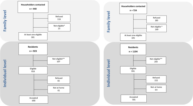
Fig 1A shows field work results in Antofagasta. Of 440 houses visited, 301 had at least one eligible subject (68.4%). A total of 923 potentially eligible subjects were identified in those households, of which 107 were not eligible. Of the total of 816 eligible subjects, 690 participated (participation rate: 84.5%). In Valdivia (Fig 1B) 724 houses were visited and 370 had at least one eligible subject (51.1%). There were 1194 potentially eligible subjects, of which 991 were eligible and 705 agreed to participate (71.1%).

Fig 1.

Fig 1

(a) Flow diagram of field work in Antofagasta. *Empty houses or no contact after several attempts. **Not eligible includes 37 subjects who did not fulfil the eligibility criteria and 70 subjects who were enumerated but not recruited because the required numbers for the corresponding categories of sex and age were already enrolled. (b) Flow diagram of field work in Valdivia. *Empty houses or no contact after several attempts. ** Not eligible includes 33 subjects who did not fulfil the eligibility criteria and 170 subjects who were enumerated but not recruited because the required numbers for the corresponding categories of sex and age were already enrolled.

Considering all ages, the sample was balanced by age group, but Antofagasta had proportionally more women than Valdivia (S1 Table). In Valdivia, there was a higher proportion of subjects of Mapuche origin while in Antofagasta there were more Aymara and more subjects of other ethnicities. Overweight and obesity, calculated only for subjects over 18, was high in both places, with approximately 75% overweight or obese.

Overall age-adjusted H. pylori ELISA seroprevalence was similar in both cities when considering ages 1-to70 (Table 1). Crude prevalence was 67% (95%CI = 63–71) in Antofagasta (lower GC risk area) and 63% (95% CI = 60–67) in Valdivia. Age-adjusted prevalence was 58% (95%CI = 51–69) in Antofagasta and 56% (95%CI = 49–66) in Valdivia. These estimates were also similar by gender with a statistically not significant higher prevalence in men than women in Valdivia (58% vs 53% in men and women, respectively, p = 0.19). Considering both sites combined, there were no clear differences in ELISA seroprevalence by gender among younger subjects, although prevalence was higher in men in all age groups over 30 years old (S1 Fig), with a p = 0.03 for all subjects over 30 combined.

Table 1. H. pylori prevalence determined by ELISA by study site (all ages).

Study site
Antofagasta Valdivia
    N = 690 (95% CI) N = 705 (95% CI)
    N (%) N (%)  
H. pylori status (crude)
Positive 432 (67) (63–71) 434 (63) (60–67)
Negative 211 (33) (29–37) 250 (37) (33–40)
Missing 47 21
H. pylori prevalence (adjusted) 58 [51–69] 56 [49–66]
Women 57 [48–70] 53 [45–62]
  Men 59 [50–69] 58 [49–70]

† Based on the Segi World Standard (Segi M, Kurihara M. Cancer mortality for selected sites in 24 Countries no. 6 (1966–1967) Nagoya, Japan Cancer Society; 1972).

In Antofagasta, but not in Valdivia, H. pylori prevalence was significantly higher in participants not born in the area compared with those born in Antofagasta (p = 0.007).

Seroprevalence of H. pylori was 20% among children under 10 years with a gradual increase thereafter (Fig 2). In Antofagasta, seroprevalence peaked at close to 90% in age group 50–59 with a subsequent decline to under 80% in subjects over 60. In Valdivia, seroprevalence peaked in the 40–49 age group with a subsequent decline to under 70% in those over 60. In the 50–59 age group, the difference between Antofagasta (prevalence = 90%, 95%CI = 83–95) and Valdivia (prevalence = 75%, 95%CI = 66–83) was statistically significant (p = 0.04).

Fig 2. H. pylori prevalence determined by ELISA by 10 years age group and site.
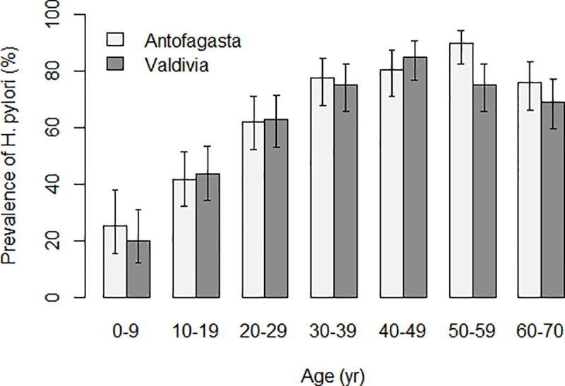
Fig 2

S2 Table compares H. pylori detection by ELISA with results based on immunoblot methods in subjects over age 40, with similar results for the two methods in both cities. The prevalence of CagA and VacA antibodies was also comparable, with estimates very similar to those of overall H. pylori positivity. Antibodies against other H. pylori antigens (37 kDa, 35 kDa, 30 kDa and 19.5 kDa) had varying levels of positivity, but in all cases were very similar in the two areas. More than 90% of H. pylori positive subjects had antibodies against CagA and VacA in both sites, and positivity of antibodies against the other antigens among the H. pylori positives was again similar by site. Only a handful of the H. pylori negatives by ELISA were positive for these antigens. The minor difference in the results between ELISA and Helicoblot assays may be explained by the fact that the latter could be a better indicator of a current H. pylori infection [23].

Fig 3 presents a forest plot with a multivariate model of the differences in prevalence of risk factors between study sites (age over 40); factors more common in Valdivia are to the right of the unity, with Antofagasta to the left. There were more men recruited in Valdivia. Chilean Hispanic ethnicity, daily intake of chili and history of parasite drug use were more common in Valdivia, the higher risk area. Age between 50–59, alcohol flush reaction, daily intake of alcohol, daily consumption of non-green vegetables (e.g., cucumber, radish, pepper), adding salt to food at the table, use of antibiotics, and history of pesticide exposure were significantly more common in Antofagasta. The strongest OR associated with the Valdivia study site was intake of chili peppers with an OR of 8.8 (95%CI = 4.2–20.6). A history of anemia was more common in Antofagasta (26% vs 16%, p = 0.002) but it was not retained in the final model. Similarly, current smoking was more common in Valdivia (34% vs 25%), but it was not retained in the final model. Education, binge drinking, consumption of salted or smoked foods, fruits and green vegetables were not different between sites in this age group.

Fig 3. Forest plot of factors associated with the Valdivia study site among participants aged 40 and over (multivariable logistic regression).
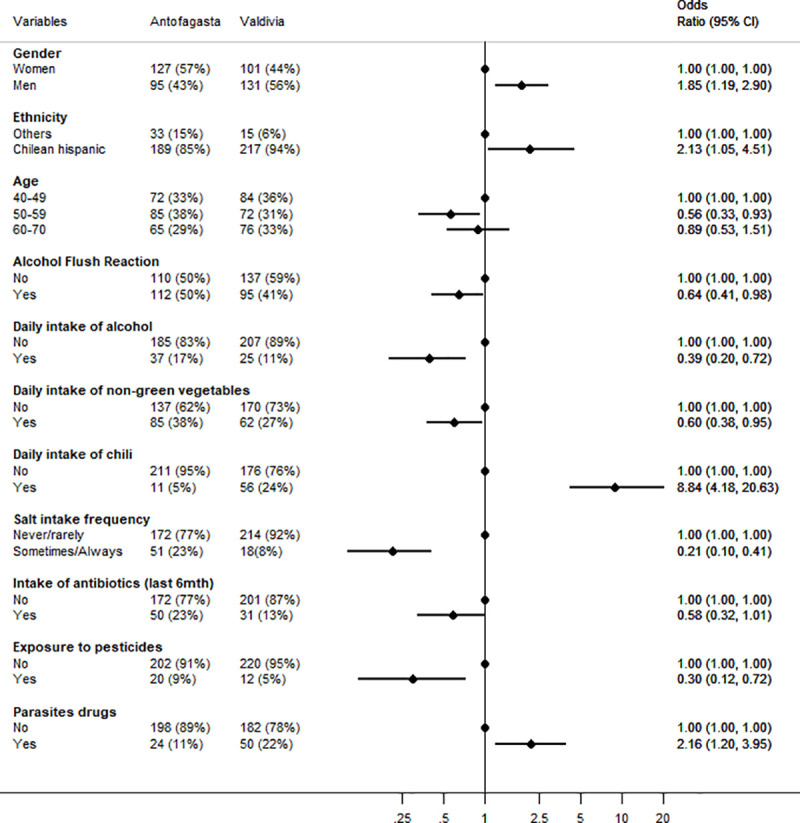
Fig 3

The first two columns present the prevalence for each site, the right part of the graph presents the adjusted odds ratio for that variable in the high-risk vs low risk site.

Prevalence of atrophy and severe atrophy, as determined by PgI and II levels, showed a significant difference between the two sites. Atrophy prevalence was significantly higher in the high risk region (31% vs 21%, p = 0.005), and this difference was stronger when restricted to H. pylori positive subjects by the Helicoblot method (p<0.001). Advanced atrophy was 11% in Valdivia and 7% in Antofagasta (p = 0.12, data not in tables). Fig 4 presents the comparison of the prevalence of atrophy by age and site. Among subjects aged 40–44 years, atrophy was less than 20% in both sites. However, while in Antofagasta (the low-risk area), prevalence remained below 20% until age 54, in Valdivia it increased to more than 30% after age 45. The prevalence of atrophy in the age group of 50–54 was more than twice higher in Valdivia (36.5%) compared with Antofagasta (15.4%) and the difference was statistically significant (P = 0.008). In Antofagasta, prevalence increased slowly with age but did not reach 30% even in the older age group. There was no difference in the prevalence of atrophy according to gender in either site. The mean of the PgI/PgII ratios was significantly lower in Valdivia (3.6) compared to Antofagasta (4.0) (p = 0.008), and there were no differences by age and sex.

Fig 4. Prevalence of atrophy (based on pepsinogen testing*) by 5 years age group and study site among 40 years and older.
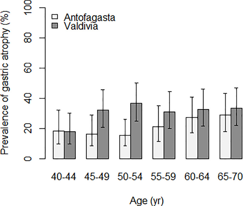
Fig 4

* Pepsinogens I and II were measured with a latex-agglutination test-system (Eiken Chemical, Tokyo, Japan) and cut off values for any gastric mucosal atrophy were PgI≤70 ng/ml and PgI/PgII≤3.

In a multivariate model of risk factors associated with atrophy (including severe atrophy) combining both sites, atrophy was positively associated with being from Valdivia, daily consumption of chili pepper and detection of antibodies against CagA while negatively associated with higher educational level (Fig 5). Smoking was not associated with risk of atrophy in this dataset.

Fig 5. Forest plot of the factors associated with atrophy among participants aged 40 and older (multivariable logistic regression).
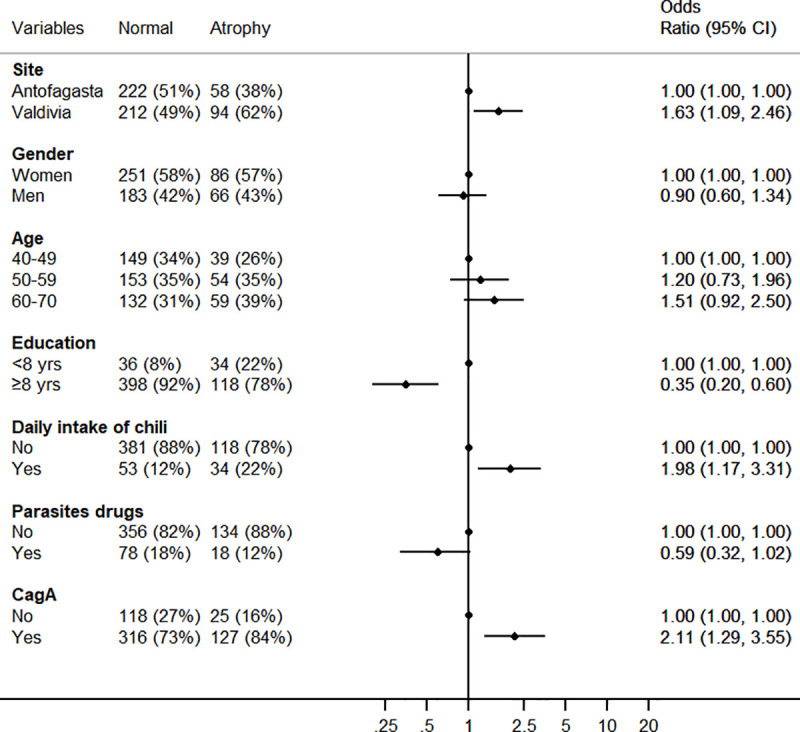
Fig 5

Discussion

This is the first of a series of IARC international prevalence surveys to clarify the epidemiology of H. pylori infection in high- and low-risk areas of GC, in an effort to predict GC incidence in young cohorts and generate hypotheses to explain its regional variations. Etiologic clues might then lead to identification of biomarkers or preventive interventions.

H. pylori infection causes 90% of non-cardia GC, and ongoing reductions in incidence are likely a result of reductions in H. pylori prevalence, which has been documented in many areas, mainly in high-income countries [9]. However, comparable data on H. pylori prevalence are scarce, as the studies have used dissimilar methodologies and laboratory methods. In addition, GC does not correlate as expected with H. pylori prevalence in adults, with many areas having high prevalence of H. pylori but low GC incidence rates. The term 'African enigma' was coined by Holcombe in 1992 [24] to describe the discrepancy between prevalence of infection and clinical manifestations of H. pylori (peptic ulcer and GC). Several authors described similar enigmas in Asia and Latin America and proposed different explanations, including variation in H. pylori strains, virulence factors or host genetics [25, 26]. Graham et al dismissed the findings as medical myths, recommending to focus on underlying patterns of gastritis (non-atrophic vs atrophic) and environmental factors [27]. In this study, we analyse both bacterial and environmental factors in conjunction with an indicator of the presence of atrophic gastritis. We believe that the same factors causing gastritis are likely to explain the regional and individual differences in GC risk.

We observed a very similar seroprevalence of H. pylori in the two communities in Chile, with an age-adjusted estimate close to 60% considering all age groups. Prevalence increased clearly with age from 20% in children under 10 to a peak at ages 40 or 50 between 80% and 90% depending on the location. Notably, H. pylori prevalence peaked earlier in the higher risk area, and declined faster and to a lower level than in the low risk area, where prevalence peaked later and at a higher level and declined less than in the high risk area. H. pylori presence in the stomach decreases with advancing atrophic changes [28], and disappearance of the bacteria results after some time in serology becoming negative [29]. We hypothesize that in the higher risk area, the earlier occurrence of atrophy might be the explanation for the earlier peak in H. pylori infection and the rapid decline afterwards. In fact, we observed that serologic evidence of atrophy occurred earlier and was higher in Valdivia than in Antofagasta (see below).

Overall, H. pylori infection was as common in men as in women (both sites combined), but higher rates were detected in men over 30 years. Given that most infections initiate in childhood, and that 90% of GCs are associated with infection, the lower prevalence in younger generations likely predicts future declines in GC incidence in Chile, as has been observed elsewhere. We plan to work on a quantitative model to estimate future risk of GC in the countries participating in ENIGMA.

In the 40+ year age group, the most likely to explain current rates of GC, we compared prevalence of H. pylori by ELISA and Helicoblot 2.1, with an 85% agreement and comparable prevalence estimates. Seroprevalence of H. pylori was around 80% among subjects over 40 years with no differences by site. Antibodies against CagA, VacA or the other antigens explored were also comparable by site. CagA and VacA antibodies were almost universal among H. pylori positive subjects and very rare in H. pylori negatives. CagA and VacA are important H. pylori virulence factors and have been associated with risk of GC [30, 31]. In some studies, CagA was more common in high- than in low-risk areas [25, 27], but not in our study and others [32, 33].

Among the interesting findings regarding differences in risk factors between the two sites was consumption of non-green vegetables, which was more common in the low risk area, but there were no significant differences for consumption of other vegetables and fruits. In the Chilean National Health Survey of 2009–2010, consuming at least five portions of fruit and vegetables daily was 32.5% in Antofagasta compared to 1.4% in the Valdivia region, but daily portions of fruits and vegetables was not significantly different by region [34].

Consumption of chili products was more common in the high risk area. Chili has been proposed as a direct irritant of the gastric mucosa but studies have been inconsistent on potential benefits or harms of its active principle capsaicin [35, 36]. López-Carrillo and colleagues explored potential interactions among capsaicin, H. pylori virulent factors and genetic factors and showed that moderate to high capsaicin consumption was associated with increased risk of GC in genetically susceptible individuals (IL1B-31C allele carriers) infected with CagA positive strains [37].

Alternatively, chili could be contaminated with aflatoxins as has been demonstrated for some chili products in Chile [38]. Aflatoxin is a known group 1 carcinogen that causes cancer of the liver [39] and its dietary consumption (by questionnaire) was associated with GC in one study [40]. We plan to explore the role of aflatoxins using our stored blood specimens.

Contrary to our expectations, addition of salt at the table was more common in Antofagasta, the low risk area. However, salt intake is a particularly challenging variable for dietary assessment given large intraindividual variation and discretionary use, often not adequately captured [41]. We will explore this further by measuring sodium excretion in urine samples.

We explored gastric atrophy as measured by reductions in the PgI/PgII ratio. Despite limited sensitivity and specificity for disease detection [42], Pgs are strongly associated with prospective GC risk [43] and in the context of this ecologic study we consider them as surrogate markers of gastric cancer risk. In Valdivia, serologic evidence of gastric atrophy was more common than in Antofagasta and occurred at earlier ages, in concordance with the proposed notion that the pattern of gastritis at the population level is an important determinant of differences in GC incidence [27]. However, the explanation of the regional differences in atrophic gastritis (an intermediate outcome) and GC risk is still to be found in bacterial, environmental or genetic factors. In the multivariable model the main risk factors for atrophy were being from Valdivia, age, chili consumption, a typical element of the diet in Southern Chile, and CagA antibodies. There have been few studies investigating factors associated with gastric atrophy, but older age and CagA seem to be consistently identified [16, 17].

The strengths of our study include recruitment of population-based samples by invitation, preventing selection based on symptoms or attendance to clinics. We used standardized methods for interviewing, specimen collection and laboratory procedures. These methods will also be used in subsequent ENIGMA studies in other countries permitting direct comparison and pooling of results.

Among study limitations, participation rates were lower in Valdivia because of idiosyncratic characteristics plus logistic and climatic reasons. Another limitation is that the difference in GC incidence between the two sites may not be sufficiently large to detect the differences in exposure to risk factors explaining it. We also had small numbers for some analyses and did not have biopsy materials to study the prevalence of histologically confirmed preneoplastic lesions or the H. pylori strains present in the two sites. The ELISA assay in this study (Biohit) is a standardized test based on purified H. pylori antigens that has been used in multiple populations in various studies. Although the use of local antigens is recommended to assure the validity of serologic methods in clinical practice, in ENIGMA we are using the same test in the different study sites to assure standardization of the results. The fact that the results of the Western Blot test were comparable to those of the ELISA is reassuring in this regard. In addition, this is an ecologic study where the areas are the unit of analysis and the study participants are not the same individuals with gastric cancer in the population, and therefore the associations described should be considered for hypothesis generating and need to be corroborated in individual-based longitudinal studies. The second component of ENIGMA (ENIGMA II) that aims to investigate the worldwide epidemiology of gastric premalignant conditions includes gastroscopy and biopsies in adult participants of the ENIGMA I studies to address these issues.

In conclusion, we found no differences in the prevalence of H. pylori or its virulence factors between the high and low risk areas in Chile. H. pylori is less common in younger generations and we should expect the trends of declining gastric cancer rates to continue rates in the future. We identified some potential risk factors that could explain both the difference in GC risk and atrophy as its surrogate. However, the role of diet, in particular chili pepper, should be explored further. Our study suggests that atrophy is more frequent and occurs much earlier in the high-risk area of GC and that one of the main risk factors for atrophic gastritis is consumption of chili pepper that could act directly or through interactions with other environmental factors. These hypotheses deserve further research as they could help design additional preventive interventions.

Supporting information

S1 Fig. H. pylori prevalence determined by ELISA by 10 years age group and gender (both sites combined).

(PNG)

S1 Table. Demographic characteristics by study site.

(DOCX)

S2 Table. H. pylori seropositivity determined by ELISA and Helicoblot and the frequency of H. pylori immunoreactive bands by study site among participants aged 40 and older.

(DOCX)

S1 File. Interview English.

(DOCX)

S2 File. Interview Spanish.

(DOCX)

S1 Data. Final dataset.

(CSV)

S1 Codebook

(XLS)

Acknowledgments

Dr Paula González, our beloved colleague and leader of cancer research in Latin America passed away before the submission of the final version of this manuscript. Rolando Herrero accepts responsibility for the integrity and validity of the data collected and analyzed.

We thank Drs Sabina Rinaldi and Maria de la Luz Hernandez for their input and support in H. pylori ELISA testing at IARC. The members of the ENIGMA Chile study group are the following: Fabio Paredes, Catterina Ferreccio (group leader) (catferre@gmail.com) from the Pontificia Universidad Católica de Chile; Janet Altamirano, Ana Wall, Maritza Corvalán, Felix Díaz, Patricia Ochoa, Francisco Mena, Eliseo Martinez from the Universidad de Antofagasta; José Ignacio Zarate, Juan Carlos Velásquez form the Registro hospitalario de cáncer Hospital Base Valdivia; Solange Vargas from the Registro Poblacional de Cancer Valdivia, SEREMI de Salud de Los Ríos; Ivo Muñoz from the Universidad Austral de Chile; Dace Rudzīte from the University of Latvia; M. Constanza Camargo from the US National Cancer Institute and Salvatore Vaccarella from the International Agency for Research on cancer (IARC).

Disclaimer: Where authors are identified as personnel of the International Agency for Research on Cancer / World Health Organization, the authors alone are responsible for the views expressed in this article and they do not necessarily represent the decisions, policy or views of the International Agency for Research on Cancer / World Health Organization.

Data Availability

All relevant data are within the paper and its Supporting Information files.

Funding Statement

The work in Chile was supported in part by a grant from Fondo Nacional de Investigación y Desarrollo en Salud (FONIS grant SA1312007) and the pepsinogen work in Latvia by a grant from the Latvian Council of Science (nr. lzp-2018/1-0135).

References

  • 1.Bray F, Ferlay J, Soerjomataram I, Siegel RL, Torre LA, Jemal A. Global cancer statistics 2018: GLOBOCAN estimates of incidence and mortality worldwide for 36 cancers in 185 countries. CA Cancer J Clin. 2018;68(6):394–424. Epub 2018/09/13. 10.3322/caac.21492 . [DOI] [PubMed] [Google Scholar]
  • 2.Bray F, Colombet M, Mery L, Piñeros M, Znaor A, Zanetti R, et al. , editors. Cancer Incidence in Five Continents, Vol. XI (electronic version). Available from: http://ci5.iarc.fr, accessed [10 September 2018]. Lyon: International Agency for Research on Cancer; 2017. [Google Scholar]
  • 3.Malekzadeh R, Derakhshan MH, Malekzadeh Z. Gastric cancer in Iran: epidemiology and risk factors. Archives of Iranian medicine. 2009;12(6):576–83. Epub 2009/11/03. . [PubMed] [Google Scholar]
  • 4.Ministry of Health of Chile. Department of Statistics and Health Information 2019 [12/11/2019]. Available from: http://www.deis.cl/series-y-graficos-de-mortalidad/.
  • 5.IARC Working Group on the Evaluation of Carcinogenic Risks to Humans. A review of human carcinogens. Part B: Biological Agents. Available from: http://monographs.iarc.fr/ENG/Monographs/vol100B/mono100B.pdf. Lyon, France: International Agency for Research on Cancer; 2012. [Google Scholar]
  • 6.Correa P, Haenszel W, Cuello C, Tannenbaum S, Archer M. A model for gastric cancer epidemiology. The Lancet. 1975;306(7924):58–60. 10.1016/S0140-6736(75)90498-5 [DOI] [PubMed] [Google Scholar]
  • 7.Plummer M, de Martel C, Vignat J, Ferlay J, Bray F, Franceschi S. Global burden of cancers attributable to infections in 2012: a synthetic analysis. Lancet Glob Health. 2016;4(9):e609–16. 10.1016/S2214-109X(16)30143-7 . [DOI] [PubMed] [Google Scholar]
  • 8.Hooi JKY, Lai WY, Ng WK, Suen MMY, Underwood FE, Tanyingoh D, et al. Global Prevalence of Helicobacter pylori Infection: Systematic Review and Meta-Analysis. Gastroenterology. 2017;153(2):420–9. 10.1053/j.gastro.2017.04.022 [DOI] [PubMed] [Google Scholar]
  • 9.Inoue M. Changing epidemiology of Helicobacter pylori in Japan. Gastric Cancer. 2017;20(1):3–7. 10.1007/s10120-016-0658-5 [DOI] [PubMed] [Google Scholar]
  • 10.Lim SH, Kim N, Kwon JW, Kim SE, Baik GH, Lee JY, et al. Trends in the seroprevalence of Helicobacter pylori infection and its putative eradication rate over 18 years in Korea: A cross-sectional nationwide multicenter study. PLOS ONE. 2018;13(10):e0204762 10.1371/journal.pone.0204762 [DOI] [PMC free article] [PubMed] [Google Scholar]
  • 11.Maucort-Boulch D, Franceschi S, Plummer M. International Correlation between Human Papillomavirus Prevalence and Cervical Cancer Incidence. Cancer Epidemiology Biomarkers & Prevention. 2008;17(3):717–20. 10.1158/1055-9965.Epi-07-2691 [DOI] [PubMed] [Google Scholar]
  • 12.Newton R, Ziegler JL, Casabonne D, Carpenter L, Gold BD, Owens M, et al. Helicobacter pylori and cancer among adults in Uganda. Infectious agents and cancer. 2006;1:5–. 10.1186/1750-9378-1-5 . [DOI] [PMC free article] [PubMed] [Google Scholar]
  • 13.Kodaman N, Pazos A, Schneider BG, Piazuelo MB, Mera R, Sobota RS, et al. Human and Helicobacter pylori coevolution shapes the risk of gastric disease. Proceedings of the National Academy of Sciences. 2014:201318093 10.1073/pnas.1318093111 [DOI] [PMC free article] [PubMed] [Google Scholar]
  • 14.Torres J, Correa P, Ferreccio C, Hernandez-Suarez G, Herrero R, Cavazza-Porro M, et al. Gastric cancer incidence and mortality is associated with altitude in the mountainous regions of Pacific Latin America. Cancer causes & control: CCC. 2013;24(2):249–56. Epub 2012/12/07. 10.1007/s10552-012-0114-8 . [DOI] [PMC free article] [PubMed] [Google Scholar]
  • 15.Fox JG, Wang TC. Inflammation, atrophy, and gastric cancer. The Journal of clinical investigation. 2007;117(1):60–9. 10.1172/JCI30111 . [DOI] [PMC free article] [PubMed] [Google Scholar]
  • 16.Adamu MA, Weck MN, Rothenbacher D, Brenner H. Incidence and risk factors for the development of chronic atrophic gastritis: five year follow-up of a population-based cohort study. Int J Cancer. 2011;128(7):1652–8. Epub 2010/05/27. 10.1002/ijc.25476 . [DOI] [PubMed] [Google Scholar]
  • 17.Eurohepygast Study Group. Risk factors for atrophic chronic gastritis in a European population: results of the Eurohepygast study. Gut. 2002;50(6):779–85. 10.1136/gut.50.6.779 . [DOI] [PMC free article] [PubMed] [Google Scholar]
  • 18.Joo Y-E, Park H-K, Myung D-S, Baik G-H, Shin J-E, Seo G-S, et al. Prevalence and risk factors of atrophic gastritis and intestinal metaplasia: a nationwide multicenter prospective study in Korea. Gut and liver. 2013;7(3):303–10. Epub 2013/05/13. 10.5009/gnl.2013.7.3.303 . [DOI] [PMC free article] [PubMed] [Google Scholar]
  • 19.Oh S, Kim N, Yoon H, Choi YJ, Lee JY, Park KJ, et al. Risk factors of atrophic gastritis and intestinal metaplasia in first-degree relatives of gastric cancer patients compared with age-sex matched controls. Journal of cancer prevention. 2013;18(2):149–60. 10.15430/jcp.2013.18.2.149 . [DOI] [PMC free article] [PubMed] [Google Scholar]
  • 20.Leja M, Camargo MC, Polaka I, Isajevs S, Liepniece-Karele I, Janciauskas D, et al. Detection of gastric atrophy by circulating pepsinogens: A comparison of three assays. Helicobacter. 2017;22(4):10.1111/hel.12393. Epub 2017/05/29. 10.1111/hel.12393 . [DOI] [PMC free article] [PubMed] [Google Scholar]
  • 21.Segi M. Cancer Mortality for Selected Sites in 24 Countries (1950–57). Sendai, Japan: Department of Public Health, Tohoku University of Medicine; 1960. [Google Scholar]
  • 22.Frank J F, Sergio M F, Svetlozar T R, Bala G A. The Basics of Financial Econometrics Appendix E: Model Selection Criterion: AIC and BIC. John Wiley & Sons, Inc; 2014. [Google Scholar]
  • 23.Ekstrom AM, Held M, Hansson LE, Engstrand L, Nyren O. Helicobacter pylori in gastric cancer established by CagA immunoblot as a marker of past infection. Gastroenterology. 2001;121(4):784–91. Epub 2001/10/19. 10.1053/gast.2001.27999 . [DOI] [PubMed] [Google Scholar]
  • 24.Holcombe C. Helicobacter pylori: the African enigma. Gut. 1992;33(4):429–31. 10.1136/gut.33.4.429 [DOI] [PMC free article] [PubMed] [Google Scholar]
  • 25.Con SA, Valerin AL, Takeuchi H, Con-Wong R, Con-Chin VG, Con-Chin GR, et al. Helicobacter pylori CagA status associated with gastric cancer incidence rate variability in Costa Rican regions. J Gastroenterol. 2006;41(7):632–7. Epub 2006/08/26. 10.1007/s00535-006-1812-3 . [DOI] [PubMed] [Google Scholar]
  • 26.Singh K, Ghoshal UC. Causal role of Helicobacter pylori infection in gastric cancer: an Asian enigma. World J Gastroenterol. 2006;12(9):1346–51. Epub 2006/03/23. 10.3748/wjg.v12.i9.1346 [DOI] [PMC free article] [PubMed] [Google Scholar]
  • 27.Graham DY, Lu H, Yamaoka Y. African, Asian or Indian enigma, the East Asian Helicobacter pylori: facts or medical myths. Journal of Digestive Diseases. 2009;10(2):77–84. 10.1111/j.1751-2980.2009.00368.x [DOI] [PMC free article] [PubMed] [Google Scholar]
  • 28.Kato I, Vivas J, Plummer M, Lopez G, Peraza S, Castro D, et al. Environmental Factors in Helicobacter pylori-Related Gastric Precancerous Lesions in Venezuela. Cancer Epidemiology Biomarkers & Prevention. 2004;13(3):468–76. [PubMed] [Google Scholar]
  • 29.Feldman M, Cryer B, Lee E, Peterson WL. Role of seroconversion in confirming cure of Helicobacter pylori infection. JAMA. 1998;280(4):363–5. Epub 1998/08/01. 10.1001/jama.280.4.363 . [DOI] [PubMed] [Google Scholar]
  • 30.Huang JQ, Zheng GF, Sumanac K, Irvine EJ, Hunt RH. Meta-analysis of the relationship between cagA seropositivity and gastric cancer. Gastroenterology. 2003;125(6):1636–44. Epub 2004/01/16. 10.1053/j.gastro.2003.08.033 . [DOI] [PubMed] [Google Scholar]
  • 31.Murphy G, Freedman ND, Michel A, Fan J-H, Taylor PR, Pawlita M, et al. Prospective study of Helicobacter pylori antigens and gastric noncardia cancer risk in the nutrition intervention trial cohort. International Journal of Cancer. 2015;137(8):1938–46. 10.1002/ijc.29543 [DOI] [PMC free article] [PubMed] [Google Scholar]
  • 32.Devi SM, Ahmed I, Francalacci P, Hussain MA, Akhter Y, Alvi A, et al. Ancestral European roots of Helicobacter pylori in India. BMC genomics. 2007;8:184 Epub 2007/06/23. 10.1186/1471-2164-8-184 [DOI] [PMC free article] [PubMed] [Google Scholar]
  • 33.Camargo MC, Beltran M, Conde-Glez CJ, Harris PR, Michel A, Waterboer T, et al. Serological response to Helicobacter pylori infection among Latin American populations with contrasting risks of gastric cancer. International journal of cancer. 2015;137(12):3000–5. Epub 2015/07/16. 10.1002/ijc.29678 . [DOI] [PMC free article] [PubMed] [Google Scholar]
  • 34.Encuesta Nacional de Salud. Chile 2009–2010. Tomo 2. Resultatos. [National Health Survey. Chile 2009–10. Volume 2 Results] http://web.minsal.cl/portal/url/item/bcb03d7bc28b64dfe040010165012d23.pdf Accessed March 30 2018. Spanish. Santiago: Ministerio de Salud, División de Planificación Sanitaria, Departamento de Epidemiología; 2011. [Google Scholar]
  • 35.López-Carrillo L, López-Cervantes M, Robles-Díaz G, Ramírez-Espitia A, Mohar-Betancourt A, Meneses-García A, et al. Capsaicin consumption, Helicobacter pylori positivity and gastric cancer in Mexico. International Journal of Cancer. 2003;106(2):277–82. 10.1002/ijc.11195 [DOI] [PubMed] [Google Scholar]
  • 36.Satyanarayana MN. Capsaicin and gastric ulcers. Critical reviews in food science and nutrition. 2006;46(4):275–328. Epub 2006/04/20. 10.1080/1040-830491379236 . [DOI] [PubMed] [Google Scholar]
  • 37.López-Carrillo L, Camargo MC, Schneider BG, Sicinschi LA, Hernández-Ramírez RU, Correa P, et al. Capsaicin consumption, Helicobacter pylori CagA status and IL1B-31C>T genotypes: a host and environment interaction in gastric cancer. Food Chem Toxicol. 2012;50(6):2118–22. Epub 2012/03/04. 10.1016/j.fct.2012.02.043 . [DOI] [PMC free article] [PubMed] [Google Scholar]
  • 38.Nogueira L, Foerster C, Groopman J, et al. Association of aflatoxin with gallbladder cancer in chile. JAMA. 2015;313(20):2075–7. 10.1001/jama.2015.4559 [DOI] [PMC free article] [PubMed] [Google Scholar]
  • 39.IARC Working Group on the Evaluation of Carcinogenic Risks to Humans. A review of human carcinogens. Volume 100F. Available from: https://monographs.iarc.fr/iarc-monographs-on-the-evaluation-of-carcinogenic-risks-to-humans-16/. 2012;100F.
  • 40.Eom S-Y, Yim D-H, Zhang Y, Yun J-K, Moon SI, Yun H-Y, et al. Dietary aflatoxin B1 intake, genetic polymorphisms of CYP1A2, CYP2E1, EPHX1, GSTM1, and GSTT1, and gastric cancer risk in Korean. Cancer Causes & Control. 2013;24(11):1963–72. 10.1007/s10552-013-0272-3 [DOI] [PubMed] [Google Scholar]
  • 41.Khaw K-T, Bingham S, Welch A, Luben R, O'Brien E, Wareham N, et al. Blood pressure and urinary sodium in men and women: the Norfolk Cohort of the European Prospective Investigation into Cancer (EPIC-Norfolk). The American Journal of Clinical Nutrition. 2004;80(5):1397–403. 10.1093/ajcn/80.5.1397 [DOI] [PubMed] [Google Scholar]
  • 42.Dinis-Ribeiro M, Yamaki G, Miki K, Costa-Pereira A, Matsukawa M, Kurihara M. Meta-analysis on the validity of pepsinogen test for gastric carcinoma, dysplasia or chronic atrophic gastritis screening. J Med Screen. 2004;11(3):141–7. Epub 2004/08/31. 10.1258/0969141041732184 . [DOI] [PubMed] [Google Scholar]
  • 43.Palli D, Masala G, Del Giudice G, Plebani M, Basso D, Berti D, et al. CagA+ Helicobacter pylori infection and gastric cancer risk in the EPIC-EURGAST study. International Journal of Cancer. 2007;120(4):859–67. 10.1002/ijc.22435 [DOI] [PubMed] [Google Scholar]

Decision Letter 0

Eric J Duell

19 Feb 2020

PONE-D-19-35057

Regional variations in Helicobacter pylori infection, gastric atrophy and gastric cancer risk: the ENIGMA study in Chile

PLOS ONE

Dear Dr Herrero,

Thank you for submitting your manuscript to PLOS ONE. After careful consideration, we feel that it has merit but does not fully meet PLOS ONE’s publication criteria as it currently stands. Therefore, we invite you to submit a revised version of the manuscript that addresses the points raised during the review process.

Please carefully address all comments raised by both reviewers, paying particular attention to statistical comparisons and the fact that this is an ecologic study and not an analytic study.  The weaknesses of the ecologic study design should be clearly stated in the discussion (e.g., that areas are the unit of analysis, and that the individuals with Hp information are not the same individuals with gastritis or gastric cancer within the study areas).  Ecologic studies may help to define hypotheses that can then be tested using analytic (observational) study designs with individual-level data.  Interventions would only be considered after analytic studies, so the final sentence of the Discussion should be revised accordingly. 

Please review the PLOS ONE data sharing policy and clearly state where the data can be accessed; and if not, the reasons should be specified.  Your revision should also be edited for English grammar. 

We would appreciate receiving your revised manuscript by Apr 04 2020 11:59PM. When you are ready to submit your revision, log on to https://www.editorialmanager.com/pone/ and select the 'Submissions Needing Revision' folder to locate your manuscript file.

If you would like to make changes to your financial disclosure, please include your updated statement in your cover letter.

To enhance the reproducibility of your results, we recommend that if applicable you deposit your laboratory protocols in protocols.io, where a protocol can be assigned its own identifier (DOI) such that it can be cited independently in the future. For instructions see: http://journals.plos.org/plosone/s/submission-guidelines#loc-laboratory-protocols

Please include the following items when submitting your revised manuscript:

  • A rebuttal letter that responds to each point raised by the academic editor and reviewer(s). This letter should be uploaded as separate file and labeled 'Response to Reviewers'.

  • A marked-up copy of your manuscript that highlights changes made to the original version. This file should be uploaded as separate file and labeled 'Revised Manuscript with Track Changes'.

  • An unmarked version of your revised paper without tracked changes. This file should be uploaded as separate file and labeled 'Manuscript'.

Please note while forming your response, if your article is accepted, you may have the opportunity to make the peer review history publicly available. The record will include editor decision letters (with reviews) and your responses to reviewer comments. If eligible, we will contact you to opt in or out.

We look forward to receiving your revised manuscript.

Kind regards,

Eric J. Duell, MS, PhD

Academic Editor

PLOS ONE

Journal Requirements:

When submitting your revision, we need you to address these additional requirements:

1. Please ensure that your manuscript meets PLOS ONE's style requirements, including those for file naming. The PLOS ONE style templates can be found at http://www.plosone.org/attachments/PLOSOne_formatting_sample_main_body.pdf and http://www.plosone.org/attachments/PLOSOne_formatting_sample_title_authors_affiliations.pdf

2. Please include additional information regarding the survey or questionnaire used in the study and ensure that you have provided sufficient details that others could replicate the analyses. For instance, if you developed a questionnaire as part of this study and it is not under a copyright more restrictive than CC-BY, please include a copy, in both the original language and English, as Supporting Information.

3. Thank you for including your ethics statement: The study was approved by IARC Ethics Committee (IEC No.14-17) and local ethical committees (Comité Ética de la Investigación, Escuela de Medicina Pontificia Universidad Católica de Chile, Comité de Ética Zona Norte, Servicio de Salud Coquimbo). Eligible adults signed informed consent. Children between 12 and 18 provided informed assent and parental informed consent, and for children under 12 only parental informed consent was obtained.

Please amend your current ethics statement to include the full name of the ethics committee/institutional review board(s) that approved your specific study.

Once you have amended this/these statement(s) in the Methods section of the manuscript, please add the same text to the “Ethics Statement” field of the submission form (via “Edit Submission”).

For additional information about PLOS ONE ethical requirements for human subjects research, please refer to http://journals.plos.org/plosone/s/submission-guidelines#loc-human-subjects-research.

4. One of the noted authors is a group or consortium (ENIGMA Chile study group). In addition to naming the author group, please list the individual authors and affiliations within this group in the acknowledgments section of your manuscript. Please also indicate clearly a lead author for this group along with a contact email address.

5. We note that you have included the phrase “data not shown” in your manuscript. Unfortunately, this does not meet our data sharing requirements. PLOS does not permit references to inaccessible data. We require that authors provide all relevant data within the paper, Supporting Information files, or in an acceptable, public repository. Please add a citation to support this phrase or upload the data that corresponds with these findings to a stable repository (such as Figshare or Dryad) and provide and URLs, DOIs, or accession numbers that may be used to access these data. Or, if the data are not a core part of the research being presented in your study, we ask that you remove the phrase that refers to these data.

6. Thank you for stating the following in the Competing Interests section:

"The authors have declared that no competing interests exist."

We note that one or more of the authors are employed by a commercial company: International Agency for Research on Cancer and Advanced Center for Chronic Diseases.

a.     Please provide an amended Funding Statement declaring this commercial affiliation, as well as a statement regarding the Role of Funders in your study. If the funding organization did not play a role in the study design, data collection and analysis, decision to publish, or preparation of the manuscript and only provided financial support in the form of authors' salaries and/or research materials, please review your statements relating to the author contributions, and ensure you have specifically and accurately indicated the role(s) that these authors had in your study. You can update author roles in the Author Contributions section of the online submission form.

Please also include the following statement within your amended Funding Statement.

“The funder provided support in the form of salaries for authors [insert relevant initials], but did not have any additional role in the study design, data collection and analysis, decision to publish, or preparation of the manuscript. The specific roles of these authors are articulated in the ‘author contributions’ section.”

If your commercial affiliation did play a role in your study, please state and explain this role within your updated Funding Statement.

b. Please also provide an updated Competing Interests Statement declaring this commercial affiliation along with any other relevant declarations relating to employment, consultancy, patents, products in development, or marketed products, etc.  

Within your Competing Interests Statement, please confirm that this commercial affiliation does not alter your adherence to all PLOS ONE policies on sharing data and materials by including the following statement: "This does not alter our adherence to  PLOS ONE policies on sharing data and materials.” (as detailed online in our guide for authors http://journals.plos.org/plosone/s/competing-interests) . If this adherence statement is not accurate and  there are restrictions on sharing of data and/or materials, please state these. Please note that we cannot proceed with consideration of your article until this information has been declared.

Please include both an updated Funding Statement and Competing Interests Statement in your cover letter. We will change the online submission form on your behalf.

Please know it is PLOS ONE policy for corresponding authors to declare, on behalf of all authors, all potential competing interests for the purposes of transparency. PLOS defines a competing interest as anything that interferes with, or could reasonably be perceived as interfering with, the full and objective presentation, peer review, editorial decision-making, or publication of research or non-research articles submitted to one of the journals. Competing interests can be financial or non-financial, professional, or personal. Competing interests can arise in relationship to an organization or another person. Please follow this link to our website for more details on competing interests: http://journals.plos.org/plosone/s/competing-interests

[Note: HTML markup is below. Please do not edit.]

Reviewers' comments:

Reviewer's Responses to Questions

Comments to the Author

1. Is the manuscript technically sound, and do the data support the conclusions?

The manuscript must describe a technically sound piece of scientific research with data that supports the conclusions. Experiments must have been conducted rigorously, with appropriate controls, replication, and sample sizes. The conclusions must be drawn appropriately based on the data presented.

Reviewer #1: Partly

Reviewer #2: Partly

**********

2. Has the statistical analysis been performed appropriately and rigorously?

Reviewer #1: No

Reviewer #2: Yes

**********

3. Have the authors made all data underlying the findings in their manuscript fully available?

The PLOS Data policy requires authors to make all data underlying the findings described in their manuscript fully available without restriction, with rare exception (please refer to the Data Availability Statement in the manuscript PDF file). The data should be provided as part of the manuscript or its supporting information, or deposited to a public repository. For example, in addition to summary statistics, the data points behind means, medians and variance measures should be available. If there are restrictions on publicly sharing data—e.g. participant privacy or use of data from a third party—those must be specified.

Reviewer #1: No

Reviewer #2: Yes

**********

4. Is the manuscript presented in an intelligible fashion and written in standard English?

PLOS ONE does not copyedit accepted manuscripts, so the language in submitted articles must be clear, correct, and unambiguous. Any typographical or grammatical errors should be corrected at revision, so please note any specific errors here.

Reviewer #1: No

Reviewer #2: Yes

**********

5. Review Comments to the Author

Please use the space provided to explain your answers to the questions above. You may also include additional comments for the author, including concerns about dual publication, research ethics, or publication ethics. (Please upload your review as an attachment if it exceeds 20,000 characters)

Reviewer #1: The authors have conducted a survey of H. pylori infection in Chile using conventional methods. Importantly, this prevalence estimates a structured population sample rather than simply controls from a case-control study or other continence sample. Overall the prevalence data on Hp seropositivity and the virulence factors was valuable. The cut points used for the presence of atrophy are OK, but it would be much better to see the actual distributions of PG1, PG2, and the PG ratio in the two communities after standardization for age sex differences in the samples. The atrophy cut points are not optimized for assessing gastric cancer risk, the main purpose of their inclusion here.

Overall, I found much of the presentation extraneous to the stated core goal of the project – the seroprevalence of Hp, CagA, VacA, and Pepsinogen. Drawing conclusions about potential dietary risk factors diminished the focus on what is most valuable about the project.

ABSTRACT

The two sentences of background in the abstract are contradictory. They properly state that the rates of gastric cancer don’t follow prevalence estimates, but then state they do the survey to explain regional differences in gastric cancer risk. The prevalence is important whether it directly explains the rates or not. According to the intro, the rates of GC in Valdivia and Antofagasta (in men) are 33.1 and 21.2, respectively. That isn’t approximately two-fold different as written, although the mortality rate is nearly 3-fold different.

• Please be more specific than ‘virulence factors’ in the methods

• Age standardized to what?

• The comments on dietary factors are supposed to inform the prevalence of Hp, gastric atrophy, or gastric cancer?

• Not clear if they create a single model for assessing risk factors for atrophy across Chile and if they did, does geographic location remain a risk factor?

The first sentence of the conclusion doesn’t seem to be paired to the data presented in the results and it isn’t clear what ‘regional variation’ refers to, gastric cancer or atrophy?

Drawing any conclusions about novel risk factors for gastric cancer, such as chili peppers, from this modest study seems a bit much in the abstract.

The abstract doesn’t present any data on prevalence! I thought that was the primary goal of the study?

MAIN TEXT

H. pylori seropositivity is highly dependent on the senstivitiy of the test. This is less important when making risk estimates comparing case and controls within a population, but here it seems all important given that event6ually they will be comparing these rates to other populations (Page 14 “This is the first of a series of IARC international prevalence surveys.”) An independent assessment of Hp would be really helpful here because there is no guarantee the test will work the same in other parts of the world that have different strains of Hp. Can the authors point to any data on the sensitivity of the Biohit test in Chilean people? It was reassuring that the Eiken and Biohit values are similar.

I am not sure how to interpret the prevalence odds ratios used in figure 3. It seems it would be simpler to just present a table of a figure with the point estimate for each location and then some measure on uncertainty around the prevalence estimate. Or maybe just the net difference in the prevalence? I also don’t know what it means to have an adjusted OR for, example, of chili intake. Chili consumption if ~5-fold more common in Valdivia (24%) than in Antofagasta (5%), so what does the OR of 8.6 mean and in what sense did the other factors in table 3 confound the chili intake estimate?

The prevalence of atrophy seems to parallel the incidence of gastric cancer quite nicely for both the lower and more stringent cut point. It would be nice if the authors showed us all values (and ratios ) for the two areas for GC incidence, GC mortality, Hp positvity, CagA, VacA, Pep1, Pep2, and PG ratio, and atrophy in a single table

Page 14, line 302 – please don’t use the term ‘developed countries’ with out a modifier such as economically-developed countries.

I don’t understand this conclusion on page 18 drawn from the analysis presented in figure 5. “…one of the main risk factors for atrophic gastritis is consumption of chili pepper.” An OR of 1.97 and a prevalence of 22% in cases seems like a modest association in a single study. In fact, chili pepper consumption would have no individual level predictive value for whether or not an individual has atrophy. In fact, lack of education has a stronger association (OR of almost 3) and the same prevalence. Why was CagA included rather than the whole cell Hp antibody? I don’t see any evidence that CagA was more predictive that just seropositivity. In some previous studies, disease risk is greatest in those that are CagA positive and whole cell negative, but it doesn’t seem that was explored here.

Finally, an established risk factor for gastric cancer, tobacco seems to be missing from the results. The methods noted that smoking was collected so it should be included in the report.

Reviewer #2: This is an ecologic study of Helicobacter pylori, gastric atrophy, selected aspects of diet and gastric from two areas of Chile with different gastric cancer mortality.

The only consistent finding is higher atrhopy in the high risk area. The others, with the exception of vegetable consumption, are difficult to explain.

Given the limitations of the study design, their interpretation therefore, should be more cautious.

**********

6. PLOS authors have the option to publish the peer review history of their article (what does this mean?). If published, this will include your full peer review and any attached files.

If you choose “no”, your identity will remain anonymous but your review may still be made public.

Do you want your identity to be public for this peer review? For information about this choice, including consent withdrawal, please see our Privacy Policy.

Reviewer #1: No

Reviewer #2: Yes: Carlo La Vecchia

[NOTE: If reviewer comments were submitted as an attachment file, they will be attached to this email and accessible via the submission site. Please log into your account, locate the manuscript record, and check for the action link "View Attachments". If this link does not appear, there are no attachment files to be viewed.]

While revising your submission, please upload your figure files to the Preflight Analysis and Conversion Engine (PACE) digital diagnostic tool, https://pacev2.apexcovantage.com/. PACE helps ensure that figures meet PLOS requirements. To use PACE, you must first register as a user. Registration is free. Then, login and navigate to the UPLOAD tab, where you will find detailed instructions on how to use the tool. If you encounter any issues or have any questions when using PACE, please email us at figures@plos.org. Please note that Supporting Information files do not need this step.

PLoS One. 2020 Sep 8;15(9):e0237515. doi: 10.1371/journal.pone.0237515.r002

Author response to Decision Letter 0


27 Jun 2020

7 April 2020

Eric J Duell

Academic Editor

PLOS ONE

PONE-D-19-35057

Regional variations in Helicobacter pylori infection, gastric atrophy and gastric cancer risk: the ENIGMA study in Chile

Dear Dr Duell:

Thanks for the careful review of our manuscript and request for revisions. Please find below our responses to the Editor and reviewers’ comments

Editor’s comment:

The weaknesses of the ecologic study design should be clearly stated in the discussion (e.g., that areas are the unit of analysis, and that the individuals with Hp information are not the same individuals with gastritis or gastric cancer within the study areas). Ecologic studies may help to define hypotheses that can then be tested using analytic (observational) study designs with individual-level data. Interventions would only be considered after analytic studies, so the final sentence of the Discussion should be revised accordingly.

The Editor’s suggestion has been considered and the following text has been added to the discussion: ‘In addition, this is an ecologic study where the areas are the unit of analysis and the study participants are not the same individuals with gastric cancer in the population. Therefore, the associations described should be considered hypothesis-generating and need to be corroborated in individual-based longitudinal studies.’

Journal requirements

1. Please ensure that your manuscript meets PLOS ONE's style requirements, including those for file naming.

The manuscript and tables have been prepared following PLOS ONE’s style requirements, including those for file naming

2. Please include additional information regarding the survey or questionnaire used in the study and ensure that you have provided sufficient details that others could replicate the analyses. For instance, if you developed a questionnaire as part of this study and it is not under a copyright more restrictive than CC-BY, please include a copy, in both the original language and English, as Supporting Information.

We have included more details in the methods section about the questionnaire and submitted the actual questionnaire in English and Spanish as supporting documentation

3. Thank you for including your ethics statement: The study was approved by IARC Ethics Committee (IEC No.14-17) and local ethical committees (Comité Ética de la Investigación, Escuela de Medicina Pontificia Universidad Católica de Chile, Comité de Ética Zona Norte, Servicio de Salud Coquimbo). Eligible adults signed informed consent. Children between 12 and 18 provided informed assent and parental informed consent, and for children under 12 only parental informed consent was obtained. Please amend your current ethics statement to include the full name of the ethics committee/institutional review board(s) that approved your specific study.

The ethics statement has been modified to include the full name of the IRBs that approved our study.

Once you heave amended this/these statement(s) in the Methods section of the manuscript, please add the same text to the “Ethics Statement” field of the submission form (via “Edit Submission”).

The modified text has been added to the Ethics Statement

4. One of the noted authors is a group or consortium (ENIGMA Chile study group). In addition to naming the author group, please list the individual authors and affiliations within this group in the acknowledgments section of your manuscript. Please also indicate clearly a lead author for this group along with a contact email address.

The authors and affiliations have been moved to the acknowledgements section and the team leader and her email are now indicated

5. We note that you have included the phrase “data not shown” in your manuscript. Unfortunately, this does not meet our data sharing requirements. PLOS does not permit references to inaccessible data. We require that authors provide all relevant data within the paper, Supporting Information files, or in an acceptable, public repository. Please add a citation to support this phrase or upload the data that corresponds with these findings to a stable repository (such as Figshare or Dryad) and provide and URLs, DOIs, or accession numbers that may be used to access these data. Or, if the data are not a core part of the research being presented in your study, we ask that you remove the phrase that refers to these data.

We have removed the reference to Data not shown, it is not necessary.

6. Thank you for stating the following in the Competing Interests section: "The authors have declared that no competing interests exist." We note that one or more of the authors are employed by a commercial company: International Agency for Research on Cancer and Advanced Center for Chronic Diseases.

None of these organizations are commercial companies. IARC is a research agency of the World Health Organization and as such it is a United Nations institution. The Advanced Center for Chronic Diseases, ACCDiS, is a research center publicly funded by the Chilean Government, after a national competition, the Direction is shared by the two main research Universities in Chile: University of Chile, and Pontificia Universidad Católica de Chile, both public non-for-profit organizations.

a. Please provide an amended Funding Statement declaring this commercial affiliation, as well as a statement regarding the Role of Funders in your study. If the funding organization did not play a role in the study design, data collection and analysis, decision to publish, or preparation of the manuscript and only provided financial support in the form of authors' salaries and/or research materials, please review your statements relating to the author contributions, and ensure you have specifically and accurately indicated the role(s) that these authors had in your study. You can update author roles in the Author Contributions section of the online submission form.

There is no commercial affiliation as described above

b. Please also provide an updated Competing Interests Statement declaring this commercial affiliation along with any other relevant declarations relating to employment, consultancy, patents, products in development, or marketed products, etc. Within your Competing Interests Statement, please confirm that this commercial affiliation does not alter your adherence to all PLOS ONE policies on sharing data and materials by including the following statement: "This does not alter our adherence to PLOS ONE policies on sharing data and materials.” (as detailed online in our guide for authors http://journals.plos.org/plosone/s/competing-interests) . If this adherence statement is not accurate and there are restrictions on sharing of data and/or materials, please state these. Please note that we cannot proceed with consideration of your article until this information has been declared. Please include both an updated Funding Statement and Competing Interests Statement in your cover letter. We will change the online submission form on your behalf.

As described in a. there are no commercial organizations involved

Reviewers' comments:

……

Review Comments to the Author

Reviewer #1: The authors have conducted a survey of H. pylori infection in Chile using conventional methods. Importantly, this prevalence estimates a structured population sample rather than simply controls from a case-control study or other continence sample. Overall the prevalence data on Hp seropositivity and the virulence factors was valuable.

We appreciate the positive comment of the reviewer

The cut points used for the presence of atrophy are OK, but it would be much better to see the actual distributions of PG1, PG2, and the PG ratio in the two communities after standardization for age sex differences in the samples.

We have included in the text a description of the mean values of the PG1/PG2 ratios, indicating that there were no differences by sex (table below for the reviewers)

Antofagasta Valdivia

N=297 N=308

Pepsinogen mean(SD) mean(SD) P values1

PGI 71.0 (44.5) 67.4 (43.2) 0.318

PGII 20.1 (13.0) 21.2 (13.3) 0.321

Ratio 4.0 (1.8) 3.6 (1.8) 0.008

1 Student t-test

Women Men

N=354 N=264

Pepsinogen mean(SD) mean(SD) P values1

PGI 71 (46.7) 66.6 (39.5) 0.2164

PGII 21.3 (14.5) 19.7 (10.9) 0.1193

Ratio 3.8 (1.9) 3.7 (1.7) 0.519

1 Student t-test with unequal variances

2 Student t-test with equal variances

The atrophy cut points are not optimized for assessing gastric cancer risk, the main purpose of their inclusion here.

We agree with the reviewer that the atrophy cutpoints are not aimed at directly estimating gastric cancer risk. Instead, they are generally accepted cutoffs to indicate the presence of gastric atrophy, which in this context represents a surrogate marker of gastric cancer risk, given the known association of atrophy with risk of gastric cancer. Additional text has been included in the discussion to clarify this aspect, as follows: ‘We explored gastric atrophy as measured by reductions in the Pg1/Pg2 ratio. Despite limited sensitivity and specificity for disease detection [42], Pgs are strongly associated with prospective GC risk [43] and in the context of this ecologic study we consider them as surrogate markers of gastric cancer risk.’

Overall, I found much of the presentation extraneous to the stated core goal of the project – the seroprevalence of Hp, CagA, VacA, and Pepsinogen. Drawing conclusions about potential dietary risk factors diminished the focus on what is most valuable about the project.

We accept the suggestion of the reviewer. Throughout the paper and following the other comments and the observation of the other reviewer, we are now emphasizing more the findings about prevalence and the differences in atrophy between the two centers than the dietary associations.

ABSTRACT

The two sentences of background in the abstract are contradictory. They properly state that the rates of gastric cancer don’t follow prevalence estimates, but then state they do the survey to explain regional differences in gastric cancer risk. The prevalence is important whether it directly explains the rates or not.

We accept the critique and modified the description of the aims as follows: ‘The IARC worldwide H. pylori prevalence surveys (ENIGMA) aim at systematically describing age and sex-specific prevalence of H. pylori infection around the world and generating hypotheses to explain regional variations in gastric cancer risk’. We think this removes the contradiction described by the reviewer.

According to the intro, the rates of GC in Valdivia and Antofagasta (in men) are 33.1 and 21.2, respectively. That isn’t approximately two-fold different as written, although the mortality rate is nearly 3-fold different.

This has been modified to avoid the inconsistency and now reads: We selected age- and sex-stratified population samples in two areas with different gastric cancer incidence and mortality in Chile.

Please be more specific than ‘virulence factors’ in the methods

We have modified the sentence and now it mentions the specific virulence factors tested for.

Age standardized to what?

To the world population, as described in the statistical methods. Modified in the abstract for clarity.

The comments on dietary factors are supposed to inform the prevalence of Hp, gastric atrophy, or gastric cancer?

It is meant to inform gastric cancer differences in the context of the ecological analysis. It was modified to read: ‘The comparison of the prevalence of known and potential cofactors of H pylori in gastric carcinogenesis between the high and the low risk area showed that consumption of chili products ….’ There is also a new section in the discussion where we discuss the ecologic nature of the study as a limitation, as proposed by the Editor.

Not clear if they create a single model for assessing risk factors for atrophy across Chile and if they did, does geographic location remain a risk factor?

Correct, we created a single model to assess risk factors for atrophy across Chile (figure 5), and geographic location remained a risk factor associated with an OR of 1.67 (95%CI=1.1-2.5). A clarification was added to the abstract.

The first sentence of the conclusion doesn’t seem to be paired to the data presented in the results and it isn’t clear what ‘regional variation’ refers to, gastric cancer or atrophy?’

This sentence was modified and does not use the phrase ‘’regional variation’’ anymore and is now more consistent with the data presented. Now it reads: ‘The prevalence of H. pylori infection and its virulence factors was similar in the high and the low risk area, but atrophy was more common and occurred at younger ages in the higher risk area’.

Drawing any conclusions about novel risk factors for gastric cancer, such as chili peppers, from this modest study seems a bit much in the abstract.

We modified the conclusion in the abstract to say just ‘dietary factors’, reducing the emphasis on chili pepper’. There is also additional paragraph in the discussion about the interpretation of the findings in an ecologic study.

The abstract doesn’t present any data on prevalence! I thought that was the primary goal of the study?

We now include more details about the prevalence of H pylori in the abstract as follows: ’H. pylori seroprevalence (age-standardized to world population) and antibodies against CagA and VacA were similar in both sites. H. pylori seroprevalence was 20% among children <10 years old, 40% among 10-19 year olds, 60% in the 20-29 year olds and close to or above 80% in those 30+ years’.

H. pylori seropositivity is highly dependent on the sensitivity of the test. This is less important when making risk estimates comparing case and controls within a population, but here it seems all important given that event6ually they will be comparing these rates to other populations (Page 14 “This is the first of a series of IARC international prevalence surveys.”) An independent assessment of Hp would be really helpful here because there is no guarantee the test will work the same in other parts of the world that have different strains of Hp. Can the authors point to any data on the sensitivity of the Biohit test in Chilean people? It was reassuring that the Eiken and Biohit values are similar.

Thanks for this important comment given the aim of the ENIGMA study is to compare seroprevalence between countries. We consider that using the same methodology offers advantages for standardization of results from country to country making them comparable. However, validation of serologic assays with local antigens has been proposed for use of serology in the clinical context. In our view, this could introduce additional variability and validation of the method in each country is at this point out of the scope of the ENIGMA studies. The ELISA method we used is based on multiple antigens from the bacterium and has been used in many epidemiologic studies in various populations. In addition, the concordance of our results between the ELISA and the Western Blot, which use antigens of different sources are reassuring as indicated by the reviewer. Additional text has been added in the discussion as part of the potential limitations of our study as follows: ‘The ELISA assay in this study (Biohit) is a standardized test based on purified H. pylori antigens that has been used in multiple studies. Although the use of local antigens is recommended to assure the validity of serologic methods in clinical practice, in ENIGMA we are using the same test in the different study sites to assure standardization. The fact that the results of the Western Blot test were comparable to those of the ELISA is reassuring in this regard.’

I am not sure how to interpret the prevalence odds ratios used in figure 3. It seems it would be simpler to just present a table of a figure with the point estimate for each location and then some measure on uncertainty around the prevalence estimate. Or maybe just the net difference in the prevalence?

Figure 3 is an effort to summarize the information: the first two columns present the prevalence for each site, the right part presents the adjusted odds ratio for that variable in the high-risk vs low risk site. We clarify this now in the footnote of the figure 3.

I also don’t know what it means to have an adjusted OR for, example, of chili intake. Chili consumption if ~5-fold more common in Valdivia (24%) than in Antofagasta (5%), so what does the OR of 8.6 mean and in what sense did the other factors in table 3 confound the chili intake estimate?

All significant factors were included in the logistic model, and we retained those significantly associated to the high-risk area. The adjusted OR was not similar to the unadjusted prevalence ratio because there were several variables that when included in the model made the association of chili pepper to Valdivia more evident and improved the fit. The OR of 8.6 indicated that if we take into account the effect of other variables and their correlations, the odds of consuming chili are much higher if you are from Valdivia. We added in the discussion that chili consumption is a typical element of the diet in Sothern CHile

The prevalence of atrophy seems to parallel the incidence of gastric cancer quite nicely for both the lower and more stringent cut point. It would be nice if the authors showed us all values (and ratios ) for the two areas for GC incidence, GC mortality, Hp positvity, CagA, VacA, Pep1, Pep2, and PG ratio, and atrophy in a single table

Thanks for the suggestion. The information about each indicator is presented in the different sections of the article following an order that we consider logical based on the importance of the markers. Including another table with the summarized information would be repetitive. On the other hand, removing the other tables where the information is now presented and using only one table is a possibility but that would somehow alter the flow of the text. We prefer to keep it the way it is now but we are totally open to changing based on the reviewer and editor’s recommendation.

Page 14, line 302 – please don’t use the term ‘developed countries’ without a modifier such as economically-developed countries.

The sentence was modified and the term was replaced by high-income countries.

I don’t understand this conclusion on page 18 drawn from the analysis presented in figure 5. “…one of the main risk factors for atrophic gastritis is consumption of chili pepper.” An OR of 1.97 and a prevalence of 22% in cases seems like a modest association in a single study. In fact, chili pepper consumption would have no individual level predictive value for whether or not an individual has atrophy. In fact, lack of education has a stronger association (OR of almost 3) and the same prevalence.

We have modified the discussion to include more details about the different factors associated with atrophy and de-emphasized the chili pepper finding as a novel association that requires further exploration.

Why was CagA included rather than the whole cell Hp antibody? I don’t see any evidence that CagA was more predictive that just seropositivity. In some previous studies, disease risk is greatest in those that are CagA positive and whole cell negative, but it doesn’t seem that was explored here.

The whole cell Hp antibody was included in the model but it was not associated with risk of atrophy, while CagA was associated. We now explain this in more detail in the results section.

Finally, an established risk factor for gastric cancer, tobacco seems to be missing from the results. The methods noted that smoking was collected so it should be included in the report.

In Chile, tobacco is expensive and low-income people (who are a high-risk group for GC) smoke less than higher income people, thus smoking is always confounded by age, sex and socioeconomic status. Current smoking was more common in Valdivia (34% vs 25%), but the association was not significant in the adjusted model. It was also not associated with atrophy in the adjusted model. We have added details of these findings in the text

Reviewer #2: This is an ecologic study of Helicobacter pylori, gastric atrophy, selected aspects of diet and gastric from two areas of Chile with different gastric cancer mortality.

The only consistent finding is higher atrophy in the high risk area. The others, with the exception of vegetable consumption, are difficult to explain.

Given the limitations of the study design, their interpretation therefore, should be more cautious.

Based on this comment and those of the other reviewer, we are now giving more importance to the finding in relation to atrophy than the dietary factors and have added several caveats about the interpretation of the findings in an ecologic study.

Attachment

Submitted filename: ENIGMA_CHILE_PlosOne_rebuttal_FINAL.docx

Decision Letter 1

Eric J Duell

29 Jul 2020

Regional variations in Helicobacter pylori infection, gastric atrophy and gastric cancer risk: the ENIGMA study in Chile

PONE-D-19-35057R1

Dear Dr. Herrero,

We’re pleased to inform you that your manuscript has been judged scientifically suitable for publication and will be formally accepted for publication once it meets all outstanding technical requirements.

Within one week, you’ll receive an e-mail detailing the required amendments. When these have been addressed, you’ll receive a formal acceptance letter and your manuscript will be scheduled for publication.

An invoice for payment will follow shortly after the formal acceptance. To ensure an efficient process, please log into Editorial Manager at http://www.editorialmanager.com/pone/, click the 'Update My Information' link at the top of the page, and double check that your user information is up-to-date. If you have any billing related questions, please contact our Author Billing department directly at authorbilling@plos.org.

If your institution or institutions have a press office, please notify them about your upcoming paper to help maximize its impact. If they’ll be preparing press materials, please inform our press team as soon as possible -- no later than 48 hours after receiving the formal acceptance. Your manuscript will remain under strict press embargo until 2 pm Eastern Time on the date of publication. For more information, please contact onepress@plos.org.

Kind regards,

Eric J. Duell, MS, PhD

Academic Editor

PLOS ONE

Additional Editor Comments (optional):

All reviewer comments have been addressed.

Reviewers' comments:

Reviewer's Responses to Questions

Comments to the Author

1. If the authors have adequately addressed your comments raised in a previous round of review and you feel that this manuscript is now acceptable for publication, you may indicate that here to bypass the “Comments to the Author” section, enter your conflict of interest statement in the “Confidential to Editor” section, and submit your "Accept" recommendation.

Reviewer #1: All comments have been addressed

Reviewer #2: All comments have been addressed

**********

2. Is the manuscript technically sound, and do the data support the conclusions?

The manuscript must describe a technically sound piece of scientific research with data that supports the conclusions. Experiments must have been conducted rigorously, with appropriate controls, replication, and sample sizes. The conclusions must be drawn appropriately based on the data presented.

Reviewer #1: Yes

Reviewer #2: Yes

**********

3. Has the statistical analysis been performed appropriately and rigorously?

Reviewer #1: Yes

Reviewer #2: Yes

**********

4. Have the authors made all data underlying the findings in their manuscript fully available?

The PLOS Data policy requires authors to make all data underlying the findings described in their manuscript fully available without restriction, with rare exception (please refer to the Data Availability Statement in the manuscript PDF file). The data should be provided as part of the manuscript or its supporting information, or deposited to a public repository. For example, in addition to summary statistics, the data points behind means, medians and variance measures should be available. If there are restrictions on publicly sharing data—e.g. participant privacy or use of data from a third party—those must be specified.

Reviewer #1: Yes

Reviewer #2: Yes

**********

5. Is the manuscript presented in an intelligible fashion and written in standard English?

PLOS ONE does not copyedit accepted manuscripts, so the language in submitted articles must be clear, correct, and unambiguous. Any typographical or grammatical errors should be corrected at revision, so please note any specific errors here.

Reviewer #1: Yes

Reviewer #2: Yes

**********

6. Review Comments to the Author

Please use the space provided to explain your answers to the questions above. You may also include additional comments for the author, including concerns about dual publication, research ethics, or publication ethics. (Please upload your review as an attachment if it exceeds 20,000 characters)

Reviewer #1: (No Response)

Reviewer #2: None additional.

None additional.

None additional.

None additional.

None additional.

None additional.

**********

7. PLOS authors have the option to publish the peer review history of their article (what does this mean?). If published, this will include your full peer review and any attached files.

If you choose “no”, your identity will remain anonymous but your review may still be made public.

Do you want your identity to be public for this peer review? For information about this choice, including consent withdrawal, please see our Privacy Policy.

Reviewer #1: No

Reviewer #2: Yes: Carlo La Vecchia

Acceptance letter

Eric J Duell

25 Aug 2020

PONE-D-19-35057R1

Regional variations in Helicobacter pylori infection, gastric atrophy and gastric cancer risk: the ENIGMA study in Chile

Dear Dr. Herrero:

I'm pleased to inform you that your manuscript has been deemed suitable for publication in PLOS ONE. Congratulations! Your manuscript is now with our production department.

If your institution or institutions have a press office, please let them know about your upcoming paper now to help maximize its impact. If they'll be preparing press materials, please inform our press team within the next 48 hours. Your manuscript will remain under strict press embargo until 2 pm Eastern Time on the date of publication. For more information please contact onepress@plos.org.

If we can help with anything else, please email us at plosone@plos.org.

Thank you for submitting your work to PLOS ONE and supporting open access.

Kind regards,

PLOS ONE Editorial Office Staff

on behalf of

Dr. Eric J. Duell

Academic Editor

PLOS ONE

Associated Data

    This section collects any data citations, data availability statements, or supplementary materials included in this article.

    Supplementary Materials

    S1 Fig. H. pylori prevalence determined by ELISA by 10 years age group and gender (both sites combined).

    (PNG)

    S1 Table. Demographic characteristics by study site.

    (DOCX)

    S2 Table. H. pylori seropositivity determined by ELISA and Helicoblot and the frequency of H. pylori immunoreactive bands by study site among participants aged 40 and older.

    (DOCX)

    S1 File. Interview English.

    (DOCX)

    S2 File. Interview Spanish.

    (DOCX)

    S1 Data. Final dataset.

    (CSV)

    S1 Codebook

    (XLS)

    Attachment

    Submitted filename: ENIGMA_CHILE_PlosOne_rebuttal_FINAL.docx

    Data Availability Statement

    All relevant data are within the paper and its Supporting Information files.


    Articles from PLoS ONE are provided here courtesy of PLOS

    RESOURCES