Vol. 219, No. 9 | 10.1083/jcb.202004191 | June 23, 2020
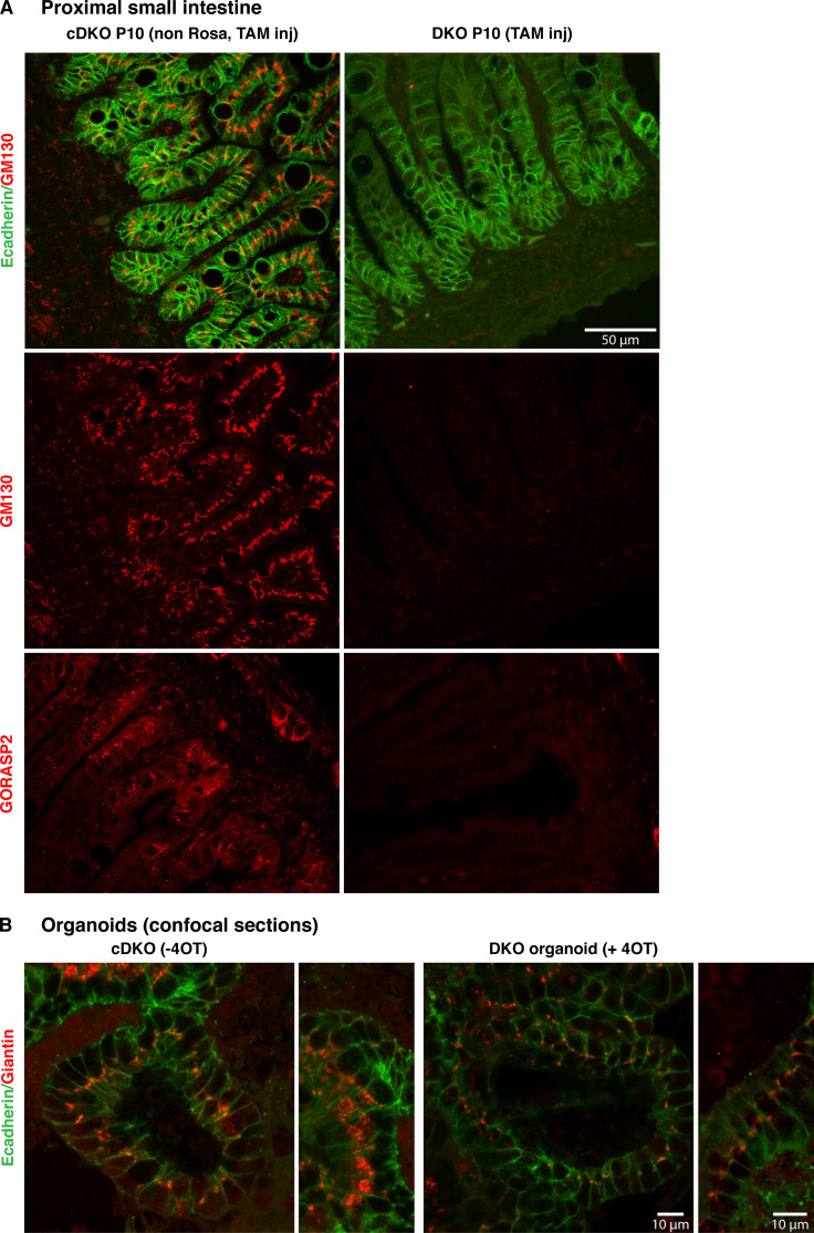
The authors noticed that they mistakenly used an incorrect image in Figure 4 A to show GORASP2 staining. The legend also lacked detail. The corrected figure and legend are shown below. The conclusions drawn from this figure are not affected by this change. This error appears only in PDF versions downloaded on or before June 26, 2020.
Figure 4.
GM130 is degraded in GORASPs knockout cells and does not affect cisternal stacking. (A) Visualization of GM130 (red) and E-cadherin (green) in section of P10 proximal small intestine in mice injected with either control (non Rosa, TAM injected, functions as control) or TAM at P1. Note that GM130 staining is no longer visible upon TAM injection at P1. GORASP2 visualization on sections from the same tissue confirm the loss of GORASP2 in TAM-injected tissue. (B) Visualization of Giantin (red) and E-cadherin (green) in budding organoids treated or not with 4OT (confocal section). Note that Giantin staining is still prominent in every cell after 4OT treatment, although the intensity of staining is decreased, possibly reflecting the Golgi ribbon unlinking and dispersion of the smaller stacks in the cell cytoplasm.


