Abstract
COVID-19 outbreak has caused a pandemonium in modern world. As the virus has spread its tentacles across nations, territories, and continents, the civilized society has been compelled to face an unprecedented situation, never experienced before during peacetime. We are being introduced to an ever-growing new terminologies: “social distancing,” “lockdown,” “stay safe,” “key workers,” “self-quarantine,” “work-from-home,” and so on. Many countries across the globe have closed their borders, airlines have been grounded, movement of public transports has come to a grinding halt, and personal vehicular movements have been restricted or barred. In the past couple of months, we have witnessed mayhem in an unprecedented scale: social, economic, food security, education, business, travel, and freedom of movements are all casualties of this pandemic. Our experience about this virus and its epidemiology is limited, and mostly the treatment for symptomatic patients is supportive. However, it has been observed that COVID-19 not only attacks the respiratory system; rather it may involve other systems also from the beginning of infection or subsequent to respiratory infection. In this article, we attempt to describe the systemic involvement of COVID-19 based on the currently available experiences. This description is up to date as of now, but as more experiences are pouring from different corners of the world, almost every day, newer knowledge and information will crop up by the time this article is published.
How to cite this article
Munjal M, Das S, Chatterjee N, Setra AE, Govil D. Systemic Involvement of Novel Coronavirus (COVID-19): A Review of Literature. Indian J Crit Care Med 2020;24(7):565–569.
Keywords: Acute respiratory distress syndrome, Bilateral predominant ground-glass opacities, COVID-19, COVID pneumonia, Elevation of cardiac troponins (cTnI), High rate of transmission, Multisystemic involvement of COVID-19, Neurological signs, Novel coronavirus, Respiratory system
Introduction
In just 4 months, the world has been turned into a tremendous turmoil by the abysmally high rate of transmission of novel coronavirus SARS-CoV-2 (COVID-19) infection. This originated in Wuhan City situated in the Hubei province of People's Republic of China. The infection has swiftly spread to the other parts of the world infecting every continent and causing thousands of deaths, serious illness, and unprecedented disruption of global economy never seen since the Second World War. Clinical experiences, as published in various frontline medical journals, confirm that COVID-19, in addition to affecting the respiratory system, may involve most of the other systems of our body. In this article, we attempt to provide comprehensive information regarding the multisystemic involvement of COVID-19 based on the evidence and experiences from the shared experiences of clinicians worldwide.
Aim
A systemwise approach of COVID-19 based on published studies and case reports.
Materials and Methods
Systemic review of published literature from February to April 15, 2020 from PubMed Central, EMBASE, Google Scholar, UpToDate, Cochrane database, and NHS England.
Central Nervous System
Involvement of central nervous system (CNS) may occur either during initial or late phase of infection. Early neurological affection in COVID-19 patients may manifest as fever with headache.1 Specific neurological symptoms related are loss of sensations (e.g., taste, smell), headache, visual impairment, dizziness, ataxia, and at times convulsions which have been reported to occur in varying frequencies in COVID-19 patients. In the beginning of infection, patients may present with loss of olfactory and gustatory senses with or without other overt neurological signs and symptoms. Some of the patients with COVID-19 infection may present with symptoms similar to intracranial infection, e.g. headache, acute change in the level of consciousness, or seizures. Neurological signs and symptoms in the COVID-19 infection might result from a combination of hypoxia, respiratory, and metabolic acidosis, usually observed at a later and advanced phase of the disease. Patients presenting with neurological symptoms in COVID-19 infection need focused and more aggressive treatment protocols targeting specific goals. Recently a case report has been published in Wuhan describing a patient presented with acute symmetrical ascending weakness resembling Guillain-Barre syndrome. The patient was tested positive for COVID-19, and he had a good response with conventional treatment (intravenous immunoglobulin G) of Guillain-Barre syndrome.2
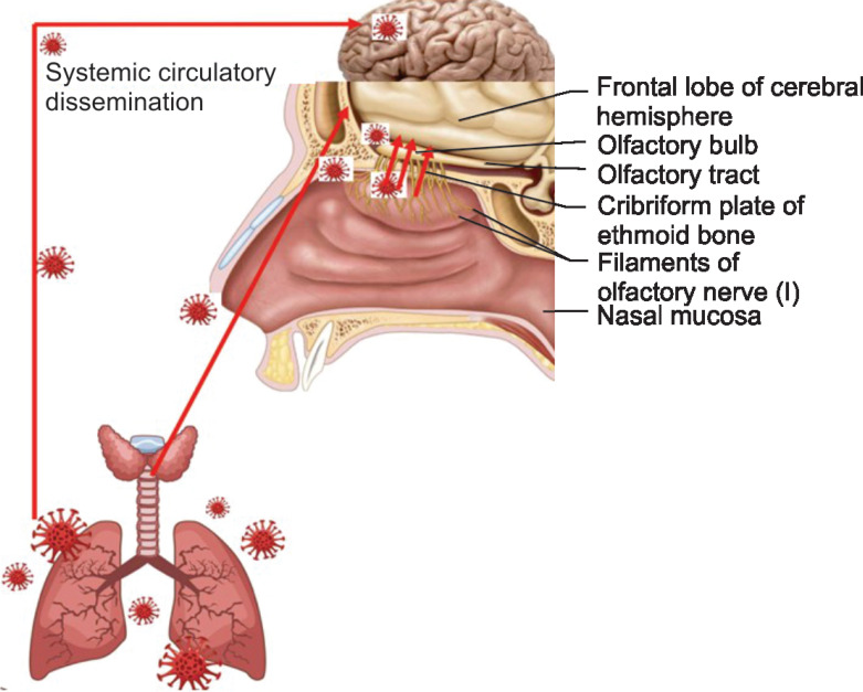
Entry of COVID-19 in the brain tissue may be through the cribriform plate of ethmoid situated in close proximity to the olfactory bulb (Fig. 1).3 Following infection of the lung, at a later stage, systemic dissemination (through circulatory system) may be a potential source of CNS infection. This might result in loss of autonomic control of breathing, leading to acute respiratory insufficiency, ultimately requiring a varying range of ventilatory support. In addition, spike protein present in SARS-CoV-2 could link with angiotensin converting enzyme 2 (ACE2) expressed in the capillary endothelium. SARs-CoV-2 could also cause damage to the blood–brain barrier and therefore enter the CNS by inflicting injuries to the vascular system.4
Fig. 1.
Entry of COVID-19 in the CNS
Respiratory System
Majority of the infected patients suffer only mild, coldlike symptoms. Signs and symptoms may appear 2–14 days from the initial exposure and present with cough (mostly dry but at times with copious sputum production), fever, sore throat, runny nose, and shortness of breath. Hospital admissions usually result due to pneumonia-like symptoms with shortness of breath and desaturation in blood oxygen levels. About 15% of COVID-19 patients develop moderate to severe disease which leads to hospitalization and oxygen support. For approximately 5% of severely symptomatic patients, intensive care unit (ICU) admission and organ supportive therapies (including intubation and ventilation) become indispensable for their care. The commonest complication for COVID-19 patients is pneumonia. Nevertheless, at times other respiratory complications, for example, acute lung injury (ALI) and acute respiratory distress syndrome (ARDS) are present in the initial stage. It is worth to note that in COVID-19 patients, pneumonia and respiratory distress fulfilling the Berlin criteria of ARDS, usually manifest as an atypical form of the syndrome. Indeed, the initially observed features show a relative dissociation between a relatively well-preserved lung and the severity of hypoxemia. Such a wide discrepancy was virtually never seen in most of the forms of ARDS reported until now.5
Chest radiography remains the primary imaging modality like any other chest conditions. However, approximately two-thirds of COVID-19 patients will have normal chest radiograph. Computed tomography (CT) scans can be considered as a primary imaging modality for suspected COVID-19 patients as it has a higher sensitivity for detecting typical features of COVID-19 pneumonia, for example, bilateral predominant ground-glass opacities in the presence or in the absence of consolidation in the peripheral lung fields.6
Cardiovascular System (CVS)
Prevalence of cardiovascular diseases (CVD) is substantial in general population and the probability of acquiring COVID-19 infection is particularly high in this subset of people. There is evidence that COVID-19 per se can deteriorate the cardiovascular function in patients without any comorbidities, resulting in acute cardiac injury manifesting at some point of time during the period of illness. Increased susceptibility to COVID-19 with a severe form of disease and poor clinical outcome has been observed in patients with coexisting CVD.7,8
In approximately 8–12% of all COVID-19 patients, a noteworthy rise of cardiac troponins (cTnI) is noted, representing acute cardiac injury. The principal mechanisms that result in such injury could be (i) viral injury to cardiac myocytes leading to direct myocardial injury and (ii) systemic inflammatory effects.9 Acute myocardial injury could be secondary to binding of SARS-CoV-2 to ACE2 and subsequent alteration of ACE2 signaling pathways. Other mechanisms include a change in myocardial demand and supply, plaque rupture and coronary thrombosis (due to inflammation and stress responses), and adverse effects of various therapies/interventions on the cardiovascular system, for example, hydroxychloroquine, antiviral drugs, corticosteroids. and electrolyte abnormalities. Common cardiac complications in COVID-19 include tachy- and brady-arrhythmias. A recent study reported clinical profile and outcomes in 138 Chinese COVID-19 patients, of whom 16.7% had cardiac arrhythmia.10
Until now, no study has reported ST segment alterations or acute myocardial infarction (MI) secondary to COVID-19. The majority of MIs are type II and related to the primary infection, hemodynamic, and respiratory derangement.
There are no reports on incidences of left ventricular (LV) dysfunction (e.g., poor LV contractility, acute LV failure, and cardiogenic shock) in COVID-19 patients. A single Chinese study described the occurrence of heart failure in COVID-19 patients.11 At this stage, it will be premature to assess the long-term implications of the consequences of COVID-19 infection of the cardiovascular system.
Gastrointestinal Tract and Liver
ACE2 is abundantly found in esophageal epithelial cells and the absorptive enterocytes that form ileum and colon. This might propose a mechanism for faucal transmission.12 Published data from Wuhan described that gastrointestinal (GI) symptoms (e.g., abdominal pain, nausea, vomiting, diarrhea, decreased appetite, and gastrointestinal bleeding) were present up to 79% of the COVID-19 patients either at the time of disease onset or later during hospitalization periods.13 However, both adults and youngsters can present exclusively with GI symptoms. Studies from China revealed that anorexia as the most common GI symptom in adults (39.9–50.2%), whereas diarrhea was the most common symptom both in adults and in children (2–49.5%). Vomiting occurred more commonly in children. GI bleeding was found only in few ventilated patients in ICU. The accuracy of faucal polymerase chain reaction (PCR) testing was almost equivalent to PCR from respiratory specimens. Recently SARS‐CoV‐2 has been reported for the first time in stool samples in United States.14
As of now, it is not possible to conclude if digestive symptoms were primary or secondary outcomes of SARS‐CoV‐2 infection in critically ill patients. Secondary to the effects of long‐term hypoxemia resulting in cell necrosis, GI mucosa may cause ulceration and bleeding. Moreover, in patients with severe illness treatments that include the use of corticosteroids and nonsteroidal anti-inflammatory drugs (NSAIDs) combined with physiological stress has the potential to affect the GI tract mucosa. Hence, the actual cause responsible for GI manifestations could be complex and multifactorial.
Mild to moderate liver injury, including elevated aminotransferases, high bilirubin, hypoproteinemia, and prolonged prothrombin time, have all been reported in COVID-19. Hepatic dysfunction in severe COVID-19 patients is associated with increased activation of coagulative and fibrinolytic pathways, comparatively decreasing platelets, increasing neutrophils, a higher neutrophil to lymphocyte ratio, and high ferritin.15
The hypothesis for liver injury by COVID-19 could be due to viral hepatitis, but there are other alternative theories. Firstly, there is a mild derangement of hepatic function. Secondly, while examining the liver function tests of different patients (with varying symptom duration), no conclusive evidence was found which positively correlates later presentation of disease and greater elevation in liver enzymes. Only one report demonstrated microvesicular steatosis from postmortem liver biopsy of a COVID-19 patient.16 However, the etiology still remains uncertain, as this is a usual finding in sepsis.
It is important to note that other respiratory viruses produce comparable derangements in biomarkers of liver function, which is considered to correlate with immune interactions (involving intrahepatic cytotoxic T cells and Kupffer cells) mediated hepatic damage. Physicians should consider drug-induced liver injury as a likely cause for abnormal liver blood test results witnessed following initiation of treatment and therapeutics. However, many COVID 19 patients present with baseline liver function derangement before the initiation of medications.
Renal System
Presently available reports indicate that the occurrence of acute kidney injury (AKI) among patients with COVID-19 is small. For illustration, in a Chinese cohort of 1,099 COVID-19 patients, 93.6% were hospitalized, 91.1% had pneumonia, 5.3% required admission to the ICU, and 3.4% had ARDS, but only 0.5% suffered from AKI.17 Studies found that the incidence of AKI was directly proportional to the baseline serum creatinine.
Increased age, greater body mass index, presence of diabetes mellitus, history of cardiac failure, higher peak airway pressure (PIP), and increased sequential organ failure assessment (SOFA) scores positively correlated with the severity of AKI. Positive end-expiratory pressure (PEEP) and prone ventilation were not found to be related with the occurrence of renal impairment, in a similar way nephrotoxic agents were not associated with clinical AKI.18
Based on the possible mechanisms of renal dysfunction, these patients can theoretically be divided into three aspects: cytokine damage, organ crosstalk, and systemic effects.
Musculoskeletal Symptoms
Although generalized fatigue and muscle pain are very common in patients with COVID-19, physicians should include the diagnosis of rhabdomyolysis in the differential for patients presented with focal myalgia and fatigue. Creatinine kinase (CK) and myoglobin levels are important markers for rhabdomyolysis. Therefore, they should be tested early to avoid acute renal failure arising from rhabdomyolysis.19 Patients who survive following a prolonged period of ventilation are likely to develop muscle atrophy and weakness during the recovery period.
Hematological Manifestations
Patients with severe COVID-19 infection are prone to develop microvascular thrombosis and disseminated intravascular coagulation (DIC) as a result of fulminant activation of coagulation cascades. This leads to a reduced platelet count, prolongation of PT/INR, PTT, elevation of D-dimer, and reduced fibrinogen levels. A peripheral blood smear may reflect microangiopathy with schistocytes.
Actively bleeding patients with DIC require platelet transfusion (one adult dose) if the platelet count falls below 50 × 109/L. Similarly, transfusion of plasma (4 units) and fibrinogen concentrate (4 g) or cryoprecipitate (10 units) is needed when INR remains more than 1.8 or fibrinogen level remains below 1.5 g/L, respectively. Established venous thromboembolism (VTE) is considered to be an indication of therapeutic anticoagulation. Ongoing therapeutic anticoagulation should continue in patients with VTE or atrial fibrillation. However, anticoagulation should be on hold if the platelet count falls below 50 × 109/L or fibrinogen is less than 1.0 g/L. Individual case to case evaluation is necessary to weigh benefits against risks regarding thrombosis and bleeding. All symptomatic patients admitted with COVID-19 infection in hospitals require a prophylactic dose of low molecular weight heparin (LMWH) even though coagulation remains abnormal if there is no active bleeding, platelets are above 25 × 109/L, or fibrinogen is above 0.5 g/L.
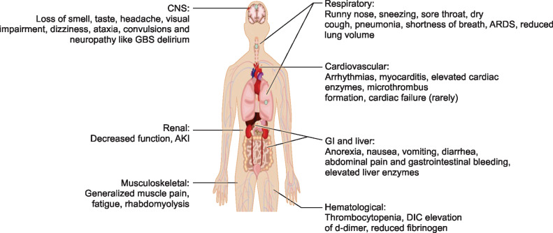
Limited evidence suggests mortality benefit in patients of severe COVID-19 infection with markedly elevated levels of D-dimer (more than 6 times the normal upper limits) treated with prophylactic doses of LMWH or unfractionated heparin (UFH). Pharmacological thromboprophylaxis is not contraindicated in patients with abnormal PT/INR or PTT. When pharmacological thromboprophylaxis is not possible, mechanical thromboprophylaxis should be used as an alternative (Fig. 2).20
Fig. 2.
Systemic involvement in COVID-19
Psychological Aspects
COVID-19 patients who spend a significant time in the hospital are more vulnerable to delirium, irrespective of ICU admission and regardless of the severity of illness. Many of them are in a state of muddled thinking, which, if remains continued in long-term, may lead to cognitive impairments (e.g., memory deficits). There are cases of mental and social health problems even among frontline health care workers.
message to Reduce the Long-Term Cognitive Impairment
Messages for the General Population
Please be empathetic to those infected with the virus. They all deserve support, compassion, and kindness.
If possible, limit the time spent on print, digital, or social media following posts or news items about COVID-19 which could cause undue anxiety or stress. For authentic information, believe only that from trusted resources.
Protect yourself and be supportive to others.
Take positive feedback from people who survived COVID-19. Learning from their experiences will provide you a positive and affirmative image.
Honor healthcare workers and supporting people.
Messages for Healthcare Workers
Mental and psychosocial well-being is important as physical health. Importance of psychological health cannot be overemphasized at the time of pandemics of such unprecedented scale.
Ensure optimum rest while working a long day or between shift duties. Take nutritious food, engage in some form of physical activity, and stay in contact with your family and friends.
If possible, keep away from tobacco, alcohol, narcotics, or other habit-forming drugs.
It is important to maintain relations with your loved ones, using digital platforms when needed.
ACKNOWLEDGMENT
Jigyasa Foundation, Jaipur, India
Footnotes
Source of support: Nil
Conflict of interest: None
Author Contributions
Samaresh Das: concept, critical review, and preparation of manuscript; Nilay Chatterjee: critical review and literature search; Manish Munjal: critical review and preparation of the manuscript; and Adarsh Eshappa Setra: literature search and critical review.
References
- 1.Baig AM. Neurological manifestations in COVID-19 caused by SARS-CoV-2. CNS Neurosci Ther. 2020;26(5):499–501. doi: 10.1111/cns.13372. DOI: [DOI] [PMC free article] [PubMed] [Google Scholar]
- 2.Zaho H, Shen D, Zhou H, et al. Guillain-Barré syndrome associated with SARS-CoV-2 infection: causality or coincidence? Lancet Neurol. 2020;19(5):383–384. doi: 10.1016/S1474-4422(20)30109-5. DOI: [DOI] [PMC free article] [PubMed] [Google Scholar]
- 3.Baig AM, Khan NA. Novel chemotherapeutic strategies in the management of primary amoebic meningoencephalitis due to Naegleria fowleri. CNS Neurosci Ther. 2014;20(3):289–290. doi: 10.1111/cns.12225. DOI: [DOI] [PMC free article] [PubMed] [Google Scholar]
- 4.Baig AM, Khaleeq A, Ali U, Syeda H. Evidence of the COVID-19 virus targeting the CNS: tissue distribution, host-virus interaction, and proposed neurotropic mechanisms. ACS Chem Neurosci. 2020;11(7):995–998. doi: 10.1021/acschemneuro.0c00122. DOI: [DOI] [PubMed] [Google Scholar]
- 5.Coppola S, Cressoni M, Busana M, Rossi S, Chiumello D. Covid-19 does not lead to a “typical” acute respiratory distress syndrome luciano gattinoni. Am J Respir Crit Care Med. 2020;201(10):1299–1300. doi: 10.1164/rccm.202003-0817LE. DOI: [DOI] [PMC free article] [PubMed] [Google Scholar]
- 6.Choi H, Qi X, Yoon SH, Park SJ, Lee KH, Kim JY, et al. Extension of coronavirus disease 2019 (COVID-19) on chest CT and implications for chest radiograph interpretation. Radiol: Cardiothora Imag. 2020;2(2):e200107. doi: 10.1148/ryct.2020200107. DOI: [DOI] [PMC free article] [PubMed] [Google Scholar]
- 7.Xiong TY, Redwood S, Prendergast B, Chen M. Coronaviruses and the cardiovascular system: acute and long-term implications. Eur Heart J. 2020;41(19):1798–1800. doi: 10.1093/eurheartj/ehaa231. DOI: [DOI] [PMC free article] [PubMed] [Google Scholar]
- 8.Li B, Yang J, Zhao F. Prevalence and impact of cardiovascular metabolic diseases on COVID-19 in China. Clin Res Cardiol. 2020;109(5):531–538. doi: 10.1007/s00392-020-01626-9. DOI: [DOI] [PMC free article] [PubMed] [Google Scholar]
- 9.Bansal M. Cardiovascular disease and COVID-19. Diabetes Metab Syndr. 2020;14(3):247–250. doi: 10.1016/j.dsx.2020.03.013. DOI: [DOI] [PMC free article] [PubMed] [Google Scholar]
- 10.Wang D, Hu B, Hu C. Clinical characteristics of 138 hospitalized patients with 2019 novel coronavirus-infected pneumonia in Wuhan, China. J Am Med Assoc. 2020;323(11):1061–1069. doi: 10.1001/jama.2020.1585. DOI: [DOI] [PMC free article] [PubMed] [Google Scholar]
- 11.Zhou F, Yu T, Du R. Clinical course and risk factors for mortality of adult inpatients with COVID-19 in Wuhan, China: a retrospective cohort study. Lancet. 2020;395(10229):1054–1062. doi: 10.1016/S0140-6736(20)30566-3. DOI: [DOI] [PMC free article] [PubMed] [Google Scholar]
- 12.Zhang H, Kang Z, Gong H, Xu D, Wang J, Li Z, et al. The digestive system is a potential route of 2019-nCov infection: a bioinformatics analysis based on single-cell transcriptomes. BioRxiv. 2020. DOI: [DOI]
- 13.Chang D, Lin M, Wei L, Xie L, Zhu G, Dela Cruz CS, et al. Epidemiologic and clinical characteristics of novel coronavirus infections involving 13 patients outside Wuhan, China. JAMA. 2020;323(11):1092. doi: 10.1001/jama.2020.1623. DOI: [DOI] [PMC free article] [PubMed] [Google Scholar]
- 14.Holshue ML, DeBolt C, Lindquist S, Lofy KH, Wiesman J, Bruce H, et al. First case of 2019 novel coronavirus in the United States. N Engl J Med. 2020;382(10):929–936. doi: 10.1056/NEJMoa2001191. DOI: [DOI] [PMC free article] [PubMed] [Google Scholar]
- 15.Wang D, Hu B, Hu C, Zhu F, Liu X, Zhang J, et al. Clinical characteristics of 138 hospitalized patients with 2019 novel coronavirus-infected pneumonia in Wuhan, China. JAMA. 2020;323(11):1061–1069. doi: 10.1001/jama.2020.1585. DOI: [DOI] [PMC free article] [PubMed] [Google Scholar]
- 16.Koskinas J, Gomatos IP, Tiniakos DG, Memos N, Boutsikou M, Garatzioti A, et al. Liver histology in ICU patients dying from sepsis: a clinico-pathological study. World J Gastroenterol. 2008;14(9):1389–1393. doi: 10.3748/wjg.14.1389. DOI: [DOI] [PMC free article] [PubMed] [Google Scholar]
- 17.Guan W, Ni Z, Hu Y, Liang W, Ou C, He J, et al. Clinical characteristics of coronavirus disease 2019 in China. N Engl J Med. 2020;382:1708–1720. doi: 10.1056/NEJMoa2002032. DOI: [DOI] [PMC free article] [PubMed] [Google Scholar]
- 18.Panitchote A, Mehkri O, Hastings A, Hanane T, Demirjian S, Torbic H, et al. Factors associated with acute kidney injury in acute respiratory distress syndrome. Ann Intensive Care. 2019;9(1):74. doi: 10.1186/s13613-019-0552-5. DOI: [DOI] [PMC free article] [PubMed] [Google Scholar]
- 19.Jin M, Tong Q. Rhabdomyolysis as potential late complication associated with COVID-centre for disease control and Prevention. 2020;26(7) doi: 10.3201/eid2607.200445. DOI: [DOI] [PMC free article] [PubMed] [Google Scholar]
- 20.https://www.hematology.org/covid-19/covid-19-and-vte-anticoagulation https://www.hematology.org/covid-19/covid-19-and-vte-anticoagulation


