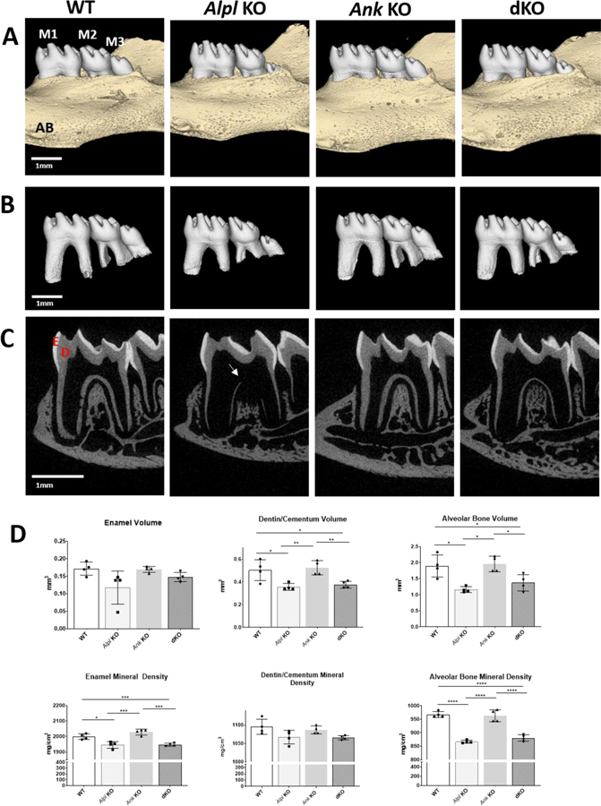
Figure 1. Ablation of Ank in Alpl KO Mice Partially Improves Dentoalveolar Mineralization in Alpl KO Mice.
Three-dimensional reconstructions of (A) mouse mandibles and (B) isolated molar teeth show comparable crown morphology and eruption pattern of mandibular first (M1) and second (M2) molars in WT, Alpl KO, Ank KO, and Alpl, Ank dKO mice at 26 dpn. Compared to WT and Ank KO, both Alpl KO and Alpl, Ank dKO mice exhibit delayed eruption and underdeveloped roots of mandibular 3rd molars (M3). Sagittal 2D images with enamel (E) in white and dentin (D) and alveolar bone (AB) in gray (C) reveal thin, hypomineralized dentin and alveolar bone mineralization defects (yellow arrow) in Alpl KO that are partially ameliorated in Alpl, Ank dKO mice. (D) Quantitative analysis indicates dentin/cementum and alveolar bone volumes and enamel and alveolar bone mineral density defects in Alpl KO molars are not corrected in Alpl, Ank dKO molars.

