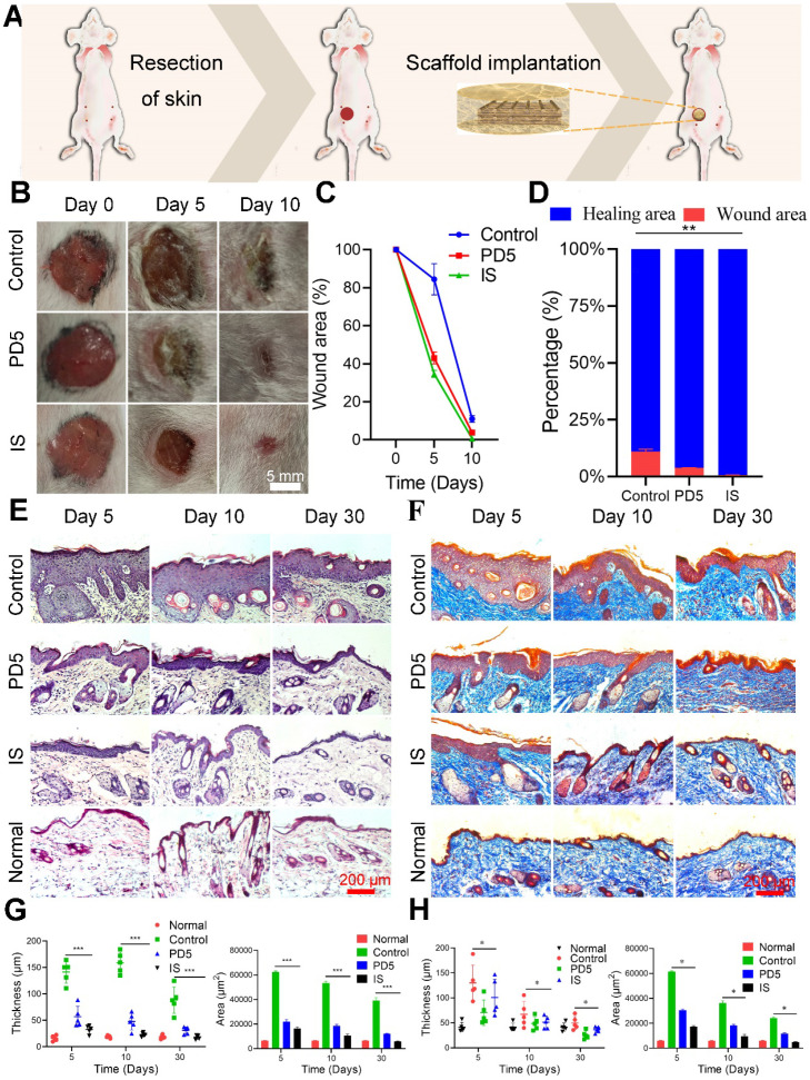
Figure 4.
Skin repair assessment using the various scaffolds. (A) Schematic diagram of the process. (B) Images of wound healing of mice treated with gauze (control), PD5 scaffold, and the intelligent scaffold (IS). (C) Quantification of wound closure within 10 days. (D) The ratios of wound closure and residual area after 30 days. (E) H&E staining images of normal skin and wound area after treatment for 5, 10, and 30 days with control, PD5 scaffold, and IS. (F) Masson staining images of normal skin and wound area after treatment for 5, 10, and 30 days with control, PD5 scaffold, and IS. (G) Quantification of the epidermal thickness and area of different groups from the H&E staining images. (H) Quantification of the epidermal thickness and area of different groups from the Masson staining images. *P < 0.05, **P < 0.01, ***P < 0.005.

