Abstract
Soil-dwelling animals are at risk of pathogen infection in soils. When choosing nesting sites, animals could reduce this risk by avoiding contact with pathogens, yet there is currently little evidence. We tested this hypothesis using Solenopsis invicta as a model system. Newly mated queens of S. invicta were found to nest preferentially in soil containing more actinobacteria of Streptomyces and Nocardiopsis and to be attracted to two volatiles produced by these bacteria, geosmin and 2-methylisoborneol. Actinobacteria-rich soil was favored by S. invicta and this soil contained fewer putative entomopathogenic fungi than adjacent areas. Queens in such soil benefited from a higher survival rate. In culture, isolated actinobacteria inhibited entomopathogenic fungi, suggested that their presence may reduce the risk of fungal infection. These results indicated a soil-dwelling ant may choose nest sites presenting relatively low pathogen risk by detecting the odors produced by bacteria with anti-fungal properties.
Author summary
Insect pathogens are widely distributed in soil. Soil-dwelling insects must overcome challenges arising from pathogens in soil. Here we report that a soil-dwelling ant may choose nest sites with lower pathogen infection risk, specifically the ant can sense the cues of some actinobacteria that can inhibit the growth of the pathogens. By choosing the sites with higher abundance of some actinobacteria, the ant can get a higher survival rate. The ant and some actinobacteria thus coordinate a specialized adaptive strategy of infection risk management, enabling the ant population to grow.
Introduction
The risk of predators, pathogen infection and abiotic condition is a major criterion used by insects when choosing nesting sites [1, 2]. In particular, for social insects that nest in soil, pathogens are a significant danger that varies greatly from place to place [3–6]. Though some insects can detect pathogens and avoid them [7], it remains unclear whether social insects can reliably detect pathogens at a potential nesting site and avoid them. Queens of wood ant were attracted to fungal pathogens during the initial stage of colony founding [8]. Pathogens manipulate uninfected insect hosts deserves further investigation since records of pathogens attracting their hosts are indeed rare, as hosts are under strong selection to resist manipulation and to avoid virulent pathogens [9, 10].
Social insects have highly developed chemical communication systems [11–15] and are sensitive to numerous cues from conspecifics, heterospecifics and the environment that alter their behavior [16–18]. Identifying these cues can facilitate the understanding of the complex behaviors of social insects and the manipulation of their abundance. Studies have shown that many volatile compounds in soil are produced by soil actinobacteria, and the soil actinobacteria can even affect the growth of underground pathogenic fungi and aboveground plants [19–21]. Recent studies indicated that some insects can be attracted or repelled by the geosmin or 2-methylisoborneol produced by the actinobacteria [22–24]. Geosmin induces aversion in Drosophila melanogaster presumably signaling the presence of harmful microbes on food [24]. In contrast to flies, geosmin is not aversive but mediated egg-laying site selection in Aedes aegypti. [23]. Streptomyces bacteria produced geosmin and 2-methylisoborneol can even attract a soil arthropod to promote spore dispersal [22]. Furthermore, the soil actinobacteria may have evolved into the defensive symbionts of soil-dwelling insects. Once associated with an insect, many actinobacteria can protect insects against pathogens by producing some antibiotics [25]. One of the most striking features of actinobacteria is that they can produce antifungal compounds [26]. Actinobacteria with antifungal activity are reported to show high species richness in certain types of soil [27–29]. Because entomopathogenic fungi are also widely distributed in soil [30–33], we hypothesize that some soil-dwelling insects may choose soil types with the highest abundance of actinobacteria for nest building to avoid being infected by some soil-derived entomopathogenic fungi.
The imported red fire ant Solenopsis invicta is a notorious invasive pest in many countries [34], where its rapid spread has caused great economic losses and ecological problems [35] and stimulated research to understand the causes of its invasiveness [36–39]. The nesting behavior of S. invicta was found to be significantly influenced by environmental conditions [40]. Studies have indicated that climate [41], habitat type [42] and soil attributes [43, 44] can affect the distribution of S. invicta. Almost five years of observations in China showed that colonies of S. invicta at relatively fine geographical scales are only distributed in certain parts of the available habitat (S1 Fig). Here, we show that soil from the favored nesting and non-favored nesting areas of S. invicta are not equally attractive to newly mated queens and workers and the possible reasons behind was tested.
Results
Solenopsis invicta ants are attracted to soil and soil volatiles from their favored nesting areas
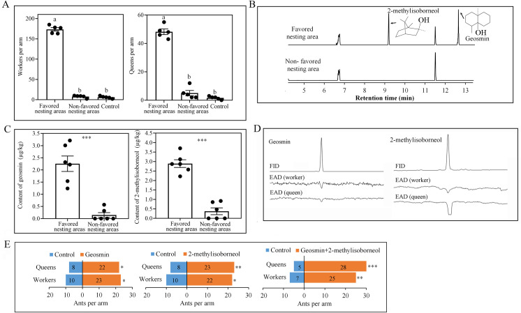
To determine whether soil from favored nesting areas in the field was more attractive to S. invicta than soil from non-favored areas, we compared the two soil types in choice assays in an olfactometer. Significantly more workers and queens were recovered from olfactometer arms connected to jars containing soil from favored nesting areas than from arms connected to jars containing soil from non-favored areas (worker: Kendall W = 0.832, χ2 = 8.316, P = 0.016; queen: Kendall W = 0.84, χ2 = 8.4, P = 0.015; Fig 1A), indicating that soil from favored nesting areas may release volatile ant attractants. To identify these attractants, soil from favored and non-favored nesting areas was analyzed by GC-MS. A marked difference between the two soils was that the norsesquiterpene geosmin and the monoterpene 2-methylisoborneol were present in soil from favored nesting areas but absent or less from soil from non-favored areas (geosmin: t = 6.334, df = 10, P < 0.01, 2-methylisoborneol: t = 9.262, df = 10, P < 0.01, independent sample t test, Fig 1B and 1C). Moreover, consistent responses of worker and queen antennae were obtained in the GC-EAD analysis (Fig 1D). To test whether geosmin and 2-methylisoborneol were indeed attractive to S. invicta, authentic standards (Sigma-Aldrich, more than 98% pure) were tested in a Y-tube olfactometer. For this purpose, 2 μg/L geosmin or 2-methylisoborneol were added as the attractants. The results showed that the olfactometer arm containing geosmin or 2-methylisoborneol attracted significantly more ants than the control arm (geosmin: χ2 = 5.121, df = 1, P = 0.024 (worker), χ2 = 6.533, df = 1, P = 0.011 (queen); 2-methylisoborneol: χ2 = 4.5, df = 1, P = 0.034 (worker), χ2 = 7.258, df = 1, P = 0.007 (queen); geosmin + 2-methylisoborneol: χ2 = 10.125, df = 1, P = 0.001 (worker), χ2 = 16.03, df = 1, P < 0.001 (queen), Fig 1E). The concentrations of geosmin and 2-methylisoborneol used in the olfactory bioassays were selected according to the concentrations in soil (Fig 1C), these results indicate that geosmin and 2-methylisoborneol likely attract S. invicta to its favored nesting areas and should be of ecological relevance.
Fig 1. Attraction of S. invicta to soil from favored nesting areas and to soil volatiles.
(A) Choices of workers and queens in regard to soil from favored or non-favored nesting areas in olfactometer bioassays (±SE, n = 5 biological replicates). Control: empty arm. (B) Typical gas chromatographic traces obtained from the soil of favored and non-favored nesting areas. (C) Content of geosmin and 2-methylisoborneol (±SE, n = 6 biological replicates, independent sample t test). (D) GC−EAD profile of geosmin and 2-methylisoborneol on worker and queen antenna. Upper trace: GC−FID. Lower trace: EAG (unit: mV). (E) Attraction of S. invicta to geosmin and 2-methylisoborneol in a Y-tube olfactometer (n ≥ 30 biological replicates, chi-square test). Control: mineral oil. Different letters above bars indicate significant differences according to Kendall’s W test at the 0.05 level. Asterisks indicate significant differences (*P < 0.05, **P < 0.01, *** P < 0.001).
Streptomycetaceae and Nocardiaceae are more abundant in soil from favored nesting areas
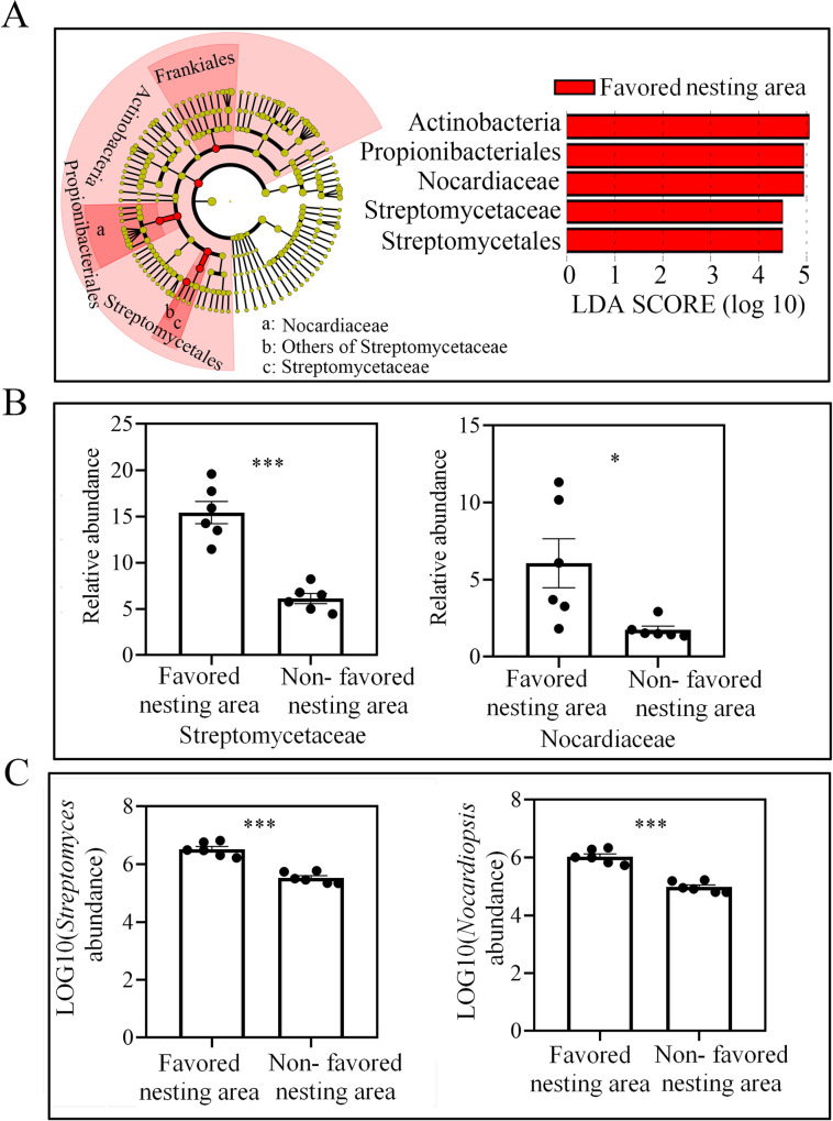
Studies have indicated that geosmin and 2-methylisoborneol can be produced as secondary metabolites by microorganisms including actinomycetes, particularly Streptomyces [45], cyanobacteria [46], myxobacteria [47], fungi [48], and even by some plants [49]. Moreover, some studies have reported that actinomycetes play an important role in the life of ants and other insects [25, 50]. Thus, we hypothesized that the presence of geosmin and 2-methylisoborneol in soil from favored nesting areas may be due to actinobacteria. Thus, the bacterial diversity in soil from favored and non-favored nesting areas was compared with 16S rRNA gene sequencing. The sequencing results indicate that there was no difference between the soils in terms of bacteria operational taxonomic units (OTU) numbers and Shannon diversity index (OTU number: t = 0.989, df = 10, P = 0.346; Bacteria Shannon index: t = 1.498, df = 10, P = 0.165, independent sample t test, S2A Fig and S2B Fig). However, comparison of the Shannon diversity index values for the actinobacteria indicated that the soil samples from favored nesting areas had a significantly lower diversity (t = 3.429, df = 10, P = 0.006, independent sample t test, S2C Fig). A separation of the samples from favored and non-favored nesting areas was also shown by the principal co-ordinates analysis (PCoA) (S2D Fig). We used the LDA Effect Size (LEfSe) method to identify bacterial OTUs that were likely to explain the major differences between the nesting and non-nesting areas. As a result, Streptomycetaceae and Nocardiaceae were identified as biomarkers of the favored nesting sites in the soil (LDA scores > 4.5) (Fig 2A), with significantly higher abundance in soil from favored vs. non-favored areas (Streptomycetaceae: t = 6.994, df = 10, P < 0.01; Nocardiaceae: t = 2.68, df = 10, P = 0.023, independent sample t test, Fig 2B). Because there was no difference in the absolute content of bacteria in soil from favored and non-favored nesting areas (t = 0.211, df = 10, P = 0.837, independent sample t test, S2E Fig), the content of Streptomycetaceae and Nocardiaceae may contribute to the differences between the two soil types. Moreover, significantly higher absolute abundances of Streptomyces and Nocardiopsis were also detected in favored soil by quantitative PCR (Streptomyces: t = 7.942, df = 10, P < 0.01; Nocardiopsis: t = 8.474, df = 10, P < 0.01, independent sample t test, Fig 2C).
Fig 2. Actinobacterial diversity in soil from favored nesting and non-favored nesting sites.
(A) Taxonomic groups responsible for the differences in microbial diversity between favored and non-favored nesting soil based on LEfSe analysis. (B) Relative abundance of Streptomycetaceae and Nocardiaceae in soil from favored vs. non-favored nesting areas (±SE, n = 6 biological replicates, independent sample t test). (C) Absolute abundance of Streptomyces and Nocardiopsis in soil achieved by qPCR (±SE, n = 6 biological replicates, independent sample t test). Asterisks indicate significant differences (*P < 0.05, ***P < 0.001).
Actinobacteria release attractive soil volatiles
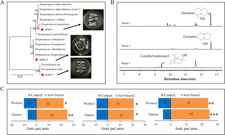
To verify whether geosmin and 2-methylisoborneol were produced by bacteria of Streptomycetaceae and Nocardiaceae, the bacterial strains of these families were isolated. With morphological identification and 16S rRNA sequencing, only two geosmin producing strains (strain 1 and strain 2) identified as Streptomyces and one 2-methylisoborneol producing strain (strain 3) identified as Nocardiopsis (99% 16S rRNA sequence similarity) were isolated from the soil samples collected from favored nesting area (Fig 3A and 3B). Volatiles produced by the isolated bacteria in culture were collected by solid-phase microextraction (SPME) and then analyzed with GC-MS. Geosmin was identified as a volatile produced by strain 1 and strain 2, and 2-methylisoborneol was identified as a volatile associated with strain 3 (Fig 3B). Moreover, the number of ants attracted to the olfactometer arms loaded with soil from non-favored nesting areas significantly increased after adding the isolated bacteria (strain 1: χ2 = 4.5, df = 1, P = 0.034 (worker), χ2 = 10.125, df = 1, P = 0.001 (queen); strain 2: χ2 = 4.8, df = 1, P = 0.028 (worker), χ2 = 4.8, df = 1, P = 0.028 (queen); strain 3: χ2 = 8, df = 1, P = 0.005 (worker), χ2 = 13.33, df = 1, P < 0.001 (queen), Fig 3C).
Fig 3. Volatiles of actinobacterial strains attract S. invicta ants.
(A) Molecular phylogenetic analysis of actinobacterial strains isolated from soil from favored nesting sites and identified based on their 16S rRNA gene sequences. Tree was constructed with a maximum likelihood estimation method. Branch support is indicated as bootstrap values (500 replicates). Red triangles indicate the isolated bacteria. (B) Typical gas chromatographic traces of the headspace of isolated bacterial cultures showing the identified volatiles. (C) Attraction of S. invicta in a Y-tube olfactometer to the isolated bacteria when added to soil from non-favored nesting areas (n ≥ 30 biological replicates, chi-square test). Control: soil from non-favored nesting areas. Asterisks indicate significant differences (*P < 0.05, **P < 0.01, ***P < 0.001).
Pathogenic fungi was less abundant in soil from favored nesting areas
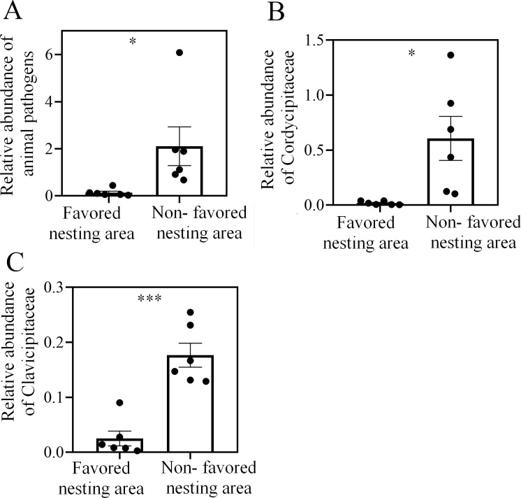
To test whether S. invicta queens choose soil with a high abundance of actinobacteria for nest building is aim to avoid being infected by soil-derived entomopathogenic fungi, fungal diversity in S. invicta-favored and non-favored soil was compared by ITS gene sequencing. There was no difference in OTU number between the favored and non-favored soil (t = 1.717, df = 10, P = 0.117, independent sample t test, S3A Fig), but the soil samples from favored nesting areas had lower Shannon diversity index values (t = 2.916, df = 10, P = 0.015, independent sample t test, S3B Fig). The PCoA also showed a separation of samples from favored and non-favored nesting areas (S3C Fig). FUNGulid (Fungi Functional Guild) classification and annotation revealed that the total relative abundance (proportion of sequencing tags in the total sequencing tags) of OTUs representing taxa that are pathogenic to insects and other animals (mainly Clavicipitaceae (Metarhizium sp.) and Cordycipitaceae (Beauveria sp.) S1 Data) was significantly lower in soil from the favored nesting areas (animal pathogen: t = 2.384, df = 10, P = 0.038; Clavicipitaceae: t = 5.899, df = 10, P < 0.001; Cordycipitaceae: t = 2.943, df = 10, P = 0.015, independent sample t test, Fig 4). Given that there was no significant difference in total fungal content between the two areas (t = 0.563, df = 10, P = 0.586, independent sample t test, S3D Fig), the absolute number of strains pathogenic to animals was likely lower in soil from the favored nesting area.
Fig 4. Diversity of fungi is greater in soil from areas not favored by nesting S. invicta queens.
(A) Relative abundance of fungi that are pathogenic to animals (±SE, n = 6 biological replicates, independent-sample t tests). (B) and (C) Relative abundance of Clavicipitaceae and Cordycipitaceae (±SE, n = 6 biological replicates, independent-sample t tests). Asterisks indicate significant differences (*P < 0.05, **P < 0.01, ***P < 0.001).
Actinobacteria can increase survival of the queens
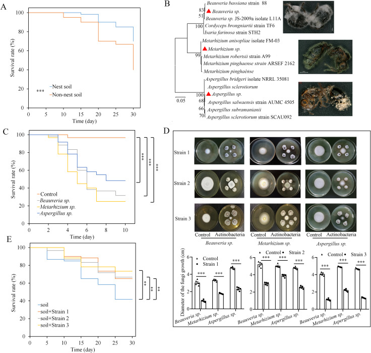
To determine whether the increased number of pathogenic fungi had any effect on S. invicta ants, the survival of newly mated queens was compared in soil from favored vs. non-favored nesting sites. Survival was significantly greater when queens built nests in soil from favored nesting areas (χ2 = 10.975, df = 1, P = 0.001, Kaplan-Meier plot, log-rank test, Fig 5A). When the dead queens from non-favored soil were collected and placed on wet filter papers, fungi grew out of their bodies after 7 days. With ITS gene sequencing, the fungi were identified as Aspergillus sp., Beauveria sp. and Metarhizium sp. (99% ITS gene sequence similarity) (Fig 5B). The numbers of queens infected by Aspergillus sp., Beauveria sp. and Metarhizium sp. were 6, 18 and 12, respectively. Though, Aspergillus sp. was not classified as “pathogenic”, reinfecting new queens with this fungi showed that it can significantly decrease the survival rate as Beauveria sp. and Metarhizium sp. did (Aspergillus sp. vs control: χ2 = 34.856, df = 1, P < 0.001; Beauveria sp. vs control: χ2 = 54.349, df = 1, P < 0.001; Metarhizium sp. vs control: χ2 = 66.243, df = 1, P < 0.001, Kaplan-Meier plot, log-rank test, Fig 5C). And no fungi were isolated from dead ants from favored soils or the fungi were saprophytic fungi. These results indicate that putative entomopathogenic fungi in the soil from non-favored nesting areas may be one reason that causes death of queens in soil. To test whether the isolated actinobacteria contributed to the lower abundance of entomopathogenic fungi in soil from favored nesting areas, we tested the antifungal activities of the previously isolated actinobacterial strains. All of the isolated actinobacteria had the ability to inhibit the growth of the isolated entomopathogenic fungi, the diameters of the fungi growth are significantly smaller than the ones in controls (Fig 5D). Moreover, the survival rate of the ant queens could be significantly increased by adding the isolated actinobacteria to the soil from non-favored nesting areas (strain 1: χ2 = 7.061, df = 1, P = 0.008; strain 2: χ2 = 6.836, df = 1, P = 0.009; strain 3: χ2 = 11.024, df = 1, P = 0.001, Kaplan-Meier plot, log-rank test, Fig 5E). Moreover, in soil added strain 1, the numbers of dead queens infected by Aspergillus sp., Beauveria sp. and Metarhizium sp. were 0, 7 and 5, respectively; in soil added strain 2, the numbers of dead queens infected by these fungi. were 1, 3 and 6, respectively; in soil added strain 3, the numbers of dead queens infected by these fungi were 0, 4 and 1, respectively; while the numbers of dead queens (in control) infected by Aspergillus sp., Beauveria sp. and Metarhizium sp. were 4, 15 and 14, respectively. These results indicate that actinobacteria may increase the survival of S. invicta queens by inhibiting the growth of putative entomopathogenic fungi in the soil.
Fig 5. Entomopathogenic fungi from soil not favored by S. invicta decreases the survival of ant queens.
(A) Kaplan-Meier plot of queen survival after they were forced to build nests in soil from nesting and non-nesting sites (log-rank test). (B) Molecular phylogenetic analysis of fungal strains isolated from soil from non-favored nesting areas based on ITS gene sequences. Tree was constructed with a maximum likelihood estimation method. Branch support is indicated as bootstrap values (500 replicates). Red triangles indicate the isolated fungi. (C) Kaplan-Meier plot of S. invicta queen survival after infection with the isolated fungi (log-rank test). (D) Inhibition effects of the isolated actinobacteria (Fig 3) on the entomopathogenic fungi. On each plate, the fungi were placed in the middle, and the actinobacteria were placed on the four corners of the fungal plug. (E) Kaplan-Meier plot of queen survival after adding the isolated strains of actinobacteria to soil from non-favored nesting sites (log-rank test). Asterisks indicate significant differences (*P < 0.05, **P < 0.01, ***P < 0.001).
Data from additional sites further confirmed nesting preference of S. invicta
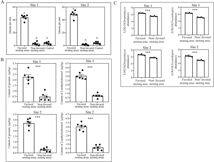
The nesting preference of S. invicta was assessed for another two field sites that are more distant from each other than the core study sites. These additional field data showed that queens preferentially selected the soil from nesting area for the two selected sites (site 1: Kendall W = 0.75, χ2 = 9, P = 0.011; site 2: Kendall W = 0.964, χ2 = 11.565, P = 0.003; Fig 6A). Geosmin and 2-methylisoborneol contents were significantly higher in soil from favored nesting areas in the two selected sites (site 1, geosmin: t = 7.235, df = 10, P < 0.001, 2-methylisoborneol: t = 11.759, df = 10, P < 0.001; site 2, geosmin: t = 13.299, df = 10, P < 0.001, 2-methylisoborneol: t = 11.589, df = 10, P < 0.001, independent sample t test, Fig 6B). qPCR assays also revealed that the abundances of Streptomyces and Nocardiopsis were significantly higher in soil from nesting area for the two selected sites (site 1, Streptomyces: t = 16.133, df = 10, P < 0.001; Nocardiopsis: t = 19.424, df = 10, P < 0.001; site 2, Streptomyces: t = 10.19, df = 10, P < 0.001; Nocardiopsis: t = 12.915, df = 10, P < 0.001, independent sample t test, Fig 6C).
Fig 6. Data from more wild sites confirmed the association between S. invicta queen nesting preference and actinobacteria.
(A) Choices of queens in regard to soil in olfactometer bioassays (±SE, n = 6 biological replicates). Control: empty arm. (B) Content of geosmin and 2-methylisoborneol (±SE, n = 6 biological replicates, independent sample t test). (C) Absolute abundance of Streptomyces and Nocardiopsis in soil achieved by qPCR (±SE, n = 6 biological replicates, independent sample t test). Different letters above bars indicate significant differences according to Kendall’s W test at the 0.05 level. Asterisks indicate significant differences (*P < 0.05, ***P < 0.001).
Discussion
In the past century, the red fire ant (S. invicta) has carried out global invasions with astonishing speed [34] and has become a dominant ant pest in many areas, causing great economic losses and ecological problems [35]. Since 2000, this species has spread to the Caribbean, Australia, New Zealand, Taiwan, Hong Kong, Macao, and China [35]. It can displace native fauna, reduce biodiversity, serve as a vector of pathogens, and impair ecosystem services [51]. Queens can fly considerable distances during mating flights [52], and many factors that affect the movement of queens have been elucidated [53, 54]. However, to the best of our knowledge, this is the first time that the chemical signals of soil microorganisms were reported to affect the nesting choice of newly mated queens. Our findings described the biochemical interactions between microorganisms and insects by microbial volatile organic compound (MVOC) production, which was often overlooked.
There are numerous instances of MVOCs being closely associated with insect feeding behaviors [55], aggregations [56], mating behaviors [57] and oviposition [58]. Emissions from microorganisms in situ may even help insect to evaluate habitat suitability or potential exposure to entomopathogens [24], however, such studies are rare and insects were repelled by MVOCs in almost all cases [59]. In contrast to this notion, our study provided a case in another way for which ant was attracted by MVOCs to select suitable habitats. The result provided considerable insight into the evolution of insect behavioral responses to MVOCs.
We demonstrated that queens of S. invicta are attracted to the volatiles emitted by actinobacteria and to soil from areas colonized by actinobacteria. The areas presented with more actinobacteria may harbor fewer putative entomopathogenic fungi. The newly mated queens are most vulnerable before the first workers emerge. Without the care of workers, queens can easily be infected by pathogens [60]. From an ecological point of view, finding a place to nest that provides protection is very important for the queen's survival. In addition, mating flights occur on sunny days (with a temperature of approximately 26°C) and are most frequent one or two days after rainy days [60]. Meanwhile, the earthy odor produced by geosmin and 2-methylisoborneol can be easily sensed at the mating flight time [61, 62]. Therefore, there may be an ecological correlation between the timing of mating flight and the release of geosmin and 2-methylisoborneol.
The factors that affect the distribution and abundance of ants have attracted wide attention from biologists during recent decades [63]. Studies have indicated that many factors may affect the demography of S. invicta. For example, climate is believed to be important in limiting fire ant expansion, but its impact at finer geographical scales is more equivocal [41]. Studies have shown that forests often have low fire ant densities, and S. invicta seems to prefer mesic habitats with high water tables over more xeric sites [42]. Russell et al. (2001) observed more fire ant nests on roadbeds than in adjacent pastures [64]. However, we observed the opposite distributional patterns at the site in our experiments. More mounds are centered in the xeric grassland than on the roadbed (S1 Fig). More generally, ant abundance also appears to be affected by soil attributes [43, 44]. Many studies have shown the effects of soil physical properties on the distribution of ants, while the effects of soil chemistry on S. invicta abundance have not yet been investigated. Our study investigated the effects of soil chemistry on the distribution of S. invicta. Our results indicate that soil volatiles produced by actinobacteria may also be one factor that affects the distribution of S. invicta at fine geographical scales. In fact, the chemical and physical properties of soil may affect one another. Ants may select a certain type of soil for nesting by sensing the chemical cues in the soil. Consistently, research has indicated that the distribution of actinobacteria can be influenced by environmental conditions, such as temperature, moisture, soil type and seasons [65]. Thus, some actinobacteria in soil may be one of the factors that can affect nesting site choice of S. invicta.
Our results showed for the first time that ants may be attracted to soil based on chemical cues produced by resident actinobacteria, which may provide protection against entomopathogenic fungi. Our finding suggested S. invicta spread at fine geographical scales may be associated with a potential factor, soil actinobacteria. These findings should prompt new studies into the ecological interactions of insects and soil microbes and the influence of microorganisms on insect distributions. And this could contribute to the management of S. invicta invasion at fine geographical scales by altering the soil microbiota.
Materials and Methods
Sampling
Sample collections were carried out in a residential area in Guangzhou, China (N23.17594389034622; E113.3647156441803). Soil samples were collected below the 15 cm of surface from both favored and non-favored nesting areas (S1 Fig). Specifically, the soil samples were collected nearby the nests in the favored nesting areas. Newly mated queens and workers were collected from the sampling site in 2017 or 2019. Briefly, newly mated queens were collected once they landed on the cement floor. The collected queens were used for experiments immediately.
Olfactometer bioassays of the soil samples
The attraction of workers and queens to volatiles from different soils was tested in an olfactometer [66] consisting of a central glass chamber (30 cm in diameter, 30 cm deep) with three equally distributed side arms. These arms connected the central chamber to three glass jars (20 cm in diameter, 20 cm deep, and containing 50 g soil) in which different samples of soils. Fresh air that had passed through an air filter with charcoal was pumped through each of the glass jars at a rate of 1.3 L/min. Five such olfactometers were prepared on a given day. Approximately 200 workers or 60 queens were released in the center of each olfactometer, from where they were free to enter the arms. One day after release, the olfactometer was disassembled, and the numbers of workers or queens in each arm were counted. Five biological replicates were performed. After each replicate, the soil in the jar was replaced with new sampled soil.
Volatile analysis
The volatile organic compounds present in the soil samples were separated by the stripping technique as described previously [67] and subsequently analyzed by gas chromatography-mass spectrometry (GC-MS). Briefly, 2 kg of soil was collected in a 4 L Erlenmeyer flask, which served as a stripping vessel. A closed stripping system was used to concentrate the volatiles on a 100-μm polydimethylsiloxane (PDMS) SPME fiber (Supelco). After 1 h of stripping, the fiber was removed, and the volatiles were analyzed by MS after thermal desorption, transfered by helium into a gas chromatograph and separation. To determine the contents of geosmin and 2-methylisoborneol in soil, a standard curve was generated with the authentic standards. The standard curves were prepared in triplicate (n = 3) using 0.02, 0.2, and 2, 20 and 200 μg/L solutions of geosmin or 2-methylisoborneol (diluted in mineral oil). And the differences of geosmin and 2-methylisoborneol in soil from two areas were compared with independent sample t test. For the analysis of volatiles from the culture media of the actinobacteria, a 100-μm polydimethylsiloxane (PDMS) SPME fiber (Supelco) was used. Compounds were analyzed by GC-MS with an Agilent 7890B Series GC system coupled to a quadrupole-type-mass-selective detector (Agilent 5977B; transfer line 230°C, source 230°C, ionization potential 70 eV). For actinobacteria volatiles, the fiber was inserted manually into the injector port (250°C) for desorption, and the volatiles were chromatographed on an HP-5 MS column (30 m, 0.25 mm internal diameter, 0.25 μm film thickness). Helium at a constant pressure of 110.9 kPa was used for carrier gas flow. After fiber insertion, the column temperature was maintained at 40°C for 2 min and then increased to 250°C at 10°C/min and maintained for 5 min. After using the full scan mode at a range of 50–300 m/z, geosmin and 2-methylisoborneol were quantified according to their main characteristic ions, 112 m/z and 95 m/z. Geosmin and 2-methylisoborneol identification was based on the comparison of their mass spectra with those listed in the NIST mass spectral library. Additionally, the identification of geosmin and 2-methylisoborneol was confirmed by comparing their retention time and mass spectra with those of authentic standards purchased from suppliers. All soil samples collected from 12 sampling sites in S1 Fig were sent for detecting geosmin and 2-methylisoborneol.
GC-EAD analysis
To determine whether geosmin and 2-methylisoborneol could elicit ant antennal responses, the GC-EAD analysis was conducted using a gas chromatograph (GC) (6890N, Agilent Technologies) equipped with a flame ionization detector (FID) and an electroantennographic detector (Syntech, Hilversum, Germany). The GC was equipped with a fused silica capillary column (30 cm ×0.25μm) coated with (5%-phenyl) dimethypolysiloxane (HP-5, 0.25μm thick, Agilent Technologies). One microliter of geosmin and 2-methylisoborneol (2μg/L) was injected. The dissected antenna was immediately attached with electrode gel (Spectra 360 Electrode Gel, Parker Laboratories, Orange, NJ) to a metal electrode with 10 × amplification (Syntech). The signal was processed through a two-channel serial-bus acquisition controller (IDAC2, Syntech) and analyzed with software (EAD ver. 2.5 Syntech). For each volatile, electroantennograms were recorded from three antennae cut from three workers or queens. Antennal responses were matched with FID signals of compounds eluting from the GC.
Olfactometer bioassays of the identified volatiles and actinobacteria
An olfactometer consisted of a Y-shaped glass tube, with a main arm (20 cm length*5 cm diameter) and two lateral arms (20 cm length,5 cm diameter) was used. The lateral arms were connected to glass chambers (20 cm diameter,45 cm height) in which the odor sources were placed. To ensure a supply of odor-free air, both arms of the olfactometer received charcoal-purified and humidified air at a rate of 1.3 L/min.
To test the attraction effect of geosmin, 2-methylisoborneol or mixture of geosmin and 2-methylisoborneol to workers or queens, 1 mL geosmin (2 μg/L diluted in mineral oil), 2-methylisoborneol (2 μg/L diluted in mineral oil) or mixture of geosmin and 2-methylisoborneol (2 μg/L diluted in mineral oil, both geosmin and 2-methylisoborneol have 2 μg in 1 L mineral oil) was placed in one odor glass chamber. In the control odor glass chamber, 1 mL mineral oil was placed. Then Newly mated queens or workers were individually released at the base of the olfactometer and allowed 5 min to show a selective response. The response was recorded when a queen/worker moved into one arm more than 3 cm and stayed for >1 min. Queens or workers that did not leave the base of the olfactometer were recorded as non-responders. Only insects that responded were included in the data analysis (for all experiments, <5% were non-responders). Odor sources were randomly placed in one arm or the other at the beginning of the bioassay and experiment was repeated ten time. The system was washed with alcohol after every experiment. More than 30 queens or workers were selected for testing and each ant was used only once for each odor.
To test the attraction effect of soil from non-favored area (sterilized before using) supplemented with the culture media of the isolated actinobacteria on workers or queens, 200 g soil mixed with 10 ml culture media of the isolated actinobacteria was placed in one odor glass chamber. In the control odor glass chamber, 200 g soil mixed with 10 mL sterilized water was placed. The culture media was prepared as following: square plugs of actinobacteria (0.6 cm on one side) were placed in the middle of the plates (Gauze’s Medium No.1), and the plates were cultured for two weeks. Then, 10 mL of sterile water was used to wash off the bacteria, which were evenly added to 200 g of soil collected from the non-favored nesting area. Then the selective behavior of workers or queens were tested as described above. Odor sources were randomly placed in one arm or the other at the beginning of the bioassay. After 10 replicates, the system was washed with alcohol. For each odor, more than 30 queens or workers were selected for testing and each ant was used only once.
Microbial diversity analysis
Microbial DNA was extracted from 5 g soil samples using the DNA extraction Kit (Omega Biotek, Norcross, GA, U.S.) according to the manufacturer’s protocols. Before sequencing, qPCR was used to estimate the differences in the absolute abundance of microorganisms. For bacteria, primers (338F: 5’-ACTCCTACGGGAGGCAGCAG-3’; 518R: 5’-ATTACCGCGGCTGCTGG-3’) [68] targeting the 16S rRNA gene were prepared, and the genomic DNA of E. coli was extracted for amplification with the primers. The amplified fragment was then cloned into the pMD 18–T vector, which was then transferred into E. coli DH5α to reproduce. The reproduced vector was then extracted with a plasmid extraction kit, and diluted in a series of 10-fold dilutions to obtain 5 different plasmid concentrations (measured by Nanodrop spectrophotometer). A standard curve for qPCR was then generated by amplifying the 16S rRNA of the plasmid. The absolute abundance of bacteria in the soil was determined by referring to the standard curve. For fungi, the same method was used to estimate the absolute abundance. Primers (NSIF: 5’-GATTGAATGGCTWAGTGAGG-3’ and 58A2R: 5’-CTGCGTTCTTCATCGAT-3’) [69] targeting the ITS gene were prepared, and Rhodosporidium YM25235 was used for amplification of the gene. To analyze the bacteria diversity, the 16S rRNA V3-V4 region was amplified with PCR (95°C for 2 min, followed by 27 cycles at 98°C for 10 s, 62°C for 30 s, and 68°C for 30 s and a final extension at 68°C for 10 min) using the primers 341F: 5’-CCTACGGGNGGCWGCAG-3’ and 806R: 5’-GGACTACHVGGGTATCTAAT-3’, where the barcode is an eight-base sequence unique to each sample. PCRs were performed in triplicate in 50 μL mixtures containing 5 μL of 10 × KOD Buffer, 5 μL of 2.5 mM dNTPs, 1.5 μL of each primer (5 μM), 1 μL of KOD Polymerase, and 100 ng of template DNA. Amplicons were extracted from 2% agarose gels and purified using the AxyPrep DNA Gel Extraction Kit (Axygen Biosciences, Union City, CA, U.S.) according to the manufacturer’s instructions and quantified using QuantiFluor -ST (Promega, U.S.).
Purified amplicons were pooled in equimolar concentrations and paired-end sequenced (2 × 250) on an Illumina platform according to standard protocols. Raw reads were removed if they contained more than 10% of unknown nucleotides (N) or fewer than 80% of bases with quality (Q-value) > 20. Paired-end clean reads were merged as raw tags using FLASH (v 1.2.11) with a minimum overlap of 10 bp and mismatch error rate of 2%. Noisy sequences of raw tags were filtered with the QIIME (V1.9.1) pipeline under specific filtering conditions to obtain high-quality clean tags. Clean tags were searched against the reference database (http://drive5.com/uchime/uchime_download.html) to perform reference-based chimera checking using the UCHIME algorithm (http://www.drive5.com/usearch/manual/uchime_algo.html). All chimeric tags were removed, and the remaining tags were subjected to further analysis (Accession number: PRJNA525653). Tags were clustered into operational taxonomic units (OTUs) of ≥ 97% similarity using the UPARSE pipeline. The tag sequence with the highest abundance was selected as a representative sequence within each cluster. The representative sequences were classified into organisms with a naive Bayesian model using the RDP classifier (version 2.2) based on the SILVA database (https://www.arb-silva.de/). Shannon index values were calculated in QIIME, out OTU rarefaction and rank abundance curves were also plotted in QIIME. Unweighted UniFrac distance matrices generated by QIIME were used to calculate the beta diversity and were visualized with principal coordinates analysis (PCoA). To identify the bacterial taxa that most likely explained differences among sites, we used the linear discriminant analysis (LDA) effect size (LEfSe) method (http://huttenhower.sph.harvard.edu/galaxy/).
For fungal diversity analysis, the primers ITS3_KYO2F: 5’-GATGAAGAACGYAGYRAA-3’ and ITS4R: 5’-TCCTCCGCTTATTGATATGC-3’, where the barcode is an eight-base sequence unique to each sample, were used. The sequencing procedures were the same as those for the actinobacterial diversity analysis (Accession number: PRJNA525653). The effective tags were clustered into operational taxonomic units (OTUs) of ≥ 97% similarity using UPARSE pipeline. The tag sequence with highest abundance was selected as reprehensive sequence within each cluster. The representative sequences were classified into organisms by a naive Bayesian model using RDP classifier (Version 2.2) based on UNITE Database (https://unite.ut.ee/). Shannon index values were calculated in QIIME, out OTU rarefaction and rank abundance curves were also plotted in QIIME. Unweighted UniFrac distance matrices generated by QIIME were used to calculate the beta diversity and were visualized with principal coordinates analysis (PCoA). The abundance statistics of each taxonomy was construction in a Perl script. Then, the functional group (guild) of the OTUs was inferred using FUNGuild (v1.0). Specifically, FUNGuild can divide the fungi into three types (Pathotroph, Saprotroph and Symbiotroph) according to the published studies. In our study, we used FUNGuild to identify the putative pathogenic fungi of the ant.
Microorganism isolation and identification
For actinobacterial isolation, 35 soil samples from the nearby the nesting area were collected (S1 Fig). Then 10 g soil of each sample was added to 90 mL of sterile water and shaken for 20 min. Then, 1 mL of liquid was added to 9 mL of sterile water and diluted to concentrations of 10−5 and 10−6. A 200 μL volume of diluted liquid was then coated onto a plate (Gauze’s Medium No.1) and cultured for 7 days. Colonies with the morphology of actinobacteria were selected for subculturing. Pure cultures were inoculated in Luria-Bertani liquid media, and the liquid cultures were stored in a 25% glycerol solution at -80°C. A Bacterial Genome DNA Extraction Kit (Tiangen, Beijing, China) was used to extract the DNA of the bacteria according to the manufacturer’s instructions. Universal primers (F: 5’-AGAGTTTCATCCTGGCTCAG-3’ and R: 5’-TACGGTTAXXTTGTTACGACTT-3’) were used to amplify the 16S rRNA. The total PCR volume was 50 μL, including 0.4 μL of DNA polymerase (5 U/μL), 5 μL of 10× PCR buffer, 4 μL of dNTPs (2.5 mM), 1 μL of each primer (10 μM), 3 μL of DNA template and 33.6 μL of ddH2O. An Eppendorf thermal cycler was used for PCR amplification, and the amplification procedure was as follows: 5 min incubation at 95°C followed by 35 cycles of 95°C for 1 min, 55°C for 1 min and 72°C for 2 min, and a final extension at 72°C for 10 min. The PCR products were confirmed by electrophoresis on a 0.8% agarose gel, and the target PCR product was sequenced (sequencing data were submitted to Figshare, https://figshare.com/articles/bacteria_and_fungi/7370435). The 16S rRNA sequence was sent for BLAST in NCBI (https://blast.ncbi.nlm.nih.gov/Blast.cgi). Based on the hits from the BLAST search, a phylogenetic tree of the identified bacteria was generated with MEGA 6.0 software. The maximum likelihood method was used to construct a phylogenetic tree based on the 16S rRNA sequences, and the phylogenetic tree was evaluated with bootstrap analysis.
For fungal identification, the queens that died in the soil from non-favored nesting areas were collected and cultivated on wet filter papers until fungi grew out from their bodies. Then, the fungi were selected for subculturing on a potato dextrose agar (PDA) plate. Pure cultures were inoculated in potato dextrose broth media, and the liquid cultures were stored in a 25% glycerol solution at -80°C. To identify the fungi, the universal primers of the ITS gene (F: 5’-TCCGTAGGTGAACCTGCGG-3’ and R: 5’- TCCTCCGCTTATTGATATGC-3’) were used. The other procedures were the same as those used for actinobacterial identification (sequencing data were submitted to Figshare, https://figshare.com/articles/bacteria_and_fungi/7370435). To confirm whether the isolated fungi were pathogenic to the queens, the fungi were used to reinfect the queens.
Quantification of Streptomyces and Nocardiopsis absolute abundance
qPCR was used to estimate the differences in the absolute abundance of Streptomyces and Nocardiopsis. For Streptomyces, primers (StrepB: 5’-ACAAGCCCTGGAAACGGGGT-3’; StrepF: 5’-ACGTGTGCAGCCCAAGACA-3’) [70] targeting the 16S rRNA gene were prepared, and the genomic DNA of strain 1 was extracted for amplification with the primers. The amplified fragment was then cloned into the pMD 18–T vector, which was then transferred into E. coli DH5α to reproduce. The reproduced vector was then extracted with a plasmid extraction kit, and the plasmid was diluted in a series of 10-fold dilutions to obtain 5 different plasmid concentrations. A standard curve for qPCR was then generated by amplifying the 16S rRNA of the plasmid. The absolute abundance of Streptomyces in the soil was determined by referring to the standard curve. For Nocardiopsis, the same method was used to estimate the absolute abundance. Primers (Nsp2: 5’-TCTCTTGGGGTTGACGGTAG-3’; Nsp1: 5’-TAAATGACCTCACATCTCT-3’) [71] targeting the 16S rRNA gene were prepared, and the genomic DNA of strain 3 was extracted for amplification with the primers.
Antifungal activity of actinobacteria
The inhibitory effect of the isolated actinobacteria on the entomopathogenic fungi (Aspergillus sp., Beauveria sp. and Metarhizium sp.) was tested on PDA plates. Square plugs containing fungi (0.6 cm on one side) were placed in the middle of the plates, and the actinobacterial plugs were placed at the four corners of the square. Then, the plates were cultured for 6 days in the incubator at 28°C. Five replicate plates were done for each fungal taxon and the diameters of fungal growth was compared between treatment and control.
Survival of queens in different soils with fungal or actinobacterial additions
To test the survival of newly mated queens in soil from favored and non-favored nesting areas, one queen was placed into a plastic square container (length×width×height: 10 cm×10 cm×10 cm) filled with 200 g of newly collected soil from the favored or non-favored nesting areas. Then, the dead queens were recorded and removed after 5, 10, 15, 20, 25 and 30 days. 120 queens were tested in the experiment (60 for favored nesting area, 60 for non-favored nesting area).
For survival tests after fungal infection, the isolated fungi were cultured in Petri dishes (9 cm diam.) containing PDA medium. The fungi were incubated in a constant-temperature incubator at 25 ± 2°C and 75 ± 5(%) RH for 10 days. The conidia were then brushed from the medium and suspended in a 0.01% aqueous solution of Tween-80. The concentration of the solution was measured using a hemocytometer. Conidial preparations were adjusted to 1*107 conidia/mL for the experiments. Then, newly mated queens were submerged in the conidial suspension for 30 s. Ants were then removed from the solution and allowed to walk freely on filter papers for 10 min to remove surface liquid before being placed back in a plastic square container (one queen in one container). Control ants were submerged in a 0.01% Tween-80 solution. Then, the dead queens were recorded and removed after 2, 4, 6, 8 and 10 days.
For survival tests in soil after the addition of the isolated actinobacteria, square plugs of actinobacteria (0.6 cm on one side) were placed in the middle of the plates (Gauze’s Medium No.1), and the plates were cultured for two weeks. Then, 10 mL of sterile water was used to wash off the bacteria, which were evenly added to 200 g of soil collected from a non-favored nesting area. Soil samples containing 10 mL sterile water were used as controls. The soil was kept at room temperature for 3 days, following which 60 newly mated queens were placed into the soil (one queen in 200 g of prepared soil), and the dead queens were recorded and removed after 5, 10, 15, 20, 25 and 30 days. Moreover, the queens that died in the experiments were collected and cultivated on wet filter papers until fungi grew out from their bodies. Then, the fungi were selected for sub culturing on a potato dextrose agar (PDA) plate. Pure cultures were inoculated in potato dextrose broth media and identified using the universal primers of the ITS gene (F: 5’-TCCGTAGGTGAACCTGCGG-3’ and R: 5’- TCCTCCGCTTATTGATATGC-3’).
Field sampling to test the association between actinobacteria cues and S. invicta nesting behavior
To further confirm the association between actinobacteria cues and S. invicta nesting behavior, soil samples were collected from another two sites where the ant nests were preferentially distributed (Site 1: N22.9788308914, E113.5229975678; Site 2: N23.176601685121355; E113.42199115344239). For each site, six soil samples (15 cm below the surface) were collected from both favored and non-favored nesting areas. Then the queen choice preference between soil samples collected from favored and non-favored nesting areas were tested as described above. Moreover, volatile contents, Streptomyces and Nocardiopsis absolute abundance were compared between favored and non-favored nesting areas for the selected two sites as described above.
Statistical analysis
Attraction of different soils to the queens or workers were determined with Kendall's W, which can be used to test the consistency of multiple related samples. A chi-square test was used to analyze the selective preference of workers or queens in the Y-shape olfactometer. Other the results for different groups were analyzed using independent sample t-test. Survival statistics were calculated using a log-rank analysis and the Kaplan-Meier test. SPSS 19.0 (IBM Corporation, USA) was used for analysis. Graphs were made using Graph-Pad Prism 8.0 (Graph-Pad Software, La Jolla, CA, USA).
Supporting information
The picture was taken by an unmanned aerial vehicle. Each detected ant colony is shown with a red dot. Non-nesting areas are indicated by red rectangles. Red pentagrams indicate the soil sampling sites.
(TIF)
(A), (B) and (C) OTU number and Shannon diversity index of bacteria and actinobacteria in soil (±SE, n = 6 biological replicates). (D) PCoA of actinobacteria community in each soil sample. (E) Absolute bacteria abundance in soil achieved by qPCR. Asterisks indicate significant differences (**P < 0.01). NS indicates no significance.
(TIF)
(A) and (B) OTU number and Shannon diversity index of fungi in soil (±SE, n = 6 biological replicates). (C) PCoA of fungal communities in each soil sample. (D) Absolute fungal abundance in soil. Asterisks indicate significant differences (*P < 0.05). NS indicates no significance.
(TIF)
(XLSX)
Acknowledgments
We thank Jiancai Li and Hetan Chang for their helpful discussions and comments on the manuscript. We are grateful to Guangzhou Genedenovo Biotechnology Co., Ltd for assisting in sequencing.
Data Availability
All relevant data are within the manuscript and its Supporting Information files.
Funding Statement
This study was supported by the National Natural Science Foundation of China (No. 31601693) and Natural Science Foundation of Guangdong Province (No. 2019A1515012191). The funders had no role in study design, data collection and analysis, decision to publish, or preparation of the manuscript.
References
- 1.Cremer S, Armitage SAO, Schmid-Hempel P. Social immunity. Current Biology. 2007;17(16):R693–R702. 10.1016/j.cub.2007.06.008 [DOI] [PubMed] [Google Scholar]
- 2.Meunier J. Social immunity and the evolution of group living in insects. Philosophical Transactions of the Royal Society B-Biological Sciences. 2015;370(1669). [DOI] [PMC free article] [PubMed] [Google Scholar]
- 3.Gurlek S, Sevim A, Sezgin FM, Sevim E. Isolation and characterization of Beauveria and Metarhizium spp. from walnut fields and their pathogenicity against the codling moth, Cydia pomonella (NL.) (Lepidoptera: Tortricidae). Egyptian Journal of Biological Pest Control. 2018;28. [Google Scholar]
- 4.Fialho VS, Rodrigues VB, Elliot SL. Nesting strategies and disease risk in necrophagous beetles. Ecology and Evolution. 2018;8(6):3296–310. 10.1002/ece3.3919 [DOI] [PMC free article] [PubMed] [Google Scholar]
- 5.Meyling NV, Thorup-Kristensen K, Eilenberg J. Below- and aboveground abundance and distribution of fungal entomopathogens in experimental conventional and organic cropping. Biological Control. 2011;59(2):180–6. [Google Scholar]
- 6.Ormond EL, Thomas APM, Pugh PJA, Pell JK, Roy HE. A fungal pathogen in time and space: the population dynamics of Beauveria bassiana in a conifer forest. Fems Microbiology Ecology. 2010;74(1):146–54. 10.1111/j.1574-6941.2010.00939.x [DOI] [PubMed] [Google Scholar]
- 7.Hendry TA, Ligon RA, Besler KR, Fay RL, Smee MR. Visual Detection and Avoidance of Pathogenic Bacteria by Aphids. Current Biology. 2018;28(19):3158–64.e4. 10.1016/j.cub.2018.07.073 [DOI] [PubMed] [Google Scholar]
- 8.Brütsch T, Felden A, Reber A, Chapuisat M. Ant queens (Hymenoptera: Formicidae) are attracted to fungal pathogens during the initial stage of colony founding. Myrmecological News. 2014;20:71–6. [Google Scholar]
- 9.George J, Jenkins NE, Blanford S, Thomas MB, Baker TC. Malaria mosquitoes attracted by fatal fungus. PLoS One. 2013;8(5):e62632 10.1371/journal.pone.0062632 [DOI] [PMC free article] [PubMed] [Google Scholar]
- 10.Poulin R, Brodeur J, Moore J. Parasite Manipulation of Host Behaviour: Should Hosts Always Lose? Oikos. 1994;70:479. [Google Scholar]
- 11.Villalta I, Abril S, Cerda X, Boulay R. Queen Control or Queen Signal in Ants: What Remains of the Controversy 25 Years After Keller and Nonacs' Seminal Paper? Journal of Chemical Ecology. 2018;44(9):805–17. 10.1007/s10886-018-0974-9 [DOI] [PubMed] [Google Scholar]
- 12.Korb J. Chemical Fertility Signaling in Termites: Idiosyncrasies and Commonalities in Comparison with Ants. Journal of Chemical Ecology. 2018;44(9):818–26. 10.1007/s10886-018-0952-2 [DOI] [PubMed] [Google Scholar]
- 13.Gruter C, Keller L. Inter-caste communication in social insects. Current Opinion in Neurobiology. 2016;38:6–11. 10.1016/j.conb.2016.01.002 [DOI] [PubMed] [Google Scholar]
- 14.Peso M, Elgar MA, Barron AB. Pheromonal control: reconciling physiological mechanism with signalling theory. Biological Reviews. 2015;90(2):542–59. 10.1111/brv.12123 [DOI] [PubMed] [Google Scholar]
- 15.Czaczkes TJ, Gruter C, Ratnieks FLW. Trail Pheromones: An Integrative View of Their Role in Social Insect Colony Organization. Annual Review of Entomology. 2015;60:581–99. 10.1146/annurev-ento-010814-020627 [DOI] [PubMed] [Google Scholar]
- 16.Villar G, Wolfson MD, Hefetz A, Grozinger CM. Evaluating the Role of Drone-Produced Chemical Signals in Mediating Social Interactions in Honey Bees (Apis mellifera). Journal of Chemical Ecology. 2018;44(1):1–8. 10.1007/s10886-017-0912-2 [DOI] [PubMed] [Google Scholar]
- 17.Mitra A, Nettimi RP, Ramachandran A, Saha P, Gadagkar R. Males and females of the social wasp Ropalidia marginata do not differ in their cuticular hydrocarbon profiles and do not seem to use any long-distance volatile mate attraction cues. Insectes Sociaux. 2015;62(3):281–9. [Google Scholar]
- 18.Galbraith DA, Wang Y, Amdam GV, Page RE, Grozinger CM. Reproductive physiology mediates honey bee (Apis mellifera) worker responses to social cues. Behavioral Ecology and Sociobiology. 2015;69(9):1511–8. [Google Scholar]
- 19.Santoyo G, Hernandez-Pacheco C, Hernandez-Salmeron J, Hernandez-Leon R. The role of abiotic factors modulating the plant-microbe-soil interactions: toward sustainable agriculture. A review. Spanish Journal of Agricultural Research. 2017;15(1). [Google Scholar]
- 20.Tyc O, Song CX, Dickschat JS, Vos M, Garbeva P. The Ecological Role of Volatile and Soluble Secondary Metabolites Produced by Soil Bacteria. Trends in Microbiology. 2017;25(4):280–92. 10.1016/j.tim.2016.12.002 [DOI] [PubMed] [Google Scholar]
- 21.Gabriel KT, Sexton DJ, Cornelison CT. Biomimicry of volatile-based microbial control for managing emerging fungal pathogens. Journal of Applied Microbiology. 2018;124(5):1024–31. 10.1111/jam.13667 [DOI] [PubMed] [Google Scholar]
- 22.Becher PG, Verschut V, Bibb MJ, Bush MJ, Molnár BP, Barane E, et al. Developmentally regulated volatiles geosmin and 2-methylisoborneol attract a soil arthropod to Streptomyces bacteria promoting spore dispersal. Nature Microbiology. 2020. [DOI] [PubMed] [Google Scholar]
- 23.Melo N, Wolff GH, Costa-da-Silva AL, Arribas R, Triana MF, Gugger M, et al. Geosmin Attracts Aedes aegypti Mosquitoes to Oviposition Sites. Current Biology. 2020;30(1):127–34.e5. 10.1016/j.cub.2019.11.002 [DOI] [PMC free article] [PubMed] [Google Scholar]
- 24.Stensmyr MC, Dweck HKM, Farhan A, Ibba I, Strutz A, Mukunda L, et al. A Conserved Dedicated Olfactory Circuit for Detecting Harmful Microbes in Drosophila. Cell. 2012;151(6):1345–57. 10.1016/j.cell.2012.09.046 [DOI] [PubMed] [Google Scholar]
- 25.Kaltenpoth M. Actinobacteria as mutualists: general healthcare for insects? Trends in Microbiology. 2009;17(12):529–35. 10.1016/j.tim.2009.09.006 [DOI] [PubMed] [Google Scholar]
- 26.Jakubiec-Krzesniak K, Rajnisz-Mateusiak A, Guspiel A, Ziemska J, Solecka J. Secondary Metabolites of Actinomycetes and their Antibacterial, Antifungal and Antiviral Properties. Polish Journal of Microbiology. 2018;67(3):259–72. 10.21307/pjm-2018-048 [DOI] [PMC free article] [PubMed] [Google Scholar]
- 27.Yang JY, Yang ZJ, Yin Y, Rao M, Liang YH, Ge M. Three novel polyene macrolides isolated from cultures of Streptomyces lavenduligriseus. Journal of Antibiotics. 2016;69(1):62–5. 10.1038/ja.2015.76 [DOI] [PubMed] [Google Scholar]
- 28.Wang X, Zhang MR, Gao JT, Pu TX, Bilal M, Wang YH, et al. Antifungal activity screening of soil actinobacteria isolated from Inner Mongolia, China. Biological Control. 2018;127:78–84. [Google Scholar]
- 29.Chen Y, Liu RH, Li TX, Huang SS, Kong LY, Yang MH. Enduspeptides A-F, six new cyclic depsipeptides from a coal mine derived Streptomyces sp. Tetrahedron. 2017;73(5):527–31. [Google Scholar]
- 30.Blond CLH, Ridgway HJ, Brownbridge M, Chapman RB, Condron LM, Saville DJ, et al. Non-host larvae negatively impact persistence of the entomopathogen Beauveria bassiana in soil. Journal of Invertebrate Pathology. 2018;156:19–28. 10.1016/j.jip.2018.07.001 [DOI] [PubMed] [Google Scholar]
- 31.Soares FB, Monteiro AC, Barbosa JC, Mochi DA. Population density of Beauveria bassiana in soil under the action of fungicides and native microbial populations. Acta Scientiarum-Agronomy. 2017;39(4):465–74. [Google Scholar]
- 32.Jacobs CGC, Wang Y, Vogel H, Vilcinskas A, van der Zee M, Rozen DE. Egg survival is reduced by grave-soil microbes in the carrion beetle, Nicrophorus vespilloides. Bmc Evolutionary Biology. 2014;14. [DOI] [PMC free article] [PubMed] [Google Scholar]
- 33.Wang HH, Lei ZR, Reitz S, Li YP, Xu XN. Production of microsclerotia of the fungal entomopathogen Lecanicillium lecanii (Hypocreales: Cordycipitaceae) as a biological control agent against soil-dwelling stages of Frankliniella occidentalis (Thysanoptera: Thripidae). Biocontrol Science and Technology. 2013;23(2):234–8. [Google Scholar]
- 34.Ascunce MS, Yang CC, Oakey J, Calcaterra L, Wu WJ, Shih CJ, et al. Global Invasion History of the Fire Ant Solenopsis invicta. Science. 2011;331(6020):1066–8. 10.1126/science.1198734 [DOI] [PubMed] [Google Scholar]
- 35.Drees BM, Calixto AA, Nester PR. Integrated pest management concepts for red imported fire ants Solenopsis invicta (Hymenoptera: Formicidae). Insect Science. 2013;20(4):429–38. 10.1111/j.1744-7917.2012.01552.x [DOI] [PubMed] [Google Scholar]
- 36.Lopez VA, Reinoso PL, Hook AW, Matthews RW. Red Imported Fire Ant Alate Corpses Opportunistically Used as a Major Food Resource by a Scavenging Solitary Wasp (Hymenoptera: Crabronidae). Journal of Entomological Science. 2014;49(4):337–41. [Google Scholar]
- 37.Yang CCS, Shoemaker DD, Wu JC, Lin YK, Lin CC, Wu WJ, et al. Successful establishment of the invasive fire ant Solenopsis invicta in Taiwan: insights into interactions of alternate social forms. Diversity and Distributions. 2009;15(4):709–19. [Google Scholar]
- 38.Yang CC, Shoemaker DD, Wu WJ, Shih CJ. Population genetic structure of the red imported fire ant, Solenopsis invicta, in Taiwan. Insectes Sociaux. 2008;55(1):54–65. [Google Scholar]
- 39.Zhang RZ, Li YC, Liu N, Porter SD. An overview of the red imported fire ant (Hymenoptera: Formicidae) in mainland China. Florida Entomologist. 2007;90(4):723–31. [Google Scholar]
- 40.Alston-Knox CL, Mengersen KL, Denham R, Strickland CM. Modelling habitat and planning surveillance using Landsat imagery: a case study using Imported Red Fire ants. Biological Invasions. 2018;20(5):1349–67. [Google Scholar]
- 41.Sutherst RW, Maywald G. A climate model of the red imported fire ant, Solenopsis invicta Buren (Hymenoptera: Formicidae): Implications for invasion of new regions, particularly Oceania. Environmental Entomology. 2005;34(2):317–35. [Google Scholar]
- 42.Marcó MVP, Larriera A, Piña CI. Red Fire Ant (Solenopsis invicta) Effects on Broad-Snouted Caiman (Caiman latirostris) Nest Success. Journal of Herpetology. 2015;49(1):70–4, 5. [Google Scholar]
- 43.Rios-Casanova L, Valiente-Banuet A, Rico-Gray V. Ant diversity and its relationship with vegetation and soil factors in an alluvial fan of the Tehuacan Valley, Mexico. Acta Oecologica-International Journal of Ecology. 2006;29(3):316–23. [Google Scholar]
- 44.Boulton AM, Davies KF, Ward PS. Species richness, abundance, and composition of ground-dwelling ants in northern California grasslands: Role of plants, soil, and grazing. Environmental Entomology. 2005;34(1):96–104. [Google Scholar]
- 45.Anuar NSS, Kassim AA, Utsumi M, Iwamoto K, Goto M, Shimizu K, et al. Characterization of Musty Odor-Producing Actinomycetes from Tropics and Effects of Temperature on the Production of Musty Odor Compounds. Microbes and Environments. 2017;32(4):352–7. 10.1264/jsme2.ME17109 [DOI] [PMC free article] [PubMed] [Google Scholar]
- 46.Suurnäkki S, Gomez-Saez GV, Rantala-Ylinen A, Jokela J, Fewer DP, Sivonen K. Identification of geosmin and 2-methylisoborneol in cyanobacteria and molecular detection methods for the producers of these compounds. Water Research. 2015;68:56–66. 10.1016/j.watres.2014.09.037 [DOI] [PubMed] [Google Scholar]
- 47.Trowitzsch W, Witte L, Reichenbach H. Geosmin from earthy smelling cultures of Nannocystis exedens (Myxobacterales). FEMS Microbiology Letters. 1981;12(3):257–60. [Google Scholar]
- 48.Mattheis JP, Roberts RG. Identification of geosmin as a volatile metabolite of Penicillium expansum. Applied and Environmental Microbiology. 1992;58(9):3170–2. 10.1128/AEM.58.9.3170-3172.1992 [DOI] [PMC free article] [PubMed] [Google Scholar]
- 49.Lu G, Edwards CG, Fellman JK, Mattinson DS, Navazio J. Biosynthetic Origin of Geosmin in Red Beets (Beta vulgaris L.). Journal of Agricultural and Food Chemistry. 2003;51(4):1026–9. 10.1021/jf020905r [DOI] [PubMed] [Google Scholar]
- 50.Mueller UG, Dash D, Rabeling C, Rodrigues A. coevolution between attine ants and actinomycete bacteria: a reevaluation. Evolution. 2008;62(11):2894–912. 10.1111/j.1558-5646.2008.00501.x [DOI] [PubMed] [Google Scholar]
- 51.Vinson SB. Invasion of the red imported fire ant (Hymenoptera: Formicidae): spread, biology, and impact. American Entomologist. 1997;43(1):23–39. [Google Scholar]
- 52.Vogt JT, Appel AG, West MS. Flight energetics and dispersal capability of the fire ant, Solenopsis invicta Buren. Journal of Insect Physiology. 2000;46(5):697–707. 10.1016/s0022-1910(99)00158-4 [DOI] [PubMed] [Google Scholar]
- 53.Hamidi R, de Biseau JC, Bourguignon T, Segundo GBM, Fontenelle M, Quinet Y. Dispersal strategies in the highly polygynous ant Crematogaster (Orthocrema) pygmaea Forel (Formicidae: Myrmicinae). Plos One. 2017;12(6). [DOI] [PMC free article] [PubMed] [Google Scholar]
- 54.Helms JA, Kaspari M. Found or Fly: Nutrient loading of dispersing ant queens decreases metrics of flight ability (Hymenoptera: Formicidae). Myrmecological News. 2014;19:85–91. [Google Scholar]
- 55.Dindonis LL, Miller JR. Onion fly and little house fly host finding selectively mediated by decomposing onion and microbial volatiles. J Chem Ecol. 1981;7(2):419–26. 10.1007/BF00995764 [DOI] [PubMed] [Google Scholar]
- 56.Wertheim B, van Baalen EJ, Dicke M, Vet LE. Pheromone-mediated aggregation in nonsocial arthropods: an evolutionary ecological perspective. Annu Rev Entomol. 2005;50:321–46. 10.1146/annurev.ento.49.061802.123329 [DOI] [PubMed] [Google Scholar]
- 57.Utrio P, Eriksson K. Volatile fermentation products as attractants for Macrolepidoptera. Annales Zoologici Fennici. 1977;14(2):98–104. [Google Scholar]
- 58.Dillon RJ, Vennard CT, Charnley AK. A note: gut bacteria produce components of a locust cohesion pheromone. J Appl Microbiol. 2002;92(4):759–63. 10.1046/j.1365-2672.2002.01581.x [DOI] [PubMed] [Google Scholar]
- 59.Davis TS, Crippen TL, Hofstetter RW, Tomberlin JK. Microbial volatile emissions as insect semiochemicals. J Chem Ecol. 2013;39(7):840–59. 10.1007/s10886-013-0306-z [DOI] [PubMed] [Google Scholar]
- 60.XU YiJuan, Huang J LU, YongYue, Zeng L, et al. Observation of Nuptial Flights of the Red Imported Fire Ant, Solenopsis invicta (Hymenoptera: Formicidae) in Mainland China. Sociobiology. 2009;330(6):526–35. [Google Scholar]
- 61.Bear IJ, Thomas RG. Nature of Argillaceous Odour. Nature. 1964;201(4923):993–5. [Google Scholar]
- 62.Joung YS, Ge Z, Buie CR. Bioaerosol generation by raindrops on soil. Nature Communications. 2016;8:14668. [DOI] [PMC free article] [PubMed] [Google Scholar]
- 63.Milks ML, Fuxa JR, Richter AR, Moser EB. Multivariate analyses of the factors affecting the distribution, abundance and social form of Louisiana fire ants, Solenopsis invicta. Insectes Sociaux. 2007;54(3):283–92. [Google Scholar]
- 64.Russell SA, Thorvilson HG, Phillips SA. Red imported fire ant (Hymenoptera: Formicidae) populations in Texas highway rights-of-way and adjacent pastures. Environmental Entomology. 2001;30(2):267–73. [Google Scholar]
- 65.Pajares S, Campo J, Bohannan BJM, Etchevers JD. Environmental Controls on Soil Microbial Communities in a Seasonally Dry Tropical Forest. Applied and Environmental Microbiology. 2018;84(17). [DOI] [PMC free article] [PubMed] [Google Scholar]
- 66.Vet L, van Lenteren J, Heymans M, Meelis E. An air-flow olfactometer for measuring olfactory responses of Hymenopterous Parasitoids and other small insects. Physiological Entomology. 1983;8:97–106. [Google Scholar]
- 67.Juttner F. monoterpenes and microbial metabolites in the soil. Environmental Pollution. 1990;68(3–4):377–82. 10.1016/0269-7491(90)90039-f [DOI] [PubMed] [Google Scholar]
- 68.Liu WT, Marsh TL, Cheng H, Forney LJ. Characterization of microbial diversity by determining terminal restriction fragment length polymorphisms of genes encoding 16S rRNA. Appl Environ Microbiol. 1997;63(11):4516–22. 10.1128/AEM.63.11.4516-4522.1997 [DOI] [PMC free article] [PubMed] [Google Scholar]
- 69.Curlevski NJA, Xu Z, Anderson IC, Cairney JWG. Soil fungal communities differ in native mixed forest and adjacent Araucaria cunninghamii plantations in subtropical Australia. Journal of Soils and Sediments. 2010;10(7):1278–88. [Google Scholar]
- 70.Rintala H, Nevalainen A, Ronka E, Suutari M. PCR primers targeting the 16S rRNA gene for the specific detection of streptomycetes. Molecular and Cellular Probes. 2001;15(6):337–47. 10.1006/mcpr.2001.0379 [DOI] [PubMed] [Google Scholar]
- 71.Salazar O, Gonzalez I, Genilloud O. New genus-specific primers for the PCR identification of novel isolates of the genera Nocardiopsis and Saccharothrix. International Journal of Systematic and Evolutionary Microbiology. 2002;52:1411–21. 10.1099/00207713-52-4-1411 [DOI] [PubMed] [Google Scholar]
Associated Data
This section collects any data citations, data availability statements, or supplementary materials included in this article.
Supplementary Materials
The picture was taken by an unmanned aerial vehicle. Each detected ant colony is shown with a red dot. Non-nesting areas are indicated by red rectangles. Red pentagrams indicate the soil sampling sites.
(TIF)
(A), (B) and (C) OTU number and Shannon diversity index of bacteria and actinobacteria in soil (±SE, n = 6 biological replicates). (D) PCoA of actinobacteria community in each soil sample. (E) Absolute bacteria abundance in soil achieved by qPCR. Asterisks indicate significant differences (**P < 0.01). NS indicates no significance.
(TIF)
(A) and (B) OTU number and Shannon diversity index of fungi in soil (±SE, n = 6 biological replicates). (C) PCoA of fungal communities in each soil sample. (D) Absolute fungal abundance in soil. Asterisks indicate significant differences (*P < 0.05). NS indicates no significance.
(TIF)
(XLSX)
Data Availability Statement
All relevant data are within the manuscript and its Supporting Information files.


