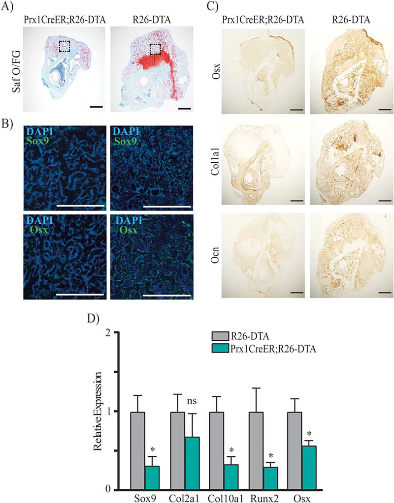
Figure 8.
Histological, immunostaining and qRT-PCR analyses of Prx1-CreER;R26-DTA calluses. (A) Representative coronal images of histological Safranin O/Fast Green (Saf O/FG) staining of fractured tibias from Prx1CreER;R26-DTA mice and R26-DTA littermate controls at 14 days after fracture. Adjacent sections were subjected respectively to: B) IF for Osterix [Osx] and Sox9 (Green) and counterstained with DAPI (Blue), areas shown correspond to the dotted squares of mineralized callus indicated in the Saf O/FG images, scale bars = 100μm; C) IHC respectively for Osx, Collagen 1 [Col1a1] and Osteocalcin [Ocn]; scale bars = 100μm. D) mRNA expression levels quantified by qRT-PCR of Sox9, Col2a1, Col10a1, Runx2 and Osx in fractured Prx1CreER;R26-DTA and R26-DTA control calluses obtained 14 days after fracture; data are reported as mean ± SD of duplicate repeats from 5 separate samples normalized to control to which was given value=1; *, p < 0.05, compared to control by unpaired two-tail t-test.

