Abstract
Background:
The mechanisms of noncontact anterior cruciate ligament (ACL) injuries are an enormously debated topic in sports medicine; however, the late phases of injury have not yet been investigated.
Hypothesis:
A well-defined posterior tibial translation can be visualized with its timing and patterns of knee flexion after ACL injury.
Study Design:
Case series.
Level of Evidence:
Level 4.
Methods:
A total of 137 videos of ACL injuries in professional male football (soccer) players were screened for a sudden posterior tibial reduction (PTR) in the late phase of noncontact ACL injury mechanism. The suitable videos were analyzed using Kinovea software for sport video analysis. The time of initial contact of the foot with the ground, the foot lift, the start of tibial reduction, and the end of tibial reduction were assessed.
Results:
A total of 21 videos exhibited a clear posterior tibial reduction of 42 ± 11 ms, after an average of 229 ± 81 ms after initial contact. The tibial reduction occurred consistently within the first 50 to 60 ms after foot lift (55 ± 30 ms) and with the knee flexed between 45° and 90° (62%) or more than 90° (24%).
Conclusion:
A rapid posterior tibial reduction is consistently present in the late phases of noncontact ACL injuries in some male soccer players, with a consistent temporal relationship between foot lift from the ground and consistent degrees of knee flexion near or above 90°.
Clinical Relevance:
This study provides insight into the late phases of ACL injury. The described mechanism, although purely theoretical, could be responsible for commonly observed intra-articular lesions.
Keywords: ACL injury, video analysis, soccer, noncontact, biomechanics
Mechanisms of noncontact anterior cruciate ligament (ACL) injuries represent an enormously debated topic in sports medicine.1,3,6,7,9-15 Many theories have been proposed, such as the quadriceps drawer mechanism (where the quadriceps muscle generates anterior shear forces on the tibia because of the patellar tendon angle),3 the internal tibial rotation on a relatively straight leg,1 knee valgus with internal rotation,14 knee valgus with external rotation,6 and tibiofemoral compression loading.12 Each of these theories has been supported using different methodologies, such as athlete interviews, clinical studies, magnetic resonance imaging evaluation of bone contusions, laboratory motion analysis, cadaveric studies, or mathematical simulations.11 One of the most authoritative theories is by Koga et al,10 who performed a 3-dimensional (3D) video analysis on real in vivo ACL injuries that occurred in handball and basketball players.10 By matching the 3D skeletal models with bidimensional images according to anatomical landmarks, they were able to demonstrate the rotational and coronal pattern of ACL rupture. However, the anteroposterior motion and the skeletal behavior in the late phases of injury have not been investigated.10 The anteroposterior translation has been assessed only in a case report of a professional soccer player during an international match in which high-definition cameras with high-speed recordings allowed an accurate motion analysis.9 The authors reported a progressive anterior subluxation that plateaued by 150 ms, followed by a posterior shift between 200 and 240 ms after initial ground contact. However, it is not clear if such an event represents a unique individual case, or a consistent and common event during ACL rupture.
Thanks to the worldwide popularity of soccer and the increasing media coverage of soccer matches, the availability of videos documenting ACL injuries is spreading and serving as a precious resource for clinicians and scientists.7,9 The traditional soccer uniform, which exposes the lower limb, allows for accurate inspection of knee landmarks during joint motion.
Therefore, the purpose of the present study was to perform a qualitative video analysis of noncontact ACL injuries in professional male soccer players with the aim to identify and describe the pattern of posterior tibial reduction. We hypothesize that a well-defined posterior tibial translation can be identified both with well-defined timing within the ACL injury dynamics and with consistent patterns of knee flexion.
Methods
Case Selection
This study is a subanalysis on a larger cohort of a systematic video analysis study on ACL injury mechanisms and situations in professional soccer players. According to our study methods, a systematic search of online database resources was performed retrospectively during a period of 10 consecutive seasons to identify ACL injuries that happened during matches belonging to the first 2 Italian soccer leagues. To identify ACL injuries, each season and each team roster were extracted from online database sources (legaseriea.it and legab.it). Then, each player was searched on Transfermarkt.de (Transfermarkt GmbH & Co KG) for injury history details. Additional online data sources (including national and local media) were used to find the ACL-injured athletes. Moreover, other online sources were screened according to Grassi et al.7
The videos of the matches were obtained from the digital platform wyscout.com (Wyscout spa and paninidigital.com, Panini Digital, Digital Soccer Project Srl). Each video was processed and cut approximately 12 to 15 seconds before and 2 to 5 seconds after the estimated injury frame using an online digital tool (Digital Log).
Two investigators performed the search, and any inconsistencies regarding the inclusion of videos in the analysis were resolved after the involvement of a third investigator.
Qualitative Video Analysis
All the videos were analyzed by Kinovea software (asso@kinovea.org) for sport video analysis. Videos were considered eligible for the evaluation of the late phase of ACL injury if:
mechanism without direct contact on injured knee was present;
the video footage presented enough quality to discern knee motion and observe posterior tibial reduction was [were] present in the video footage;
both knee and foot were assessable on the same video captions;
both moment of initial contact of the foot on the ground and the moment when the foot was lifted from the ground were present in the video footage.
When a video met the aforementioned criteria, it was evaluated using all the available views, replays, and slow motion footage to allow a better understanding of the relationship between body segments. When the evaluation was performed in slow motion sequences, synchronization was activated to match the gap of milliseconds between the original speed and the slow-motion sequence. This was performed by measuring the length between the same well-defined visual landmarks (eg, contact with ball, foot strike, etc) in both normal speed and slow motion replays. The ratio between the lengths of the same action (at the 2 different speeds) represented the amount of slow-down. All the videos had a frame rate of 25 to 50 frames per second, which means 1 frame every 40 to 20 ms at normal speed. Based on the speed of the slow motion sequence (from 2× to 5×), it was possible to analyze up to 1 frame every 4 ms.
According to Koga et al,10 the player maneuver during injury was classified as (1) cutting or (2) single-leg landing, while indirect contact with a hit on body part other than lower extremity was defined as (1) present or (2) absent. Finally, based on video examination, the following time points were obtained:
Initial contact: the moment when the foot of the injured leg struck the ground
Foot lift: the moment when the foot of the injured leg started to lose contact with the ground after the initial contact
Start of tibial reduction: the moment when the tibia started the posterior reduction
End of tibial reduction: the moment when tibial reduction ended and no further anteroposterior movements were noted
Furthermore, the knee flexion at the time of initial contact and start of the tibial reduction were estimated using a custom software (GPEM Screen Editor; GPEM Srl), which allows a quantification of joint angles on the desired frame. Angles were then grouped into 4 categories: extension or hyperextension, from >0° to 45°, from 45° to 90°, and >90°.
All the videos were reviewed and analyzed independently by 2 examiners; then, the videos were reviewed simultaneously by the 2 examiners to obtain consensus regarding all the items evaluated after discussion. In the case of disagreement, a third examiner was involved.
Statistical Analysis
Values are expressed as means with standard deviations. The values of initial contact, foot lift from the ground, start of tibial reduction and end of tibial reduction were plotted graphically as well.
Results
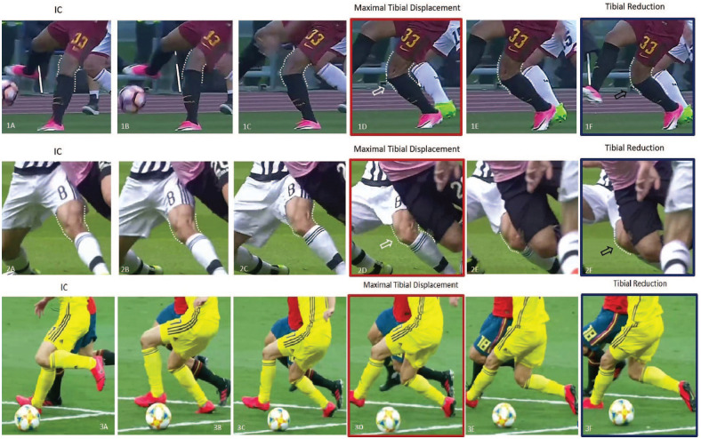
Overall, 137 videos were screened for inclusion, and a clear posterior tibial reduction was identified in 27 ACL injuries. Four videos were excluded because it was not possible to correctly identify all the time landmarks of injury dynamics (initial contact, foot release, or start of tibial reduction), while 2 other videos were excluded because of a direct contact. Therefore, 21 videos showing posterior tibial reduction were included in the final video analysis (Figure 1).
Figure 1.
Three cases of indirect contact anterior cruciate ligament injury with observable tibial reduction. The red-framed pictures (1D, 2D, and 3D) represent the observed maximal anterior tibial displacement, while the blue-framed pictures are the predicted end frames of tibial reduction (1F, 2F, and 3F). IC, initial contact.
All injuries occurred in male soccer players (2 goalkeepers, 5 defenders, 8 midfielders, 6 forwards) without a direct contact on the injured leg. Of the injuries, 14 (67%) had a cutting maneuver and 7 (33%) occurred during a single-leg landing, while an indirect contact with a hit on the upper body before ground contact was present only in 5 cases (24%) (Table 1).
Table 1.
Description of injury situation and mechanism from the 21 included videos
| Player | Role | Injured Leg | Maneuver | Indirect Contact |
|---|---|---|---|---|
| Player 1 | Defender | L | Cutting | No |
| Player 2 | Defender | R | Single-leg landing | Yes |
| Player 3 | Defender | L | Cutting | No |
| Player 4 | Forward | R | Single-leg landing | Yes |
| Player 5 | Midfielder | L | Cutting | No |
| Player 6 | Forward | R | Single-leg landing | No |
| Player 7 | Midfielder | R | Cutting | No |
| Player 8 | Forward | L | Cutting | No |
| Player 9 | Forward | R | Cutting | No |
| Player 10 | Midfielder | R | Cutting | No |
| Player 11 | Forward | R | Single-leg landing | No |
| Player 12 | Defender | L | Single-leg landing | Yes |
| Player 13 | Goalkeeper | L | Cutting | No |
| Player 14 | Forward | R | Cutting | No |
| Player 15 | Midfielder | L | Cutting | No |
| Player 16 | Midfielder | L | Cutting | Yes |
| Player 17 | Goalkeeper | R | Single-leg landing | No |
| Player 18 | Midfielder | R | Cutting | No |
| Player 19 | Defender | L | Single-leg landing | Yes |
| Player 20 | Midfielder | R | Cutting | No |
| Player 21 | Midfielder | R | Cutting | No |
L, Left; R, right.
Video Analysis
According to the analysis of the 21 videos, foot lift occurred an average of 173 ± 73 ms (range, 33-373 ms) after initial contact, while tibial reduction started an average of 55 ± 30 ms (range, 27-180 ms) after foot lift and took an average of 42 ± 11 ms (range, 18-57 ms) to end. Based on these measurements, tibial reduction occurred on average 229 ± 81 ms (range, 76-423 ms) after initial foot contact (Table 2; Video Supplement 1, available in the online version of this article).
Table 2.
Details from the analysis of the 21 included videos, with the timing of the different phases of injury mechanism
| Player | Initial Contact to Foot Lift, ms | Foot Lift to Start Reduction, ms | Start Reduction to End Reduction, ms | Initial Contact to Start Reduction, ms | Knee Flexion at Initial Contact, deg | Knee Flexion at Start Reduction, deg |
|---|---|---|---|---|---|---|
| Player 1 | 200 | 58 | 42 | 258 | 0-45 | 0-45 |
| Player 2 | 260 | 58 | 43 | 319 | 0-45 | 45-90 |
| Player 3 | 166 | 47 | 47 | 213 | 0-45 | 45-90 |
| Player 4 | 227 | 45 | 23 | 273 | 0-45 | 45-90 |
| Player 5 | 206 | 46 | 57 | 251 | 0-45 | >90 |
| Player 6 | 33 | 44 | 55 | 76 | 0-45 | 45-90 |
| Player 7 | 169 | 46 | 46 | 215 | 0-45 | 45-90 |
| Player 8 | 63 | 63 | 42 | 126 | 0-45 | >90 |
| Player 9 | 166 | 41 | 31 | 207 | 0-45 | >90 |
| Player 10 | 220 | 40 | 40 | 260 | 0-45 | 45-90 |
| Player 11 | 206 | 54 | 40 | 260 | 0-45 | 0-45 |
| Player 12 | 133 | 38 | 47 | 171 | 0-45 | 45-90 |
| Player 13 | 217 | 27 | 27 | 244 | 0-45 | 45-90 |
| Player 14 | 127 | 51 | 51 | 178 | 0-45 | 45-90 |
| Player 15 | 124 | 50 | 50 | 174 | 0-45 | 45-90 |
| Player 16 | 208 | 180 | 55 | 388 | 0-45 | >90 |
| Player 17 | 373 | 50 | 50 | 423 | 0-45 | 45-90 |
| Player 18 | 132 | 66 | 44 | 197 | 0-45 | >90 |
| Player 19 | 183 | 41 | 18 | 223 | 0-45 | 45-90 |
| Player 20 | 80 | 53 | 53 | 133 | 0-45 | 0-45 |
| Player 21 | 151 | 62 | 27 | 213 | 0-45 | 45-90 |
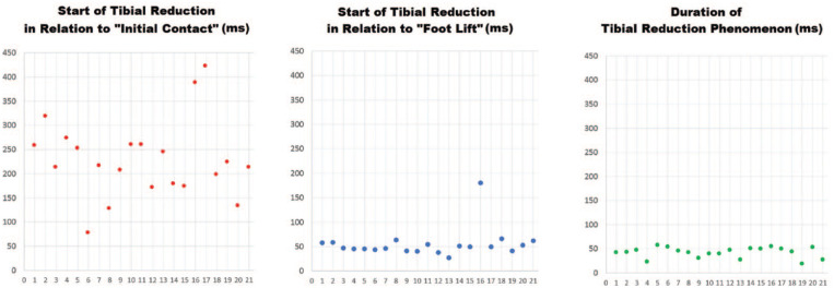
From the scatterplots, it is possible to observe the consistent occurrence of tibial reduction around the first 50 ms after foot lift in most of the cases, while the timing of tibial reduction in relation to initial contact appears scattered more chaotically (Figure 2). Only 1 case had an outlier behavior, with tibial reduction occurring 180 ms after foot lift. If this patient were excluded from analysis, the mean value and variability would decrease to 49 ± 10 ms. The duration of tibial reduction was extremely consistent as well, and did not exceed the value of 60 ms.
Figure 2.
Temporal relationship between initial contact with the ground and start of tibial reduction (red dots), between the foot lift and start of tibial reduction (blue dots), and the length of tibial reduction (green dots).
Regarding knee flexion during ACL injury, all players (100%) had the knee flexed between 0° and 45° at the time of initial contact; in other words, when tibial reduction occurred, the knee was flexed between 45° and 90° in 13 cases (62%), more than 90° in 5 cases (24%), and between 0° and 45° in only 3 cases (14%).
Discussion
The most important finding of the present study was that in a series of 21 videos of noncontact ACL injuries in professional soccer players, it was possible to identify a clear posterior tibial reduction with a specific and redundant pattern. Specifically, a sudden posterior movement that lasted 40 to 50 ms was noted. This movement had no temporal correlation with initial ground contact. On the contrary, a remarkably consistent temporal relationship was instead found with respect to the injured leg lift, after approximately 40 to 60 ms. Finally, most of the posterior tibial reductions (86%) occurred with more than 45° of knee flexion. These values are extremely similar to what was reported in a model-based image-matching analysis of a single patient.9 In that case report, a posterior tibial reduction that lasted 40 ms was identified.9 It started 200 ms after initial foot contact with the ground, with knee flexion exceeding 90°.
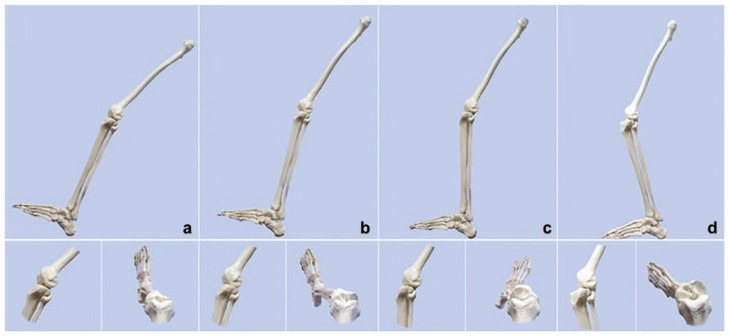
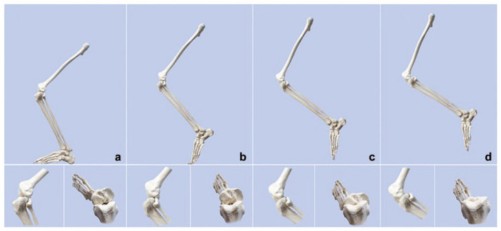
From a theoretical standpoint, the findings of the present study seem to complete the noncontact ACL injury pattern introduced by Koga et al,9,10 which consists of 3 phases: (1) a knee valgus load is applied, the medial collateral ligament becomes taut, and lateral compression occurs; (2) the compressive force, coupled with anterior force vector caused by quadriceps contraction, causes a displacement of the femur relative to the tibia where the lateral femoral condyle shifts posteriorly, and the tibia translates anteriorly and rotates internally, resulting in ACL rupture; and (3) once the ACL is torn, the primary restraint to tibial anterior translation is gone, which causes the medial femoral condyle to be displaced posteriorly, resulting in external rotation of the tibia.10 This external rotation may be exacerbated by the typical movement pattern when the athlete starts changing directions and by the larger anteroposterior depth of the medial tibial plateau in comparison with the lateral tibial plateau (Figure 3). At this point, the posterior reduction described in the present study could occur as follows in 3 further steps: (4) due to the inertia of the motor task that caused injury (eg, jump landing, cutting), the knee increases its flexion, while anterior translation reaches its plateau; (5) since the subluxated tibia is not able to support the body weight, the player reacts by lifting the foot from the ground; and (6) when the anteriorly directed force that caused ACL rupture is dissipated and the tibia has no constraint with the ground, a sudden and rapid tibial reduction occurs due to tibiofemoral congruity, soft tissue elasticity, and hamstring contraction (Figure 4).
Figure 3.
Mechanism of anterior cruciate ligament (ACL) injury according to Koga et al.10 (a) Knee in neutral position before ground stroke; (b) with ground contact, when the valgus load is applied, the medial collateral ligament becomes taut and lateral compression occurs; (c) the compressive force, coupled with an anterior force vector caused by quadriceps contraction, causes a displacement of the femur relative to the tibia where the lateral femoral condyle shifts posteriorly and the tibia translates anteriorly and rotates internally, resulting in ACL rupture; (d) once the ACL is torn, the primary restraint to tibial anterior translation is gone and this causes the medial femoral condyle to also be displaced posteriorly, resulting in external rotation of the tibia.
Figure 4.
Mechanism of tibial posterior reduction. (a) Because of the inertia of the motor task that caused injury (eg, jump landing, cutting movement) the knee increases its flexion, while anterior translation reaches its plateau; (b) since the subluxated tibia is not able to support the body weight, the player lifts the foot from the ground, with a timing based on the ongoing task and the possibility of subtracting weight from the injured leg; (c) when the anteriorly directed force that caused anterior cruciate ligament (ACL) rupture is dissipated and the tibia has no restrains with the ground, a sudden and rapid posterior tibial reduction occurs; (d) when the posterior tibial reduction concludes, normal tibiofemoral congruency is restored.
The above-described mechanism, although purely theoretical, conveys the awareness of a sudden tibial reduction in the late phases of ACL injury and offers new insights. As an example, such an event could be hypothetically responsible for intra-articular lesions, such as bone contusions, osteochondral injuries, or meniscal ruptures. In fact, Kaplan et al8 suggested a backward countercoup etiology for the medial compartment bone bruises that are found in magnetic resonance imaging after ACL rupture. If bone bruising occurs in a late phase of the ACL injury mechanism (eg, during tibial reduction), their role in ACL rupture should be revisited.13,16
Unfortunately, the bidimensional and qualitative evaluation performed in the present study does not allow quantification of the posterior reduction or the rotational behavior.
Another relevant consideration is the timing of posterior reduction. In fact, it does not seem related to the initial ground contact and therefore with the time of ACL rupture (estimated within the first 40 ms). Rather, it occurs with extreme consistency a few milliseconds after the foot is lifted from the ground. In this context, the action performed by the player at the time of ACL injury thus represents a crucial factor for the late phase of the injury dynamic. If the player is performing a task involving a rapid monopodalic load (eg, rapid change of direction), a rapid foot lifting is possible, thus allowing the player to subtract weightbearing and allow tibial reduction. In other words, if the player is performing a task involving progressively increased weightbearing (eg, landing from jump), the player is unable to lift the foot from the ground after the initial subluxation. Therefore, the reaction is to unload the weightbearing leg, possibly causing dramatic collapse.
The knowledge of the posterior tibial reduction in ACL injury videos could also help on-field clinicians to suspect an ACL injury in the case of knee sprains. This would allow preventive retirement of players from the field to avoid further injuries. However, the reliability of such an approach should be validated in further studies.
Last, the posterior tibial reduction could also explain the common “pop” sensation described by the patients experiencing ACL rupture.2,4,5 It is possible that such a sound derives from a bone-on-bone knock rather than ligament stretching and rupture.
The main limitations of the present study are the low number of cases evaluated and the very specific population regarding patient features and injury mechanism. First of all, it is not easy to obtain ACL injury videos with sufficient quality and characteristics to appreciate a rapid event such as tibial reduction. Moreover, it was not the purpose of this study to document the frequency of the phenomenon, but rather to describe its occurrence and main characteristics. Second, the choice to assess only noncontact injuries in male soccer players allowed us to better analyze the phenomenon; on the other hand, the findings of the present study remain limited to this subtype of injury and population, thus hampering its generalizability to other sports, as well as to female athletes and direct injuries. The last limitation is the lack of model-based image-matching analysis,9,10 which could have allowed the precise estimation of tibial reduction and rotation. Despite our findings being extremely consistent with what was reported when using more complex technologies,9,10 the timing estimation described in the present study could be imprecise due to the gross method utilized. Therefore, the comparisons done with the articles from Koga et al9,10 are currently only theories. However, it was not the aim of this study to analyze ACL injuries in depth, but rather to highlight the tibial reduction event and its gross role in the injury mechanism, providing a base for future research. The definition of knee flexion could be biased due to the assessment on bidimensional images. However, the choice to categorize knee flexion in broad categories (0°-45°, 45°-90°, and >90°) allowed a straightforward evaluation. Regarding the more precise model-based image-matching analysis, it is very selective in terms of video characteristics and extremely time-consuming (up to 2 months for each video). Therefore, with the current materials, such analysis was precluded.
Conclusion
This study provides insight into the late phases of ACL injury with the awareness of a sudden tibial reduction. A rapid posterior tibial reduction was consistently present in the late phases of noncontact ACL injuries in male soccer players, with a consistent temporal relationship with foot lifting from the ground and consistent degrees of knee flexion near or >90°. The described mechanism, although purely theoretical, could be responsible for commonly observed intra-articular lesions such as bone contusions, osteochondral injuries, or meniscal ruptures.
Acknowledgments
The authors want to thank Margaux Hylton for her contribution in the language revision of the manuscript.
Footnotes
The authors report no potential conflicts of interest in the development and publication of this article.
References
- 1. Arnold JA, Coker TP, Heaton LM, Park JP, Harris WD. Natural history of anterior cruciate tears. Am J Sports Med. 1979;7:305-313. [DOI] [PubMed] [Google Scholar]
- 2. Ayre C, Hardy M, Scally A, et al. The use of history to identify anterior cruciate ligament injuries in the acute trauma setting: the “LIMP index.” Emerg Med J. 2017;34:302-307. [DOI] [PubMed] [Google Scholar]
- 3. DeMorat G, Weinhold P, Blackburn T, Chudik S, Garrett W. Aggressive quadriceps loading can induce noncontact anterior cruciate ligament injury. Am J Sports Med. 2004;32:477-483. [DOI] [PubMed] [Google Scholar]
- 4. Feagin JA, Jr, Curl WW. Isolated tear of the anterior cruciate ligament: 5-year follow-up study. Am J Sports Med. 1976;4:95-100. [DOI] [PubMed] [Google Scholar]
- 5. Fetto JF, Marshall JL. The natural history and diagnosis of anterior cruciate ligament insufficiency. Clin Orthop Relat Res. 1980;147:29-38. [PubMed] [Google Scholar]
- 6. Fung DT, Zhang LQ. Modeling of ACL impingement against the inter-condylar notch. Clin Biomech (Bristol, Avon). 2003;18:933-941. [DOI] [PubMed] [Google Scholar]
- 7. Grassi A, Smiley SP, Roberti di, Sarsina T, et al. Mechanisms and situations of anterior cruciate ligament injuries in professional male soccer players: a YouTube-based video analysis. Eur J Orthop Surg Traumatol. 2017;27:967-981. [DOI] [PubMed] [Google Scholar]
- 8. Kaplan PA, Gehl RH, Dussault RG, Anderson MW, Diduch DR. Bone contusions of the posterior lip of the medial tibial plateau (contrecoup injury) and associated internal derangements of the knee at MR imaging. Radiology. 1999;211:747-753. [DOI] [PubMed] [Google Scholar]
- 9. Koga H, Bahr R, Myklebust G, Engebretsen L, Grund T, Krosshaug T. Estimating anterior tibial translation from model-based image-matching of a noncontact anterior cruciate ligament injury in professional football: a case report. Clin J Sport Med. 2011;21:271-274. [DOI] [PubMed] [Google Scholar]
- 10. Koga H, Nakamae A, Shima Y, et al. Mechanisms for noncontact anterior cruciate ligament injuries: knee joint kinematics in 10 injury situations from female team handball and basketball. Am J Sports Med. 2010;38:2218-2225. [DOI] [PubMed] [Google Scholar]
- 11. Krosshaug T, Andersen TE, Olsen OE, Myklebust G, Bahr R. Research approaches to describe the mechanisms of injuries in sport: limitations and possibilities. Br J Sports Med. 2005;39:330-339. [DOI] [PMC free article] [PubMed] [Google Scholar]
- 12. Meyer EG, Haut RC. Anterior cruciate ligament injury induced by internal tibial torsion or tibiofemoral compression. J Biomech. 2008;41:3377-3383. [DOI] [PubMed] [Google Scholar]
- 13. Patel SA, Hageman J, Quatman CE, Wordeman SC, Hewett TE. Prevalence and location of bone bruises associated with anterior cruciate ligament injury and implications for mechanism of injury: a systematic review. Sports Med. 2014;44:281-293. [DOI] [PMC free article] [PubMed] [Google Scholar]
- 14. Speer KP, Spritzer CE, Bassett FH, 3rd, Feagin JA, Jr, Garrett WE., Jr. Osseous injury associated with acute tears of the anterior cruciate ligament. Am J Sports Med. 1992;20:382-389. [DOI] [PubMed] [Google Scholar]
- 15. Waldén M, Krosshaug T, Bjørneboe J, Andersen TE, Faul O, Hägglund M. Three distinct mechanisms predominate in non-contact anterior cruciate ligament injuries in male professional football players: a systematic video analysis of 39 cases. Br J Sports Med. 2015;49:1452-1460. [DOI] [PMC free article] [PubMed] [Google Scholar]
- 16. Zhang L, Hacke JD, Garrett WE, Liu H, Yu B. Bone bruises associated with anterior cruciate ligament injury as indicators of injury mechanism: a systematic review. Sports Med. 2019;49:453-462. [DOI] [PubMed] [Google Scholar]


