Abstract
Objective
To assess and compare volumetric and shape changes of the orbital cavity in patients treated with tooth-borne (TB) and bone-borne (BB) rapid maxillary expansion (RME).
Study design
Forty adolescents with bilateral maxillary cross-bite received tooth-borne (TB group = 20; mean age 14.27 ± 1.36 years) or bone-borne (BB group = 20; mean age of 14.62 ± 1.45 years) maxillary expander. Cone-beam computed tomography (CBCT) were taken before treatment (T1) and 6-month after the expander activation (T2). Volumetric and shape changes of orbital cavities were detected by referring to a specific 3D digital technology involving deviation analysis of T1/T2 CBCT-derived models of pulp chamber. Student’s t tests were used to 1) compare T1 and T2 volumes of orbital cavities in TB and BB groups, 2) compare volumetric changes and the percentage of matching of 3D orbital models (T1-T2) between the two groups.
Results
Both TB and BB groups showed a slight increase of the orbital volume (0.64 cm3 and 0.77 cm3) (p < 0.0001). This increment were significant between the two groups (p < 0.05) while no differences were found in the percentage of matching of T1/T2 orbital 3D models (p > 0.05). The areas of greater changes were detected in the proximity of the frontozygomatic and frontomaxillary sutures.
Conclusion
TB-RME and BB-RME would not seem to considerably affect the anatomy or the volume of the orbital cavity in adolescents.
Introduction
Rapid Maxillary expansion (RME) is the most efficient treatment for the correction of maxillary transverse deficiency [1]. Findings from the highest levels of evidence confirm that this protocol accomplishes transverse expansion of maxillary arch through skeletal and a dentoalveolar effects [1–3]. However, previous studies based on finite element analyses (FEM) showed that RME increases the stress levels on neighboring structures and on the cranial base due to the cumulative forces released during the repeated activations of the expansion screw[4, 5]. It would seem that this cumulative force pattern can be responsible for the widening of the spheno-occipital synchondrosis in young subjects underwent RME [6]. Concerning the circummaxillary structures, post-treatment changes were found at the level of the zygomatic process, the frontozygomatic suture and the frontal process of the maxilla [1, 7–9], all these structures being part of the orbital cavity. In this regard, previous evidence suggested that maxillary expansion may alter the volume of the orbital cavity [10], however this aspect has not been studied extensively nor it has been determined if volumetric changes of the orbital cavity caused by RME may interfere with its normal growth pattern and physiology. This aspect could be of clinical relevance considering that a positive correlation was found between the increment of orbital volumes and the degree of dysmorphology, enophtalmos, orbitopathy [11].
Skeletal anchorage has been proposed to increase the skeletal effect of RME, limiting dento-alveolar effects and widening the spectrum of patients to late adolescent and adults [12–14]. In this regard, bone-borne RME (or miniscrew-assisted RME) has showed greater skeletal effect compared to the tooth-borne RME [14–16]. As consequence, it could be postulated that the greater effectiveness of miniscrew-assisted RME could be associated with a greater effect on surrounding structures, such as the orbital cavity. Considering the prevalence of transverse maxillary deficiency among children and adolescent [17], awareness of the risks associated with the use of a tooth-borne and a bone-borne RME appears clinically relevant.
Nowadays, progress in radiographic techniques and 3D imaging software allows a more accurate evaluation and comparison of the morphological changes of anatomical structure. In particular, specific anatomical structures can be reconstructed (3D rendering) from CBCT scans, and superimposed to accurately evaluate growth or treatment changes [18]. Meanwhile, deviation analysis can be used to detect shape differences between two CBCT-derived anatomical models as well as to obtain precise dimensional information [19–21].
In this respect, the aim of the present study was to assess volumetric as well as morphological surface changes of the orbital cavity in patients treated with both tooth-borne and bone-borne rapid maxillary expansion, by referring to the surface-to-surface technique and deviation analysis of CBCT-derived 3D rendered models of the orbital cavity. The null hypothesis was the absence of significant changes in the orbital cavity architecture between the two different protocols of skeletal maxillary expansion.
Materials and methods
The study obtained the approval of the Institutional Review Board of Indiana University–Purdue University (IRB protocol number: Pro00075765) and included a sample of adolescents with a diagnosis of skeletal transverse deficiency and who completed the orthodontic treatment at the Orthodontic Clinic of the University of Alberta (Edmonton, Canada, USA). The study sample was obtained from previously published materials [22], in order to avoid unnecessary or additional radiation exposure to the patients. All subjects were diagnosed with a bilateral maxillary cross-bite involving at least the two maxillary first molars and were treated with a tooth-borne (TB group) or a bone-borne (BB group) RME as part of their comprehensive orthodontic therapy. Eligibility criteria were as follow: 1) age between 11 and 15 years, 2) CBCT scans of good quality taken prior to the placement of the maxillary expander (T1) and after its removal (T2), 3) no artifact due to restorative materials present, 4) no previous orthodontic treatment, 5) no systemic disease or usage of medication, 6) no craniofacial syndromes or anomalies.
The characteristics of the RME protocol used in this study have been previously described [22, 23]. Briefly, in the TB group, 20 subjects (12 female and 8 male, with a mean age of 14.27 ± 1.36 years) received the Hyrax-type expander with bands on the permanent first molars and first premolars (Fig. 1a). In the BB group, 20 subjects (15 female and 5 male, with a mean age of 14.62 ± 1.45 years) received two miniscrews in the palate between the permanent first molar and the second premolar (length: 12 mm; diameter: 1.5 mm; Straumann GBR System, Andover, MA) that were connected with the expander (Palex II Extra-Mini Expander, Summit Orthodontic Services, Munroe Falls, OH) (Fig. 1b). In both groups, the activation rate of the jackscrew was 0.25 mm/turn and the RME protocol included two turns per day, for a total of 0.5 mm/d. Clinical assessment of successful expansion protocol was achieved by visual inspection of the opening of the interincisal diastema. Activations were stopped once overexpansion was achieved, i.e., when the mesiopalatal cusps of the maxillary first permanent molars were in contact with the buccal cusps of the mandibular first permanent molars. At the end of the activation period, the expansion screw was blocked with flowable composite and the appliance was kept in place for 6 months as retention; during this period, patients did not receive any other type of orthodontic treatment.
Fig. 1.
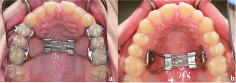
Tooth-borne (a) and Bone-borne (b) palatal expander used in this study. See the interincisal diastema after maxillary expansion
Cone beam computed tomography (CBCT) was performed on all subjects before the expansion (T1) and after the 6 months of retention (T2). Patients were scanned with the same iCAT CBCT Next Generation Unit (Imaging Sciences International, Hartfield, PA). The setting protocol included 0.3 voxel, 8.9 seconds, large field of view (17 x 23 cm) at 120 kV and 20 mA. The distance between 2 slices was 0.3 mm which provided accuracy in anatomic registration. The protocols used in this study for segmentation, model rendering and deviation analysis were previously validated and clearly described [18, 24].
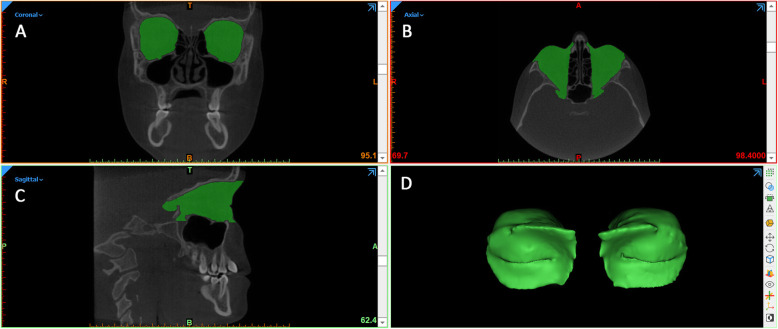
Step 1- Generating the preliminary segmentation masks. The first step was performed by using Slicer 3D software (http://www.slicer.org), to develop the segmentation masks of soft tissues from CBCT. The 'fast marching' algorithm [25] was used to accurately segment the mask of the orbital cavity, in particular this algorithm allowed the initial marked regions to propagate toward regions with similar gray levels intensity, excluding bone structures as well as cavities full of air (Fig. 2). Afterwards, the soft tissue segmentation mask was rendered into a 3D surface models (Standard Triangulation Language - .stl).
Fig. 2.
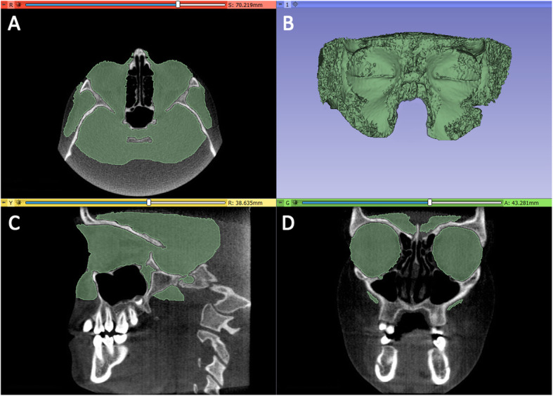
Segmentation mask of soft tissues from CBCT using Slicer 3D software. The ‘fast marching’ algorithm allowed the initial marked regions to propagate toward regions with similar gray levels intensity, excluding bone structures as well as cavities full of air
Step 2 - Removal of outside regions and cropping of T1 - orbit 3D models. The 3D model of orbital soft tissue and outside regions and the CBCT scans were imported onto Mimics Research software (vr. 21.0.0.406, Materialise NV, Liege, Belgium) with the same coordinates system (Fig. 3). A new segmentation mask was obtained from the original .stl file and the 'edit mask' function was used to cleaned up the orbital cavity (removal of outside regions). To improve quality and contour delineation, the mask was smoothed and finely adjusted by using the interactive ‘Contour Edit’ function. Afterwards, the edited mask was converted and rendered to a 3D model.
Fig. 3.
Once imported into Mimics Research software, the soft tissues mask was cleaned up to keep the soft tissues of the orbital region (removal of outside regions)
Lastly, two planes cut were selected in order to accurately and reliably define anterior and posterior boundaries of the orbital cavity (Figs. 4, 5). Anterior plane was constructed by selecting two points on the coronal view, respectively the fronto-zygomatic suture at the external rim of the orbit (FZS) and the supraorbital foramen (SF), and the anterior lacrimal crest (ALC) on the axial view. Posterior plane was constructed by selecting two points on the axial view, respectively the lateral (OC1) and medial (OC2) walls of the optic foramen taken at the most caudal scan, and the most cranial point of the lateral walls (OC3) of the optic foramen on the sagittal view.
Fig. 4.
The anterior plane was constructed by selecting two points on the coronal view, i.e., the fronto-zygomatic suture at the external rim of the orbit (FZS) and supraorbital foramen (SF) and by selecting the anterior lacrimal crest (ALC) point on the axial view
Fig. 5.
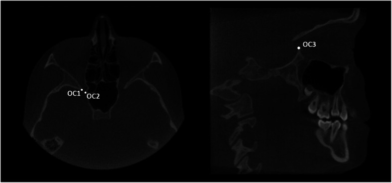
The posterior plane was constructed by selecting the lateral (OC1) and medial (OC2) walls of the optic foramen taken at the most caudal axial view and the most cranial point of the lateral walls (OC3) of the optic foramen taken on the sagittal view
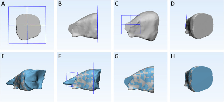
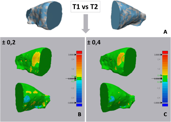
Step 3 – Superimposition and cropping of T2 - orbit 3D models. The 3D model of each orbit (T1 and T2) were imported onto 3-matic Medical software (vr. 13.0.0.188, Materialise NV, Liege, Belgium) with the same coordinates system. Firstly, three points were randomly selected on the anterior and posterior surfaces of T1-3D model, in order to replicate the same planes cut of the segmentation masks (Fig. 6a-d). Using a specific function of the software (‘global registration’), the T1 and T2 3D models of orbital cavity were superimposed using a surface-based registration, with the former used as fixed entity (Fig. 6e). The software automatically calculates the best fitting between the two models. Finally, the cut planes previously selected were used to cut the T2 orbit model (Fig. 6f-h). Step 4 - 3D Deviation analysis. After surface-based superimposition, the registered T1 and T2 models of the orbital cavity underwent deviation analysis by using the Geomagic Control X software (version 2017.0.0, 3D Systems, Santa Clara, CA 95054, USA). The analysis was complemented by visualization of the 3D color-coded maps set with a range of tolerance of 0.20 mm and 0.40 mm (Fig. 7). After the deviation analysis, percentages of all the distance values within the tolerance range were calculated.
Fig. 6.
The 3D model of each orbit (T1 and T2) were imported onto 3-matic Medical software. Construction of two cutting planes, one on the anterior surface of T1- orbit 3D model (a, b) and one on the posterior surface (c, d) (this planes replicated the planes cut of the segmentation masks); (e) surface-based registration between T1 and T2 3D models of orbital cavity, using T1 model as fixed entity; (f-h) the cut planes previously selected were used to cut the T2 orbit model
Fig. 7.
Example of deviation analysis between the T1 and T2 orbital cavity obtained in both tooth-borne and bone-borne expander groups. The colored map shows the deviations (negative blue, positive red) between the mesh models. a models superimposition, b deviations analysis set at 0.2 mm of tolerance; c deviations analysis set at 0.4 mm of tolerance
Step 5 - 3D Matching percentage calculation and volumetric assessment. Once the deviation analysis was carried out, the percentages (%) of all the distance values within the tolerance ranges were calculated. These values represented the degree of matching between the pairs of 3D orbital cavity models (T1 and T2).
The amount of skeletal maxillary expansion was also recorded in each group by calculating the maxillary palatal width. This measure connected the junctions of the hard palate and lingual alveolar bone on the coronal section passing through the maxillary right first molar furcation [22].
The entire digital workflow was performed by a single expert examiner (A.L.G.) who processed only 2 CBCT scans each day to avoid fatigue. All images were standardized for brightness and contrast, and magnified by the same amount. Since post-treatment CBCTs were acquired after removal of the palatal expander, it was possible to blind the examiner regarding the type of appliance used. CBCT records of 10 subjects were randomly selected 4 weeks after the last examination to calculate the intra-observer and inter-observer variability and the method error, assuring that both examiners (A.L.G. and L.R.) were not aware of data of the first measurements.
Statistics
The Shapiro-Wilk Normality Test was preliminarily performed to test the normality of the data. Since the data showed homogeneous variance, parametric tests were used to evaluate and compare measurements. In this respect, the paired Student’s t-test was used to compare T1 and T2 volumetric measurements of the orbital cavity in both TB and BB groups. The unpaired Student’s t test was used to assess if the post-treatment volumetric changes and the percentage of matching between the pre and post-treatment orbit models significantly differed between the two groups. The unpaired Student’s t test was also used to assess if the post-treatment volumetric changes (T1-T2) of the orbital cavity were different between right and left side, in both TB and BB groups. Since no differences were detected (p > 0.05), volumetric data of both sides were merged, thus the final study sample included 40 orbital 3D models in each group, increasing the robustness of the study.
Linear regression was performed to investigate a cause-effect relationship between skeletal expansion (independent variable) and orbital volume changes (dependent variable). Intra-examiner and inter-examiner reliability were assessed using intraclass correlation coefficient (ICC) while assessment of the method error was performed using Dahlberg’s formula [26]. Data sets were analyzed using SPSS® version 24 Statistics software (IBM Corporation, 1 New Orchard Road, Armonk, New York, USA).
Results
Pre and post-treatment volumetric measurements of the orbital cavity are reported in Table 1 along with inferential statistics. In both groups, there was a slight increment of the orbital volume (TB = 0,64 cm3 ± 0,07; BB = 0,77 cm3 ± 0,22) that was statistically significant (p < 0.0001) (Table 2). Such volumetric increment was statistically significant were between TB and BB groups, with mean difference value of 0, 13 cm3 (p < 0.05). Considering that the sample size included 40 orbital cavities for each group and the significance level set at p < 0.05, the present investigation was adequately powered (94.5 %) to distinguish a mean difference of 0.13 cm3 in the post-treatment volumetric changes of the orbital cavities between TB and BB groups.
Table 1.
Data of orbital cavity volumes recorded in both study groups (data included right/left side) and comparison of volumetric changes in orbital cavity between groups
| TB Group | BB Group | ||||||||||||||
|---|---|---|---|---|---|---|---|---|---|---|---|---|---|---|---|
| Sample | Orbital volume T1 (cm3) | SD | Orbital volume T2 (cm3) | SD | Significance | Orbital volume T1 (cm3) | SD | ||||||||
| 40 | 24,04 | 1,71 | 24,68 | 1,74 | p < 0.0001 | 23,65 | 1,58 | 24,41 | 1,66 | p < 0.0001 | 0,64 | 0,07 | 0,77 | 0,22 | p < 0.05 |
T1 = before maxillary expansion, T2 = 6 months after maxillary expansion. TB = Tooth-borne, BB = Bone-borne
P values are reported according to the paired Student’s t Test for intra-group assessment and according to the unpaired Student’s t Test for comparison of mean differences between groups
NS not significant
Table 2.
Comparison of matching percentage of pre and post-treatment orbital 3D models between tooth-borne (TB) and bone-borne (BB) group
| TB Group | BB group | |||||
|---|---|---|---|---|---|---|
| Sample size | Matching | SD | Matching | SD | Significance | |
| Percentage | Percentage | |||||
| (T1-T2) | (T1-T2) | |||||
| Tolerance A | 40 | 83,84% | 3,67% | 81,87% | 2,70% | p > 0.05 |
| Tolerance B | 40 | 94,46% | 2,97% | 92,89% | 2,65% | p > 0.05 |
T1 = before maxillary expansion, T2 = 6 months after maxillary expansion. P values are reported according to the unpaired Student’s t Test
Tolerance A = 0,20 mm; Tolerance B = 0,40 mm. SD = standard deviation
No differences were found in the percentage of matching of T1/T2 orbital 3D models between TB and BB groups (p > 0.05), at both tolerance A (TB = 83,84% ± 3,67%; BB = 81,87% ± 2.70%) and tolerance B (TB = 94,46% ± 2,97%; BB = 92,89% ± 2.65%) (Table 3).
Table 3.
Linear regression tests model using maxillary expansion as indipendent variable (predictor) and orbital volume as dependent variable
| Groups | Maxillary expansion (mm) | Orbital volume (cm3) | Dependent Variables | Predictor Variables | R | R squared | Coefficients | 95% Interval Coefficient (B) | ||
|---|---|---|---|---|---|---|---|---|---|---|
| Beta | Standard error | Lower Limit | Upper Limit | |||||||
| TB | 1.83 ⏈ 0.42 | 0.64 | Maxillaryexpansion (mm) | Orbital volume (cm3) | 0.551 | 0.319 | 0.551 | 0.087 | 0.061 | 0.454 |
| BB | 2.2 ⏈ 0.33 | 0.77 | 0.582 | 0.325 | 0.582 | 0.136 | 0.088 | 0.635 | ||
Additionally, the colour map analysis of the superimposition of the T1-T2 orbital shells showed red tone (positive mismatching) in a small area located in the anterior upper lateral side of the orbital surface model. Blue tone (negative mismatching) was shown in a small area located in the anterior lower median side of the orbital surface (Fig. 6). The remaining areas demonstrated a prevalence of green, which indicates no significant differences.
The amount of skeletal maxillary expansion recorded was respectively 1.83 mm (± 0.42) in the TB group and 2.2 mm (± 0.33) in the BB group. The post-treatment increment of the orbital volume showed no significant correlations with the amount of skeletal expansion obtained, being the values of R (linear regression) 0.051 in the TB and 0.582 in the BB group respectively (Table 3).
Concerning the reliability of the methodology, no differences were found between the two readings of the orbital volumes with a correlation indexes of 0,933 and 0,894 respectively for intra-operator and inter-operator variability. According to the Dahlberg’s formula, the random error for the orbital volumetric measurements was 0,63 cm.3
Discussion
Previous studies have deeply investigated the effects of tooth-borne maxillary expansion confirming that its effects are not limited to the maxilla but also involve adjacent structures, in particular the bones delimiting the nasal cavities and the circummaxillary sutures [1–9, 22, 27]. Among these, the zygomatic process, the frontozygomatic suture and the frontal process of the maxilla are part of the orbital cavity. In this respect, it has been reported that subjects underwent RME could feel sensation of pressure in the maxillary, nasal, or orbital areas [28, 29]. Two studies [30, 31] respectively reported the case of an adolescent with neurologic and ophthalmic symptoms (e.g. headache and diplopia) related to tooth-borne maxillary expansion, with the assumption of a potential association of RME and pseudotumor cerebri syndrome (PTCS) related manifestations. Particularly, in the latter study [31] the patient showed a protrusion of the optic nerve head and enlarged perioptic subarachnoid space confirming the diagnosis of PTCS, as assessed via magnetic resonance imaging (MRI). However, the interruption of the maxillary expansion protocol led to the complete resolution of clinical symptoms in 1 week. The authors assumed that PTCS may be a potential complication of RME due to an alteration of the cerebral venous circulation with subsequent impaired venous drainage and increased intracranial pressure. However, it must be underlined that these two findings were case reports with the intrinsic limitations in terms of scientific evidence.
A previous CT study [10] assessed the volumetric dimension and the anterior aperture width of the orbital cavity before and after RME. It was found a slight significant increment of the orbital volume as well as of the anterior width, however authors concluded that such modifications, despite statistically significant, were not so considerable as to affect the architecture of the orbits or the craniofacial anatomy.
In the present study, we investigated the volumetric and morphological changes of the orbital cavity after tooth-borne and bone-borne RME basing on the assumption that skeletal anchorage, being more effective in obtaining skeletal improvement of maxillary transversal deficiency, could also induce greater side-effects. To the best of our knowledge, this is the first study in literature for this purpose. We refer to a specific 3D imaging technology involving the fast marching algorithm procedure that allowed the 3D rendering of complex structures [25] such as the orbital cavity.
According to our findings, both TB and BB groups showed a slight increment of the orbital volume at T2 (respectively 0,64 cm3 and 0,77 cm3) that was statistically significant; this is in agreement with previous findings showing that the orbital volume increased of 0.72 cm3 after treatment with maxillary expander. Due to the absence of a control group, these findings should be taken with some caution since part of this volumetric increment could be attributed to the potential residual grow of this region that reach almost 90% at the age of almost 17 years [32, 33] (slight above the mean age of the subjects included in this study). Despite we accepted the null hypothesis of the study since the volumetric increment of the orbital cavity was statistically significant greater in the BB group, it remains questionable if the mean difference of post-treatment orbital changes between the two groups, i.e. 0,13 cm3, is of clinical relevance. In this regard, we are conscious that our study provides some new evidence as well as new unanswered questions and further studies are certainly required.
The volumetric assessment of the orbits provides a quantitative evaluation of the potential orbital modification. In order to achieve a qualitative evaluation of these changes and to assess the morphological modification of the orbits, we used the deviation surface analysis between pre and post treatment 3D rendered orbital models. Both TB and BB groups showed a percentage of matching higher than 80% at 0.2 mm of tolerance and higher than 90% at 0.4 mm of tolerance which suggest minimal changes in the shape of the orbital cavity. Interestingly, in both groups, the color-coded map showed a small area of increment (red-to-yellow tones) in the proximity of the inner limit of the fronto-zygomatic suture and a small area of reduction in the proximity of the lateral aspect of the fronto-maxillary sutures. The remaining areas demonstrated a prevalence of green, which indicates no significant differences or potential minimal differences that were within the ranges of tolerance.
Previous evidence [1, 2, 16, 27–29] confirmed the wedge shape opening of the maxilla in the frontal plane with the fulcrum of the rotation approximately located at the frontomaxillary suture. Basing on these findings, it could be postulated that the small areas of morphological modification of the orbits detected in the present study are the consequence of: 1) a lateral stretching of the inner part of the fronto-zygomatic suture, b) a lateral stretching of the fronto-maxillary suture. These changes are likely the consequence of the stress induced by the orthopedic forces accumulated during the expansion period and a potential relapse during retention period cannot be excluded. In this respect, the subjects included in the present study were all in the adolescence stage and previous studies confirmed that at this stage there is an increased rigidity of the facial skeleton and of the zygomaticotemporal, zygomaticofrontal, and zygomaticomaxillary sutures that can represent the primary anatomic sites of resistance to RME [34, 35].
Finally, considering the potential effects of rapid maxillary expansion at level of circummaxillary structures, we postulated that the amount of skeletal expansion obtained with both TB and BB expander could influence the post-treatment increment of the orbital volume found in the present study. However, we found no significant correlations between skeletal expansion and orbital volume changes. This finding could be explained considering that the amount of skeletal maxillary expansion and the orbital volume changes were notably homogeneous among individuals, as showed by the small values of standard deviation, and the limited study sample may have not allowed to detect a causative relationship between the two variables.
Limitations
Data from the BB group of the present study are limited to a specific skeletal anchorage system with two miniscrews placed in the palate between the permanent first molar and the second premolar. Since fulcrum position may vary depending on the design of the expander, further studies including different skeletal anchorage systems, for example the maxillary skeletal expander (MSE) [36, 37], are required to evaluate potential side-effects of bone-borne RME on the craniofacial components.
In the present study, CBCT examinations were acquired after 6 month of appliance retention, limiting our observations to a short-term period. In this regard, studies with longer follow-up could be required along with a multidisciplinary approach involving ophtalmologists, for evaluating the potential clinical relevance of the findings.
Conclusion
According to the results of the present study:
tooth-borne (TB) and bone-borne (BB) rapid maxillary expansion caused a slight increase of the orbital volume
this volumetric increment was significantly greater in the BB group compared to the TB group, thus the null hypothesis of the study was accepted
the area most involved by post-treatment changes were that close to the inner limit of the fronto-zygomatic suture and that close to the lateral aspect of the fronto-maxillary sutures
it is questionable if the increment of orbital volume and the morphological changes detected in the present study are of relevance from a clinical perspective.
Acknowledgements
Not applicable.
Authors’ contributions
Antonino Lo Giudice: methdology, validation, reviewing & editing; Lorenzo Rustico: writing - original draft; Vincenzo Ronsivalle: software, formal analysis; Carmelo Nicotra: data curation, formal analysis; Manuel Lagraverè: conceptualization; Cristina Grippaudo: supervision, reviewing & editing. All authors have read and approved the manuscript.
Funding
None to declare.
Availability of data and materials
The datasets used and/or analyzed during the current study are available from the corresponding author on reasonable request.
Ethics approval and consent to participate
The study obtained the approval of the Institutional Review Board of Indiana University–Purdue University (IRB protocol number: Pro00075765). All patients were fully informed about the nature of this study and signed an informed consent form for this treatment.
Consent for publication
The authors obtained written consent for publication from the patients enrolled in this study.
Competing interests
The authors declare that they have no competing interests.
Footnotes
Publisher’s Note
Springer Nature remains neutral with regard to jurisdictional claims in published maps and institutional affiliations.
Contributor Information
Antonino Lo Giudice, Email: nino.logiudice@gmail.com.
Lorenzo Rustico, Email: lorenzo.rustico@gmail.com.
Vincenzo Ronsivalle, Email: ronsiba7@gmail.com.
Carmelo Nicotra, Email: gnicotra@unict.it.
Manuel Lagravère, Email: manuel@ualberta.ca.
Cristina Grippaudo, Email: cristina.grippaudo@policlinicogemelli.it.
References
- 1.Bucci R, D'Anto V, Rongo R, Valletta R, Martina R, Michelotti A. Dental and skeletal effects of palatal expansion techniques: a systematic review of the current evidence from systematic reviews and meta-analyses. J Oral Rehabil. 2016;43(7):543–564. doi: 10.1111/joor.12393. [DOI] [PubMed] [Google Scholar]
- 2.Zhou Y, Long H, Ye N, Xue J, Yang X, Liao L, Lai W. The effectiveness of non-surgical maxillary expansion: a meta-analysis. Eur J Orthod. 2014;36(2):233–242. doi: 10.1093/ejo/cjt044. [DOI] [PubMed] [Google Scholar]
- 3.Lo Giudice A, Fastuca R, Portelli M, Militi A, Bellocchio M, Spinuzza P, Briguglio F, Caprioglio A, Nucera R. Effects of rapid vs slow maxillary expansion on nasal cavity dimensions in growing subjects: a methodological and reproducibility study. Eur J Paediatr Dent. 2017;18(4):299–304. doi: 10.23804/ejpd.2017.18.04.07. [DOI] [PubMed] [Google Scholar]
- 4.Holberg C, Rudzki-Janson I. Stresses at the cranial base induced by rapid maxillary expansion. Angle Orthod. 2006;76:543–550. doi: 10.1043/0003-3219(2006)076[0543:SATCBI]2.0.CO;2. [DOI] [PubMed] [Google Scholar]
- 5.Gautam P, Valiathan A, Adhikari R. Stress and displacement patterns in the craniofacial skeleton with rapid maxillary expansion: a finite element method study. Am J Orthod Dentofac Orthop. 2007;132:5e1–e11. doi: 10.1016/j.ajodo.2006.09.044. [DOI] [PubMed] [Google Scholar]
- 6.Leonardi R, Cutrera A, Barbato E. Rapid maxillary expansion affects the spheno-occipital synchondrosis in youngsters. A study with low-dose computed tomography. Angle Orthod. 2010;80(1):106–110. doi: 10.2319/012709-56.1. [DOI] [PMC free article] [PubMed] [Google Scholar]
- 7.Jafari A, Shetty KS, Kumar M. Study of stress distribution and displacement of various craniofacial structures following application of transverse orthopedic forces–a three-dimen- sional FEM study. Angle Orthod. 2003;73:12–20. doi: 10.1043/0003-3219(2003)073<0012:SOSDAD>2.0.CO;2. [DOI] [PubMed] [Google Scholar]
- 8.Bazargani F, Feldmann I, Bondemark L. Three-dimensional analysis of effects of rapid maxillary expansion on facial sutures and bones. Angle Orthod. 2013;83:1074–1082. doi: 10.2319/020413-103.1. [DOI] [PMC free article] [PubMed] [Google Scholar]
- 9.Leonardi R, Sicurezza E, Cutrera A, Barbato E. Early post-treatment changes of circumaxillary sutures in young patients treated with rapid maxillary expansion. Angle Orthod. 2011;81(1):36–41. doi: 10.2319/050910-250.1. [DOI] [PMC free article] [PubMed] [Google Scholar]
- 10.Sicurezza E, Palazzo G, Leonardi R. Three-dimensional computerized tomographic orbital volume and aperture width evaluation: a study in patients treated with rapid maxillary expansion. Oral Surg Oral Med Oral Pathol Oral Radiol Endodontics. 2011;111(4):503–7. 10.1016/j.tripleo.2010.11.022. [DOI] [PubMed]
- 11.Forte AJ, Steinbacher DM, Persing JA, Brooks ED, Andrew TW, Alonso N. Orbital Dysmorphology in untreated children with Crouzon and Apert syndromes. Plast Reconstr Surg. 2015;136(5):1054–1062. doi: 10.1097/PRS.0000000000001693. [DOI] [PubMed] [Google Scholar]
- 12.Lin L, Ahn HW, Kim SJ, Moon SC, Kim SH, Nelson G. Tooth-borne vs bone-borne rapid maxillary expanders in late adolescence. Angle Orthod. 2015;85(2):253–262. doi: 10.2319/030514-156.1. [DOI] [PMC free article] [PubMed] [Google Scholar]
- 13.Celenk-Koca T, Erdinc AE, Hazar S, Harris L, English JD, Akyalcin S. Evaluation of miniscrew-supported rapid maxillary expansion in adolescents: a prospective randomized clinical trial. Angle Orthod. 2018;88(6):702–709. doi: 10.2319/011518-42.1. [DOI] [PMC free article] [PubMed] [Google Scholar]
- 14.Khosravi M, Ugolini A, Miresmaeili A, Mirzaei H, Shahidi-Zandi V, Soheilifar S, Karami M, Mahmoudzadeh M. Tooth-borne versus bone-borne rapid maxillary expansion for transverse maxillary deficiency: a systematic review. Int Orthod. 2019;17(3):425–436. doi: 10.1016/j.ortho.2019.06.003. [DOI] [PubMed] [Google Scholar]
- 15.Lee KJ, Park YC, Park JY, Hwang WS. Miniscrew-assisted nonsurgical palatal expansion before orthognathic surgery for a patient with severe mandibular prognathism. Am J Orthod Dentofac Orthop. 2010;137:830–839. doi: 10.1016/j.ajodo.2007.10.065. [DOI] [PubMed] [Google Scholar]
- 16.Krusi M, Eliades T, Papageorgiou SN. Are there benefits from using bone-borne maxillary expansion instead of tooth-borne maxillary expansion? A systematic review with meta-analysis. Prog Orthod. 2019;20:9. doi: 10.1186/s40510-019-0261-5. [DOI] [PMC free article] [PubMed] [Google Scholar]
- 17.Da Silva Filho OG, Santamaria M, Jr, Capelozza Filho L. Epidemiology of posterior crossbite in the primary dentition. J Clin Pediatr Dent. 2007;32(1):73–78. doi: 10.17796/jcpd.32.1.h53g027713432102. [DOI] [PubMed] [Google Scholar]
- 18.Leonardi R, Muraglie S, Lo Giudice A, Aboulazm KS, Nucera R. Evaluation of mandibular symmetry and morphology in adult patients with unilateral posterior crossbite: a CBCT study using a surface-to-surface matching technique. Eur J Orthod. 2020. 10.1093/ejo/cjz106. [DOI] [PubMed]
- 19.Bazina M, Cevidanes L, Ruellas A, Valiathan M, Quereshy F, Syed A, Wu R. Palomo JM precision and reliability of dolphin 3-dimensional voxel-based superimposition. Am J Orthod Dentofac Orthop. 2018;153(4):599–606. doi: 10.1016/j.ajodo.2017.07.025. [DOI] [PubMed] [Google Scholar]
- 20.Leonardi R, Lo Giudice A, Rugeri M, Muraglie S, Cordasco G, Barbato E. Three-dimensional evaluation on digital casts of maxillary palatal size and morphology in patients with functional posterior crossbite. Eur J Orthod. 2018;40(5):556–562. doi: 10.1093/ejo/cjx103. [DOI] [PubMed] [Google Scholar]
- 21.Ong SC, Khambay BS, McDonald JP, Cross DL, Brocklebank LM, Ju X. The novel use of three-dimensional surface models to quantify and visualise the immediate changes of the mid-facial skeleton following rapid maxillary expansion. Surgeon. 2015;13(3):132–138. doi: 10.1016/j.surge.2013.10.012. [DOI] [PubMed] [Google Scholar]
- 22.Kavand G, Lagravere M, Kula K, Stewart K, Ghoneima A. Retrospective CBCT analysis of airway volume changes after bone-borne vs tooth-borne rapid maxillary expansion. Angle Orthod. 2019;89:566–574. doi: 10.2319/070818-507.1. [DOI] [PMC free article] [PubMed] [Google Scholar]
- 23.Luebbert J, Ghoneima A, Lagravère MO. Skeletal and dental effects of rapid maxillary expansion assessed through three-dimensional imaging: a multicenter study. Int Orthod. 2016;14(1):15–31. doi: 10.1016/j.ortho.2015.12.013. [DOI] [PubMed] [Google Scholar]
- 24.Lo Giudice A, Ronsivalle V, Grippaudo C, Lucchese A, Muraglie S, Lagravère MO, Isola G. One step before 3D printing-evaluation of imaging software accuracy for 3-dimensional analysis of the mandible: a comparative study using a surface-to-surface matching technique. Materials (Basel) 2020;13(12):2798. doi: 10.3390/ma13122798. [DOI] [PMC free article] [PubMed] [Google Scholar]
- 25.Forcadel N, Guyader C, Gout C. Generalized fast marching method: applications to image segmentation. Numerical Algorithms. 2008;48(1–3):189–211. doi: 10.1007/s11075-008-9183-x. [DOI] [Google Scholar]
- 26.Dahlberg G. Statistical Methods for Medical and Biological Students. London: George Allen and Unwin; 1940. pp. 122–132. [Google Scholar]
- 27.Cordasco G, Nucera R, Fastuca R, Matarese G, Lindauer SJ, Leone P, Manzo P, Martina R. Effects of orthopedic maxillary expansion on nasal cavity size in growing subjects: a low dose computer tomography clinical trial. Int J Pediatr Otorhinolaryngol. 2012;76(11):1547–1551. doi: 10.1016/j.ijporl.2012.07.008. [DOI] [PubMed] [Google Scholar]
- 28.Ghoneima A, Abdel-Fattah E, Hartsfield J, El-Bedwehi A, Kamel A, Kula K. Effects of rapid maxillary expansion on the cranial and circummaxillary sutures. Am J Orthod Dentofac Orthop. 2011;140:510–519. doi: 10.1016/j.ajodo.2010.10.024. [DOI] [PMC free article] [PubMed] [Google Scholar]
- 29.Haas AJ. Palatal expansion: just the beginning of dentofacial orthopedics. Am J Orthod. 1970;57:219–255. doi: 10.1016/0002-9416(70)90241-1. [DOI] [PubMed] [Google Scholar]
- 30.Timms DJ. Rapid maxillary expansion. Chicago: Quintessence Publishing Co Incorporation; 1986. [Google Scholar]
- 31.Romeo AC, Manti S, Romeo G, Stroscio G, Dipasquale V, Costa A, De Vivo D, Morabito R, David E, Savasta S, Granata F. Headache and Diplopia after Rapid Maxillary Expansion: A Clue to Underdiagnosed Pseudotumor Cerebri Syndrome? J Pediatr Neurol. 2015;13(1):31–34. doi: 10.1055/s-0035-1555148. [DOI] [Google Scholar]
- 32.Tasman W, Jaeger EA. eds. Embryology and Anatomy of the Orbit and Lacrimal System. Duane's Ophthalmology. Lippincott Williams & Wilkins. 2007. ISBN 978-0-7817-6855-9.
- 33.Osaki TH, Fay A, Mehta M, Nallasamy N, Waner M, De Castro DK. Orbital development as a function of age in indigenous north American skeletons. Ophthalmic Plast Reconstr Surg. 2013;29(2):131–136. doi: 10.1097/IOP.0b013e3182831c49. [DOI] [PubMed] [Google Scholar]
- 34.Lines PA. Adult rapid maxillary expansion with corticotomy. Am J Orthod. 1975;67:44–56. doi: 10.1016/0002-9416(75)90128-1. [DOI] [PubMed] [Google Scholar]
- 35.Bell WH, Epker BN. Surgical-orthodontic expansion of the maxilla. Am J Orthod. 1976;70:517–528. doi: 10.1016/0002-9416(76)90276-1. [DOI] [PubMed] [Google Scholar]
- 36.Lo Giudice A, Quinzi V, Ronsivalle V, Martina S, Bennici O, Isola G. Description of a digital work-flow for CBCT-guided construction of micro-implant supported maxillary skeletal expander. Materials. 2020;13(8):1815. doi: 10.3390/ma13081815. [DOI] [PMC free article] [PubMed] [Google Scholar]
- 37.Cantarella D, Savio G, Grigolato L, Zanata P, Berveglieri C, Lo Giudice A, Isola G, Del Fabbro M, Moon W. A New Methodology for the Digital Planning of Micro-Implant-Supported Maxillary Skeletal Expansion. Med Devices (Auckl) 2020;13:93–106. doi: 10.2147/MDER.S247751. [DOI] [PMC free article] [PubMed] [Google Scholar]
Associated Data
This section collects any data citations, data availability statements, or supplementary materials included in this article.
Data Availability Statement
The datasets used and/or analyzed during the current study are available from the corresponding author on reasonable request.


