Abstract
Objectives:
The aim of this study was to develop fluticasone propionate (FP)-loaded solid lipid nanoparticle (SLN) formulations by using factorial design approach.
Materials and Methods:
Tristearin percentages (X1) (1%, 2%, and 4%) and homogenization cycles (X2) (2, 4, and 8 cycles) were selected as independent variables in the factorial design. SLN formulations were optimized by multiple linear regression (MLR) to evaluate the influence of the selected process and formulation independent variables on SLNs’ characteristics, namely as encapsulation efficiency (Q1) and particle size (Q2). The polydispersity index and surface charge of the SLNs were also evaluated in this research. Moreover, transmission electron microscopy, differential scanning calorimetry, and in vitro drug release studies were carried out on the optimum SLN formulation.
Results:
The MLR analysis indicated that as the homogenization cycle (X2) increased in the production process, the mean particle size decreased.
Conclusion:
This research showed that FP-encapsulated SLNs with desired characteristics can be produced by varying the production and content variables of the formulations.
Keywords: Experimental design, fluticasone propionate, nanoparticles
INTRODUCTION
Topical corticosteroids are regularly used drugs in the practice of dermatology, especially for the treatment of inflammatory skin disorders. However, the long-term application of them is restricted due to their local or systemic adverse effects. Several studies have been performed to enhance the anti-inflammatory efficiency of these active substances and to reduce their side effects.1,2,3
Fluticasone propionate (FP), which is a potent ant-iinflammatory, immunosuppressive, and antiproliferative drug, is a synthetic trifluorinated topical corticosteroid and is used for the therapy of skin conditions like atopic dermatitis and psoriasis.4,5 It is a highly lipophilic substance and highly glucocorticoid receptor binding and activation is its main characteristic.2 FP is available in 0.005% ointment and 0.05% cream formulations for the treatment of inflammatory skin disorders that are responsive to corticosteroids.5,6
The purpose of dermal drug delivery is to deliver the active molecules to the skin layers with minimum systemic absorption. One of the most important issues regarding the therapy of skin disorders such as atopic dermatitis, psoriasis, and skin cancer is the accumulation of the active substances in skin layers.7,8 In other words, drugs should reach the skin layers at a sufficient concentration and stay there for a particular duration. However, the stratum corneum, the outermost layer of the epidermis, considerably restricts the penetration of active substances into the skin.9 Nanosized drug delivery systems come into play at this point since they offer several advantages for dermal drug application. These advantages can be summarized as improving the skin penetration and reducing the adverse effects of active substances, achieving site-specific drug targeting into the skin, providing sustained and/or controlled drug release, and enhancing the chemical stability of molecules.9,10,11 Dermal drug delivery by liposomes,12 niosomes,13 nanoemulsions,14 polymeric nanoparticles,15 and lipid nanoparticles16,17,18 has been extensively researched by several groups.
Solid lipid nanoparticles (SLNs) were investigated at the beginning of the 90s for the elimination of the drawbacks of pre-existing colloidal systems such as nanoemulsions, liposomes, and polymeric nanoparticles.19,20 SLNs are produced by physiologically tolerated lipids or a mixture of lipids that are in solid form at body and room temperature. SLNs have several advantages like biocompatibility, protection of drugs against degradation, modification of the drug release rate, and the possibility of large scale production without the use of organic solvents. Moreover, the structural similarity and interactions between the epidermal lipids and the lipid matrix of SLNs could enhance the skin permeation of encapsulated drugs. The nanosize, narrow size distribution, and greater surface area of SLNs also facilitate drug penetration into the skin.21,22,23 The controlled release of drugs can be achieved because of the considerably lower mobility of drug in a solid matrix than in a droplet. Several types of solid lipids including fatty acids, triglycerides, partial glycerides, waxes, and steroids can be used as the main ingredients of SLNs. The most frequently used surfactants are nonionic triblock copolymers of polyoxypropylene and polyoxyethylene, nonionic surfactant, and emulsifiers such as polysorbates, lecithins, or polyvinyl alcohol for providing the stabilization of nanodispersions.24,25
There are various methods for the production of SLNs such as high pressure homogenization, microemulsion, high shear homogenization and/or ultrasonication, solvent emulsification/evaporation, solvent emulsification/diffusion, electrospraying, solvent injection, and the use of membrane contactors and supercritical fluids. High pressure homogenization is the most desirable production technique for SLNs since it exhibits several advantages compared to the other techniques, such as suitability for large scale industrial production, the possibility of avoiding use of organic solvents, and the quite short processing time.26,27
Factorial design is an approach that provides a statistical perspective to determine the effects and effect levels of input factors on the final product. The main objective in the factorial design approach is to obtain the maximum information between the minimum sample size and the cause-effect relationship for optimization of the formulation.28 For this purpose, the factorial design approach makes controlled changes in input variables. The factorial design helps to scale the replies of the dependent variables based on the defined goals. Response surface methodology provides a graphical evaluation of the effects of input variables on response variables.29,30 The aim of the present investigation was to develop FP-loaded SLNs using the factorial design approach. A 32 full factorial design through design expert 6.0.8 software was used to optimize the various physico-chemical characteristics of SLNs. Two formulation parameters, tristearin percentages (1%, 2%, and 4%) and homogenization cycles (2, 4, and 8 cycles) were chosen as input factors. The output (response) factors selected to evaluate the particles in vitro were the encapsulation efficiency percentage (Q1) and particle size (Q2) of the SLNs.
MATERIALS AND METHODS
Materials
FP was kindly supplied as a gift from Deva Drug Company (İstanbul, Turkey). Tristearin was obtained from Sigma Aldrich (USA). Tween 80 was purchased from Fluka (USA). All other materials were of analytical grade.
Analytical validation of the high performance liquid chromatography (HPLC) method
The FP was analyzed by HPLC and the method was validated by means of the linearity, accuracy, precision, limit of detection (LOD), and limit of quantification (LOQ).
Optimization by 32 factorial design
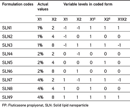
Multiple linear regression (MLR) was carried out to examine the variables influencing the final characteristics of SLNs.31 Nine SLN formulations were produced as per a 32 factorial design to investigate the effect of two input factors, namely tristearin percentages (X1) and homogenization cycle (X2), on the two output factors, namely entrapment efficiency percentage (Q1) and mean particle size (Q2), of the FP-loaded SLNs. Three levels were determined in order to evaluate each factor: -1, 0, and 1. The fitted models’ regression equation for the output variables is presented in Equation 1 below.
Q= bo+b1X1+b2X2+b3X12+b4X22+b5X1X2 (Equation 1)
In the model, Q is the output factor, bo is the arithmetic average value of the tests, and b1, b2, b3, b4, and b5 are the forecasted coefficients for the factors X1 and X2. Nonlinearity is analyzed through the polynomial terms (X12 and X22). The outcomes were investigated statistically using analysis of variance (ANOVA)4. The variable levels and the actual values are tabulated in Table 1.
Table 1. Actual values and variable levels designed through 32 factorial design of FP-loaded SLNs.
Preparation of solid lipid nanoparticle formulations
FP-loaded SLN formulations were manufactured using a high pressure homogenizer. Tristearin was melted and 50 mg of FP was added to the melted lipid. Aqueous Tween 80 (1%) solution was also heated to the same temperature. After an Ultraturrax T25 (IKA, Germany) at 13.500 rpm was used to mix the lipid phase and aqueous phase for 3 min, the hot pre-emulsion was subsequently homogenized by a Microfluidics M110L (USA) at a pressure of 18.000 Psi. Three different tristearin percentages (1%, 2%, and 4%) and three different cycle numbers (2, 4, and 8 cycles) were investigated based on two responses: encapsulation efficiency (Q1) and particle size (Q2). SLN dispersions were centrifuged using a Vivaspin (MWCO=10.000) at 4500 rpm for 30 min (Sigma 3K30, Germany) and then lyophilized.
Particle size and zeta potential analysis
Particle size measurements were obtained by a dynamic light scattering technique. For this purpose, a Malvern Zetasizer (Malvern, UK) was used to measure the mean particle size, polydispersity index, and the zeta potential values of FP-loaded SLNs. Dry powder of lipid nanoparticles was dispersed in ultrapure water before analysis.
Determination of encapsulation efficiency
For the determination of the encapsulation efficiency of SLN formulations, 10 mg of SLN was dissolved in methanol at 75°C. The solution was stirred using a magnetic stirrer in a tightly sealed vial. After that, the solution was ultrasonicated with 50% power for 5 min (Bandelin Sonoplus HD 2070, Germany) and then cooled to room temperature. It was centrifuged at 26.000 rpm for 20 min at 4°C (Sigma 3K30, Germany) and then the supernatant was filtered using a 0.22 µm cellulose acetate membrane filter. The amount of FP was determined using an HPLC system (Agilent 1260 Infinity). Separation was carried out using a NovaPak® C18 column (4 µm, 150x3.9 mm) (Waters, Ireland). The column temperature was set to 35°C. The mobile phase composition was acetonitrile:water (60:40 v/v) and the flow rate was 1 mL/min. A 10-µL sample was injected into the system and the samples were analyzed at a wavelength of 236 nm.
In vitro drug release study
The dialysis bag method was used to determine the in vitro release profile of FP from the SLN formulation. SLN formulation corresponding to 5 mg of FP was placed into hydrated dialysis membranes (MWCO=12-14 kDa, Spectrapor-2). A mixture of 100 mL of phosphate buffered saline pH 7.4 and ethanol (70:30) was used as dissolution medium at 37°C and under constant stirring (100 rpm). The samples were taken at particular times over 24 h. The medium was completely removed and replaced with 100 mL of fresh dissolution medium at each time point to provide a sink condition. Samples taken were filtered through 0.45 µm regenerated cellulose membrane filters and the amount of FP was determined by HPLC.
Transmission electron microscopy (TEM) analysis
The shapes of the lipid nanoparticles were examined by FEI Tecnai G2 S Twin TEM (Osaka, Japan) at an acceleration voltage of 120 kV. After dry powder of lipid nanoparticles was dispersed in ultrapure water the dispersion was dropped on a copper grid.
Differential scanning calorimetry (DSC) analysis
The thermal properties and crystallinity of pure FP, bulk lipid, and FP-loaded SLNs were determined by DSC (Shimadzu DSC-60, Japan). Five milligrams of sample was placed in hermetically sealed aluminum pans and a DSC thermogram was obtained at a scanning rate of 5°C/min while the samples were heated from room temperature to 300°C. Moreover, for the calibration of the instrument indium was used as a reference.
Statistical analysis
All results were statistically analyzed using one-way ANOVA through design expert 6.0.8 software.
RESULTS AND DISCUSSION
Analytical validation of the HPLC method
Linearity
After analyzing the eight different concentration of FP six times by HPLC, the average peak areas were plotted against concentrations. A linear relationship between peak area and concentration was observed. The best linearity was obtained between concentrations of 0.25 and 10 µg/mL in methanol since the correlation coefficient was found to be 0.9996.
Accuracy
After FP solutions in methanol at concentrations of 0.5 µg/mL, 4 µg/mL, and 10 µg/mL were injected 6 times as a test sample, the detector responses were used to calculate the concentrations of FP. The accuracy of the analytical method was determined with the help of the variation coefficient [relative standard deviation (RSD)] of the percent recovery values. Since the RSD values obtained were close to or less than 2%, the method was assumed to be accurate (Table 2).
Table 2. The RSD % values obtained for the analytical validation parameters.

Precision
The repeatability and intermediate precision of the method were evaluated. The repeatability of the method was determined by analysis of 6 repetitive injections of FP-methanol solutions and was shown as the RSD of measured concentrations. The RSD values were less than 2% as can be seen in Table 2. The intermediate precision of the HPLC method was defined by the RSD value of 12 injections performed on two different days and the RSD values were less than 2% (Table 2). On the other hand, there was no statistically significant difference between the means of the measured concentrations obtained on two different days for each FP solution (p>0.05).
LOD and LOQ values
The LOD and LOQ values were calculated in accordance with the equations below. The standard deviation (s) of the response and the slope (m) of the calibration curve were used. While the LOD value was 0.09 µg/mL, the LOQ value was 0.28 µg/mL.
LOD=3.3×s/m
LOQ=10×s/m
Formulation optimization by 32 factorial design
Tristearin percentages were varied (1%, 2%, and 4%). These three different ratios were tested at three different numbers of homogenization cycles: 2, 4, and 8. In this way, nine SLNs were produced as per the 32 factorial design. The magnitude and sign of the main influence indicate the relative effect of each factor on the response by means of polynomial equations. Table 3 gives the predicted and the observed values of responses (Q1, Q2). The predicted values were derived from the equations and the observed values were determined from experimental results.
Table 3. Observed and predicted responses of FP-loaded SLNs.
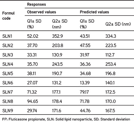
Table 4 shows the results of model coefficients estimated by MLR and the ANOVA of the investigated model for all responses. The quality of the model developed was evaluated based on the regression coefficient values. The determination coefficient (r2 value) for the response Q2 was nearer to 1, indicating that there was a good correlation between the observed and the response measures from the model. The negative sign in front of the coefficients indicated that the response of the nanoparticles increased when the independent factor was decreased, and the positive sign for the coefficients showed the positive effect of the independent factors on the observed replies. The model F-value of 3.87 for Q1 response implied there was a 5.30% probability that a “Model F-Value” of this magnitude could be caused by noise. On the other hand, the “Model F-value” of 57.71 for Q2 response indicated that the model was statistically meaningful. The possibility of such a large “Model F-Value” due to noise is only 0.01%.28,31,32
Table 4. Results of model coefficients estimated by MLR and the ANOVA of the fitted model for all responses.
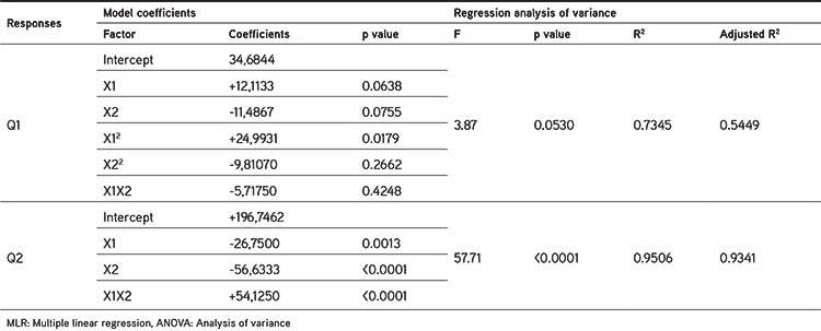
Figure 1 shows the linearity plots between the Q1 and Q2 values. The correlation graphs that show linearity between actual and predicted response variables indicated that the fit to the model was at an excellent level for Q2 (p<0.05), whereas the linear correlation plots showed a low compliance to the model for Q1 (p>0.05) (Figure 1). This situation is also evidenced by the F-value calculated for the Q1 model. The F-value of the Q1 model (F=3.87) is smaller than the tabulated F-value (F tab=4.46). This situation indicates statistical nonsignificance of the model (Figure 1 and also the p values in Table 4).
Figure 1.
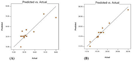
Linearity correlation graphs between actual and predicted values of (A) Q1, (B) Q2
As seen from Table 3, drug entrapment efficiency of all factorial formulations was produced within a broad range of 27.07-94.65%. Drug entrapment efficiency was not affected significantly by the level of X1 or X2 (p>0.05). Generally, as seen in p values that indicated the significance of the coefficients (Table 4), neither of the independent factors (X1 and X2) had a strong effect on the drug entrapment efficiency (Q1) (p>0.05).
When the average size of the SLNs was investigated depending on the variation in homogenization cycles (X2) at each tristearin percentage (X1), it was observed that as X2 increased from 2 to 8 the mean particle size decreased significantly (p<0.05). The average particle size of SLNs ranged from 130.9±3.30 to 352.9±10.93 nm. Generally, from the p values of the coefficients presented in Table 4, it was concluded that both of the investigated variables (X1 and X2) had a major influence on the output Q2 (p<0.05). The biggest average size was observed in the lowest level of X1 (1%) and the lowest level of X2 (2 cycles) in factorial formulation SLN1.
PDI, which is the indicator of homogeneity of the size distribution in colloidal drug delivery systems, is generally expressed as less than 0.3 for narrow size distribution.28,32 The PDI values of all factorial formulations were between 0.181 and 0.497 (Figure 2). It was observed that the factorial formulations that contain tristearin with a percentage of 1 or 2 showed a wide size distribution (PDI >0.2) based on the homogenization cycles investigated except in the formulations that contained 2% tristearin at homogenization cycle 8 (SLN6 coded formulation). As tristearin percentage increased from 1% or 2% to 4%, the PDI values were less than 0.3, indicating a uniform size distribution.
Figure 2.
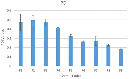
PDI values of the FP-loaded SLN formulations
PDI: Polydispersity index, FP: Fluticasone propiyonat, SLN: Solid lipid nanoparticle
The surface charge of nanosized particles is the potential at the hydrodynamic shear plane and indicates the particle stability in dispersions.31 All of the SLNs exhibited negative surface charge between -19.5 and -29.7 mV. The surface charge of SLNs was not affected significantly by the variation in tristearin percentages or the homogenization cycles (Figure 3).
Figure 3.
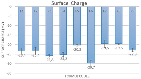
Surface charge of the FP-encapsulated SLN formulations
FP: Fluticasone propiyonat, SLN: Solid lipid nanoparticle
Simplified models were also utilized to draw contour plots for analyzing the effect of independent variables. The contour plots give a diagrammatical demonstration of the values of the response. Since the contour plot of Q1 (Figure 4A) was nonlinear, it demonstrates a nonlinear relationship between input factors. As can be seen from the contour plot of Q2 (Figure 4B), the indicator of the linear relationship between X1 and X2 input factors is the linearity of the graph.
Figure 4.
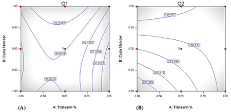
Contour plots of FP-loaded SLNs showing the influence of X1 and X2 on A) Q1, B) Q2
FP: Fluticasone propiyonat, SLN: Solid lipid nanoparticle
According to the release profile study of FP-loaded SLNs as shown in Figure 5, prolonged release was obtained without any initial burst effect. The nature of the lipid matrix affects the release profile of the active substance. It is thought that FP-loaded SLNs formed in a core-shell model with a drug-enriched core. This may be responsible for the slow release.
Figure 5.
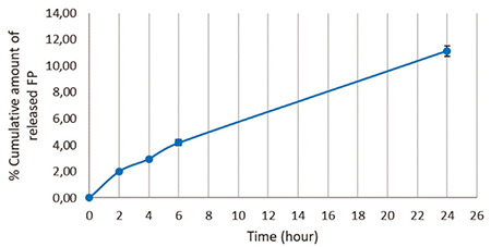
In vitro drug release profile of FP-loaded SLNs
FP: Fluticasone propiyonat, SLN: Solid lipid nanoparticle
TEM micrographs of FP-loaded SLNs are shown in Figure 6. TEM analysis confirmed the colloidal sizes of the FP-loaded SLNs with spherical shapes.
Figure 6.
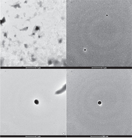
TEM micrograph of the optimal formulation
TEM: Transmission electron microscopy
Differential scanning calorimetry analysis
The DSC thermograms given in Figure 7 show that pure FP is decomposed by a small exothermic peak at 271.72°C. This outcome is in agreement with previous research by El-Gendy et al.33 and Dai et al.34 The peak of the active agent thus observed also indicated that the FP was a crystal structure. When the thermogram of the pure form of tristearin was evaluated, it was seen that tristearin produced a small exothermic shoulder peak at 49.89°C at first and then it gave a large endothermic peak at 60.73°C, which indicated the presence of a crystal structure in tristearin.35 When the thermogram of the optimum SLN was examined, it was seen that the exothermic peak of FP disappeared. This indicated that FP’s crystal structure was turned into an amorphous structure within the SLN matrix. When the optimum formulation’s thermogram was examined for tristearin peaks, it was seen that the exothermic shoulder peak of tristearin disappeared where the main endothermic main peak at 57.02°C remained the same shape with the same sharpness. This situation was interpreted as showing that tristearin in the SLN formulation preserved a large proportion of its crystal structure.
Figure 7.
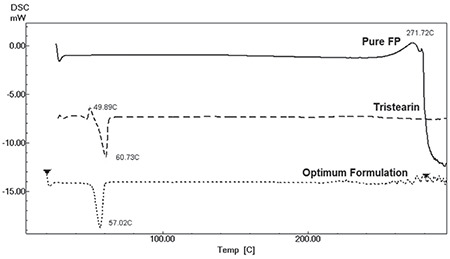
DSC thermograms of pure FP, tristearin, and optimum formulation
DSC: Differential scanning calorimetry, FP: Fluticasone propiyonat
CONCLUSION
FP-loaded SLNs were successfully fabricated using high pressure homogenization. A 32 experimental design and contour plot analysis were used with software to set up the best formulation conditions with a limited number of experiments. This study showed that tristearin percentages and the number of homogenization cycles used in the SLN formulations significantly affected the physico-chemical characteristics of FP-loaded SLNs. According to the factorial design study performed in this research, the optimum formulation could be achieved with the content of 4% tristearin and 4 homogenization cycles.
Footnotes
Conflicts of interest: No conflict of interest was declared by the authors. The authors alone are responsible for the content and writing of the paper.
References
- 1.Brazzini B, Pimpinelli N. New and established topical corticosteroids in dermatology: clinical pharmacology and therapeutic use. Am J Clin Dermatol. 2002;3:47–58. doi: 10.2165/00128071-200203010-00005. [DOI] [PubMed] [Google Scholar]
- 2.Roeder A, Schaller M, Schäfer-Korting M, Korting HC. Safety and efficacy of fluticasone propionate in the topical treatment of skin diseases. Skin Pharmacol Physiol. 2005;18:3–11. doi: 10.1159/000081680. [DOI] [PubMed] [Google Scholar]
- 3.Gual A, Pau-Charles I, Abeck D. Topical corticosteroids in dermatology: from chemical development to galenic innovation and therapeutic trends. J Clin Exp Dermatol Res. 2015;6:1–5. [Google Scholar]
- 4.Doktorovová S, Araújo J, Garcia ML, Rakovský E, Souto EB. Formulating fluticasone propionate in novel PEG-containing nanostructured lipid carriers (PEG-NLC) Colloids Surf B Biointerfaces. 2010;75:538–542. doi: 10.1016/j.colsurfb.2009.09.033. [DOI] [PubMed] [Google Scholar]
- 5.Korting HC, Schöllmann C. Topical fluticasone propionate: intervention and maintenance treatment options of atopic dermatitis based on a high therapeutic index. J Eur Acad Dermatol Venereol. 2012;26:133–140. doi: 10.1111/j.1468-3083.2011.04195.x. [DOI] [PubMed] [Google Scholar]
- 6.Spencer CM, Wiseman LR. Topical fluticasone propionate: a review of its pharmacological properties and therapeutic use in the treatment of dermatological disorders. BioDrugs. 1997;7:318–334. doi: 10.2165/00063030-199707040-00006. [DOI] [PubMed] [Google Scholar]
- 7.Brown MB, Martin GP, Jones SA, Akomeah FK. Dermal and transdermal drug delivery systems: current and future prospects. Drug Deliv. 2006;13:175–187. doi: 10.1080/10717540500455975. [DOI] [PubMed] [Google Scholar]
- 8.Badilli U, Gumustas M, Uslu B, Ozkan SA. Lipid Based Nanoparticles for Dermal Drug Delivery, In: Grumezescu AM, Andrew W, eds. Organic Materials as Smart Nanocarriers for Drug Delivery; Applied Science Publishers-Elsevier; 2018;9:369–413. [Google Scholar]
- 9.Gupta M, Agrawal U, Vyas SP. Nanocarrier-based topical drug delivery for the treatment of skin diseases. Expert Opin Drug Deliv. 2012;9:783–804. doi: 10.1517/17425247.2012.686490. [DOI] [PubMed] [Google Scholar]
- 10.Papakostas D, Rancan F, Sterry W, Blume-Peytavi U, Vogt A. Nanoparticles in dermatology. Arch Dermatol Res. 2011;303:533–550. doi: 10.1007/s00403-011-1163-7. [DOI] [PubMed] [Google Scholar]
- 11.Zhang Z, Tsai PC, Ramezanli T, Michniak-Kohn BB. Polymeric nanoparticles-based topical delivery systems for the treatment of dermatological diseases. Wiley Interdiscip Rev Nanomed Nanobiotechnol. 2013;5:205–218. doi: 10.1002/wnan.1211. [DOI] [PMC free article] [PubMed] [Google Scholar]
- 12.López-Pinto JM, González-Rodríguez ML, Rabasco AM. Effect of cholesterol and ethanol on dermal delivery from DPPC liposomes. Int J Pharm. 2005;298:1–12. doi: 10.1016/j.ijpharm.2005.02.021. [DOI] [PubMed] [Google Scholar]
- 13.Manconi M, Sinico C, Valenti D, Lai F, Fadda AM. Niosomes as carriers for tretinoin. III. A study into the in vitro cutaneous delivery of vesicleincorporated tretinoin. Int J Pharm. 2006;311:11–19. doi: 10.1016/j.ijpharm.2005.11.045. [DOI] [PubMed] [Google Scholar]
- 14.Shakeel F, Haq N, Al-Dhfyan A, Alanazi FK, Alsarra IA. Double w/o/w nanoemulsion of 5-fluorouracil for self-nanoemulsifying drug delivery system. Journal of Molecular Liquids. 2014;200:183–190. [Google Scholar]
- 15.Alvarez-Román R, Naik A, Kalia YN, Guy RH, Fessi H. Skin penetration and distribution of polymeric nanoparticles. J Control Release. 2004;99:53–62. doi: 10.1016/j.jconrel.2004.06.015. [DOI] [PubMed] [Google Scholar]
- 16.Souto EB, Wissing SA, Barbosa CM, Müller RH. Development of a controlled release formulation based on SLN and NLC for topical clotrimazole delivery. Int J Pharm. 2004;278:71–77. doi: 10.1016/j.ijpharm.2004.02.032. [DOI] [PubMed] [Google Scholar]
- 17.Gainza G, Pastor M, Aguirre JJ, Villullas S, Pedraz JL, Hernandez RM, Igartua M. A novel strategy for the treatment of chronic wounds based on the topical administration of rhEGF-loaded lipid nanoparticles: In vitro bioactivity and in vivo effectiveness in healing-impaired db/db mice. J Control Release. 2014;185:51–61. doi: 10.1016/j.jconrel.2014.04.032. [DOI] [PubMed] [Google Scholar]
- 18.Pradhan M, Singh D, Murthy SN, Singh MR. Design, characterization and skin permeating potential of Fluocinolone acetonide loaded nanostructured lipid carriers for topical treatment of psoriasis [published correction appears in Steroids. 2016 Feb;106:93] Steroids. 2015;101:56–63. doi: 10.1016/j.steroids.2015.05.012. [DOI] [PubMed] [Google Scholar]
- 19.Gasco MR. Method for producing solid lipid microspheres having a narrow size distribution. US Patent. 1993;5:250–236. [Google Scholar]
- 20.Müller RH, Radtke M, Wissing SA. Solid lipid nanoparticles (SLN) and nanostructured lipid carriers (NLC) in cosmetic and dermatological preparations. Adv Drug Deliv Rev. 2002;54(Suppl 1):S131–S155. doi: 10.1016/s0169-409x(02)00118-7. [DOI] [PubMed] [Google Scholar]
- 21.Zhai Y, Zhai G. Advances in lipid-based colloid systems as drug carrier for topic delivery. J Control Release. 2014;193:90–99. doi: 10.1016/j.jconrel.2014.05.054. [DOI] [PubMed] [Google Scholar]
- 22.Sala M, Diab R, Elaissari A, Fessi H. Lipid nanocarriers as skin drug delivery systems: Properties, mechanisms of skin interactions and medical applications. Int J Pharm. 2018;535:1–17. doi: 10.1016/j.ijpharm.2017.10.046. [DOI] [PubMed] [Google Scholar]
- 23.Garcês A, Amaral MH, Sousa Lobo JM, Silva AC. Formulations based on solid lipid nanoparticles (SLN) and nanostructured lipid carriers (NLC) for cutaneous use: A review. Eur J Pharm Sci. 2018;112:159–167. doi: 10.1016/j.ejps.2017.11.023. [DOI] [PubMed] [Google Scholar]
- 24.Gastaldi L, Battaglia L, Peira E, Chirio D, Muntoni E, Solazzi I, Gallarate M, Dosio F. Solid lipid nanoparticles as vehicles of drugs to the brain: current state of the art. Eur J Pharm Biopharm. 2014;87:433–444. doi: 10.1016/j.ejpb.2014.05.004. [DOI] [PubMed] [Google Scholar]
- 25.Ganesan P, Narayanasamy D. Lipid nanoparticles: Different preparation techniques, characterization, hurdles, and strategies for the production of solid lipid nanoparticles and nanostructured lipid carriers for oral drug delivery. Sustainable Chemistry and Pharmacy. 2017;6:37–56. [Google Scholar]
- 26.Mehnert W, Mader K. Solid lipid nanoparticles: production, characterization and applications. Advanced Drug Delivery Reviews. 2012;64:83–101. doi: 10.1016/s0169-409x(01)00105-3. [DOI] [PubMed] [Google Scholar]
- 27.Muller RH, Mader K, Gohla S. Solid lipid nanoparticles (SLN) for controlled drug delivey: a review of the state of the art. Eur J Pharm Biopharm. 2000;50:161–177. doi: 10.1016/s0939-6411(00)00087-4. [DOI] [PubMed] [Google Scholar]
- 28.Sengel-Turk CT, Hascicek C. Design of lipid-polymer hybrid nanoparticles for therapy of BPH: Part I. Formulation optimization using a design of experiment. J Drug Deliv Sci Technol. 2017;39:16–27. [Google Scholar]
- 29.Blasi P, Giovagnoli S, Schoubben A, Puglia C, Bonina F, Rossi C, Ricci M. Lipid nanoparticles for brain targeting I. Formulation optimization. Int J Pharm. 2011;419:287–295. doi: 10.1016/j.ijpharm.2011.07.035. [DOI] [PubMed] [Google Scholar]
- 30.Kumar Das S, Yuvaraja K, Khanam J, Nanda A. Formulation development and statistical optimization of Ibuprofen-loaded polymethacrylate microspheres using response surface methodology. Chem Eng Res Des. 2015;96:1–14. [Google Scholar]
- 31.Gu B, Burgess DJ. Prediction of dexamethasone release from PLGA microspheres prepared with polymer blends using a design of experiment approach. Int J Pharm. 2015;495:393–403. doi: 10.1016/j.ijpharm.2015.08.089. [DOI] [PMC free article] [PubMed] [Google Scholar]
- 32.Sengel Turk CT, Oz UC, Serim TM, Hascicek C. Formulation and optimization of nonionic surfactants emulsified nimesulide-loaded PLGA-based nanoparticles by design of experiments. AAPS PharmSciTech. 2014;15:161–176. doi: 10.1208/s12249-013-0048-9. [DOI] [PMC free article] [PubMed] [Google Scholar]
- 33.El-Gendy N, Pornputtapitak W, Berkland C. Nanoparticle agglomerates of fluticasone propionate in combination with albuterol sulfate as dry powder aerosols. Eur J Pharm Sci. 2011;44:522–533. doi: 10.1016/j.ejps.2011.09.014. [DOI] [PMC free article] [PubMed] [Google Scholar]
- 34.Dai J, Ruan BH, Zhu Y, Liang X, Su F, Su W. Preparation of Nanosized Fluticasone Propionate Nasal Spray With Improved Stability and Uniformity. Chem Ind Chem Eng. Q. 2015;21:457–464. [Google Scholar]
- 35.Amasya G, Badilli U, Aksu B, Tarimci N. Quality by design case study 1: Design of 5-fluorouracil loaded lipid nanoparticles by the W/O/W double emulsion - Solvent evaporation method. Eur J Pharm Sci. 2016;84:92–102. doi: 10.1016/j.ejps.2016.01.003. [DOI] [PubMed] [Google Scholar]


