Abstract
Background
People with HIV (PWH) taking antiretroviral therapy (ART) may experience weight gain, dyslipidemia, increased risk of non-AIDS comorbidities, and long-term alteration of the gut microbiota. Both low CD4/CD8 ratio and chronic inflammation have been associated with changes in the gut microbiota of PWH. The antidiabetic drug metformin has been shown to improve gut microbiota composition while decreasing weight and inflammation in diabetes and polycystic ovary syndrome. Nevertheless, it remains unknown whether metformin may benefit PWH receiving ART, especially those with a low CD4/CD8 ratio.
Methods
In the Lilac pilot trial, we recruited 23 nondiabetic PWH receiving ART for more than 2 years with a low CD4/CD8 ratio (<0.7). Blood and stool samples were collected during study visits at baseline, after a 12-week metformin treatment, and 12 weeks after discontinuation. Microbiota composition was analyzed by 16S rDNA gene sequencing, and markers of inflammation were assessed in plasma.
Results
Metformin decreased weight in PWH, and weight loss was inversely correlated with plasma levels of the satiety factor GDF-15. Furthermore, metformin changed the gut microbiota composition by increasing the abundance of anti-inflammatory bacteria such as butyrate-producing species and the protective Akkermansia muciniphila.
Conclusions
Our study provides the first evidence that a 12-week metformin treatment decreased weight and favored anti-inflammatory bacteria abundance in the microbiota of nondiabetic ART-treated PWH. Larger randomized placebo-controlled clinical trials with longer metformin treatment will be needed to further investigate the role of metformin in reducing inflammation and the risk of non-AIDS comorbidities in ART-treated PWH.
Keywords: HIV, metformin, microbiota, nondiabetic, weight
The ability to keep an HIV elite controller status is not permanent. We showed that, independently to protective MHC-I HLA alleles, elevated level of anti-CMV IgG is linked to CD4 T-cell decay in a Canadian cohort of elite controllers.
Isolated from French lilac in the 1920s, metformin has been used for decades to treat type 2 diabetes mellitus (DM2). In diabetic people, metformin promotes euglycemia without the risk of hypoglycemia. Metformin also reduces inflammation, a feature not observed with other antidiabetic drugs such as insulin or sulfonylurea [1]. In addition, metformin has been shown to be beneficial beyond a glucose-lowering effect [2–7]. In women with polycystic ovary syndrome, metformin increased fertility while lowering the rate of the inflammatory cytokines interleukin (IL)-6 and tumor necrosis factor (TNF)–α [6]. In patients with advanced lung adenocarcinoma, a combination therapy of metformin and epidermal growth factor receptor–tyrosine kinase inhibitor (EGFR-TKI) increased survival compared with EGFR-TKIs alone [7]. Metformin has an anti-aging effects in animal models [8, 9]. Lastly, metformin improves the gut microbiota in diabetic rats and has a minimal effect on glucose level when administered intravenously [10, 11]. Depommier et al. showed that metformin was associated with variation in gut microbiota composition, with increased abundances of Escherichia and Akkermansia muciniphila in diabetic subjects [12–16]. Furthermore, in nondiabetic men, metformin was also associated with an increased abundance of Escherichia/Shigella and a decrease of Intestinibacter and Clostridium in stool, without changing glycemic levels [17].
HIV targets and rapidly depletes mucosal CD4 T cells, and as a result, people with HIV (PWH) have impaired gut barrier integrity, leading to translocation of microbial products and systemic inflammation [18–20]. Epithelial gut immune damage also induces modification of defensin and mucosal barrier composition, contributing to a modification of the gut microbiota composition called dysbiosis [21–23]. Gut damage, dysbiosis, and the accompanying persistent inflammation are not resolved by antiretroviral therapy (ART) and contribute to risk of developing non-AIDS comorbidities including dyslipidemia, fatty liver, cardiovascular disease, and cancer [24–26]. Recently, integrase inhibitor–based ART has been shown to contribute to weight gain and development of DM2 through the cytochrome P450 pathway [27]. Non-AIDS comorbidities and weight gain while on ART were more frequently observed in PWH with a low CD4 cell count and low CD4/CD8 ratio [28].
Similarly, gut dysbiosis is associated with a low CD4 count in PWH on ART [29–34]. In PWH, dysbiosis is characterized by an increase of Proteobacteria, Enterobacteria, and Fusobacteria abundances and a decrease in Ruminococcaceae and Lachnospiraceae abundances compared with uninfected controls, Independently of age, sex, and sexual practice [26, 30]. As observed in obese people, a decrease of the gut-protective bacteria A. muciniphila was observed in both ART-naïve and ART-treated PWH [34, 35]. Moreover, gut microbiota–derived metabolites such as short-chain fatty acids (SCFAs) have been shown to protect the gut epithelial barrier and reduce inflammation levels in PWH receiving ART [36–38]. Composition of the gut microbiota has been associated with disease outcome in ART-treated PWH [26, 31, 33, 39, 40].
These findings advocate in favor of repurposing metformin to restore gut microbiota composition while decreasing inflammation in nondiabetic PWH. Given the effect of metformin on the gut microbiota and inflammation in nondiabetic people and its low toxicity, we hypothesize that metformin could decrease weight, improve gut microbiota composition, and reduce inflammation in ART-treated PWH with a low CD4/CD8 ratio [41, 42].
METHODS
Study Design
As part of the Lilac study (CIHR/CTN PT027), 23 PWH on ART were recruited in Montréal and Ottawa (Canada), following the protocol previously reported by our group [41]. In brief, all participants had an HIV RNA plasma viral load <50 copies/mL for a minimum of 3 years. Participants with a CD4/CD8 ratio <0.7 were selected for their increased risk of inflammatory non-AIDS comorbidities [43]. All participants were nondiabetic (HbA1c <6.4%). Participants received 850 mg of metformin (Glucophage; Sanofi-Aventis Canada Inc., Canada) twice daily, except for those taking dolutegravir, who received 500 mg twice daily due to a known drug–drug interaction [41, 44]. Blood and stool samples were collected at baseline (V1), after 12 weeks of metformin intake (V2), and 12 weeks after metformin discontinuation (V3). Clinical data were extracted from study charts, including participants’ demographics, weight and waist circumference at study visits, and clinical lab measurements, including CD4 and CD8 count, fasting glucose, and percentage of HbA1c. Plasma and serum were isolated by centrifugation and frozen at –80°C until used. Stools were collected by participants, kept at 4°C for a maximum of 24 hours, and then frozen at –80°C until used.
Clinical Assessments
HIV viral load (VL) in plasma was quantified by the Abbott RealTime HIV-1 assay (Abbott Laboratories, Abbott Park, IL, USA).
Quantification of Soluble Markers of Gut Damage and Inflammation
Plasma levels of I-FABP, REG3α, sCD14, Lipopolysaccharide (LPS) binding protein (LBP), GDF-15, and Ghrelin were quantified by enzyme-linked immunosorbent assay (ELISA; Hycult Biotech, Uden, the Netherlands, or R&D Systems, Minneapolis, MN, USA). LPS was measured by ELISA (Cusabio, Wuhan, China). (1→3)-β-D-glucan (βDG) was measured using the Fungitell assay (Associates of Cape Cod, Inc., East Falmouth, MA, USA). All measures were performed in duplicate as per manufacturers’ instructions.
Microbiota Composition Analysis in Stools
DNA was extracted from ~1 g of stool from each sample using a commercialized kit (Qiagen, Toronto, ON, Canada) adding Mutanolysin and Lysosyme to the lysis buffer (Sigma Aldrich, Oakville, ON, Canada) in the presence of silica beads (Biospec/Cole parmer Canada, Montréal, QC, Canada) for 1 hour at 37°C. Samples were then lysed mechanically on a bead beater at 2000 rpm for 2 minutes. DNA extraction was then performed according to the provided Qiagen protocol.
Extracted DNA samples were used for 16S rRNA gene amplification of the V3–V4 region, adapted to incorporate the transposon-based Illumina Nextera adapters (Illumina, San Diego, CA, USA) and a sample barcode, as previously described [45]. Cutadapt was used to trim forward and reverse primers from 16 S rRNA gene amplicons [46]. Paired-end reads generated from 16S rRNA gene sequencing were filtered and analyzed using DADA2 (version 1.10.1) [47]. Taxonomic assignment of amplicon sequence variants (ASVs) was performed against the Silva database 132 [48] using the RDP classifier algorithm (version 2.2) [49]. Samples were rarefied to an even sampling depth of 14 405 sequences in order to normalize sampling effort.
SCFA Composition Analysis
Serum levels of SCFA were analyzed by automated online solid phase extraction high-performance liquid chromatography–tandem mass spectrometry as previously reported [50].
Statistical Analysis
Descriptive analyses were conducted using GraphPad Prism 8.3 (La Jolla, CA, USA). Nonparametric 1-way analyses of variance were performed using Friedman’s test. Multivariate analyses were performed with SPSS (IBM).
Microbiota composition analysis was performed in R (http://www.R-project.org). α-diversity was assessed using Shannon’s and Simpson’s reciprocal indexes, calculated using the phyloseq package (version 1.22) [51]. Statistical significance of differentially abundant features was assessed with a differential abundance analysis (DESeq2) R package (Benjamin-Hochberg procedure) [52] and a linear discriminant analysis effect size (LEfSe) [53]. Comparison of log2 abundances and soluble factors was performed using Spearman’s tests.
A P value of <.05 and a linear discriminant analysis (LDA) score >2.5 were considered statistically significant.
Patient Consent Statement
This study was approved by the Research Ethics Boards of the Research Institute of the McGill University Health Centre (MUHC; number MP-37-2016-2456) and by the Health Canada Therapeutic Products Directorate. The study was also approved by the Internal Review Board (IRB) of the Ottawa Hospital Research Institute, Ontario, Canada (IRB No. 20160433-01H) and CHUM Research Centre, Montréal, Québec, Canada (IRB No. 17.074). This study was conducted in accordance with the Declaration of Helsinki. Each participant provided written informed consent before entering the study (Canadian CIHR/CTN protocol CTNPT027; trial registration: NCT02659306).
RESULTS
Participant Characteristics
From a total of 23 participants, 2 were female and 21 were male. The median age was 56 years, ranging from 41 to 69. Eight out of the 23 participants defined themselves as of African origin, while the others defined themselves as Caucasian. The median CD4 count (range) was 435 (141–1082) cells/mm3 of blood, the median CD8 count was 729 (351–1867) cells/mm3, and the median CD4/CD8 was 0.6 (0.2–0.7). All participants were receiving ART for a median (range) of 10 (3–25) years and had a viral load <50 copies/mL (Supplementary Table 1). No participants were treated with antibiotics, and no changes in ART or other medications occurred during the study.
Metformin Use Appeared Safe in Nondiabetic PWH
Out of the 23 participants, no serious adverse events were reported. One participant presented with stomach cramps, generalized muscle aches, and loose stools 2 weeks after starting metformin and chose to discontinue medication (Supplementary Table 1).
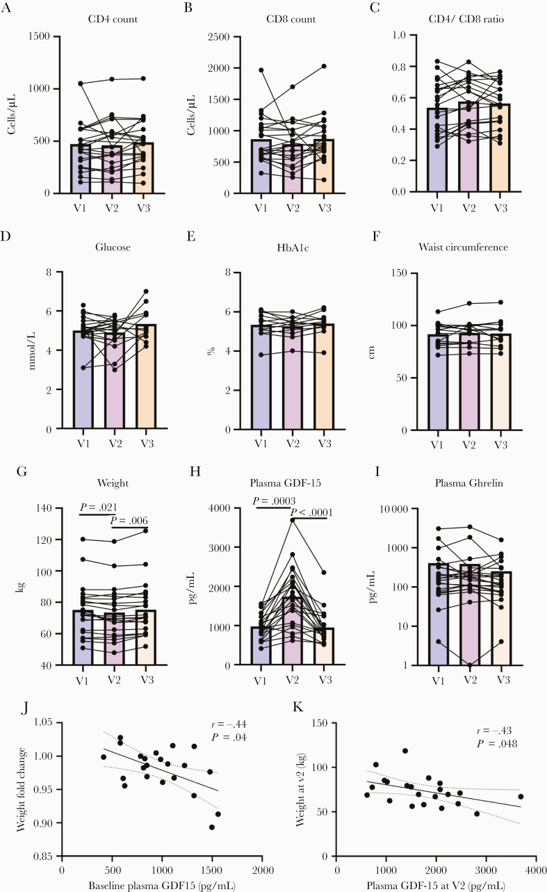
In the 22 participants who completed the study, CD4 count, CD8 count, and CD4/CD8 ratio were not influenced by metformin (Figure 1A–C). All participants had a viral load <50 copies/mL at all study visits.
Figure 1.
Metformin decreased participant weight in association with an increase in plasma GDF15 levels without modifying glucose levels, CD4 T-cell counts, or CD8 T-cell counts. CD4 (A) and CD8 (B) T-cell counts and CD4/CD8 ratio (C) were measured at each study visit. Fasting glucose levels (D) and percentage of glycosylated hemoglobin (HbA1c) (E) were quantified in blood. Waist circumference (F) and weight (G) were measured at each visit. GDF-15 (H) and Ghrelin levels (I) were quantified in plasma. Weight change after metformin correlated inversely with baseline plasma GDF-15 (J) (Spearman’s test). After metformin treatment, weight correlated inversely with plasma GDF-15 levels (K) (Spearman’s test). Bars represent mean values. Abbreviations: V1, baseline; V2, after 12 weeks of metformin treatment; V3, 12 weeks after metformin discontinuation.
Metformin Decreased Participant Weight Independent of a Glucose-Lowering Effect, in Association With Increased Plasma GDF-15 Levels
Fasting glucose, percentage of HbA1c, and waist circumference were not affected by metformin (Figure 1D–F). However, participants experienced weight loss after metformin intake (median loss [range], 1.6 [–8 to +2.3]; P = .021; 1.4% of participants’ weight) (Figure 1G; Supplementary Figure 1). Weight returned to the baseline level 3 months after metformin discontinuation. Weight variation was observed in absence of changes in participants’ diet and physical activity. After metformin treatment, plasma levels of GDF-15 were increased (fold change, 1.8; P = .0003) (Figure 1H) and returned to baseline levels after discontinuation. Conversely, plasma levels of the “hunger hormone” Ghrelin remained stable throughout the study (Figure 1I). Interestingly, baseline plasma levels of GDF-15 but not Ghrelin negatively correlated with weight loss after metformin use (r = –.44; P = .04) (Figure 1J; Supplementary Figure 2). Moreover, plasma levels of GDF-15 but not Ghrelin after metformin treatment (V2) negatively correlated with weight at the same study visit (r = –.43; P = .048) (Figure 1K; Supplementary Figure 2). Multivariate analysis showed that the weight loss associated with GDF-15 increase was independent of age, sex, and use of integrase inhibitors. Hence, metformin induced weight loss in participants independent of a glucose-lowering effect in association with a GDF-15 increase in nondiabetic PWH.
Metformin Modified Gut Microbiota Composition
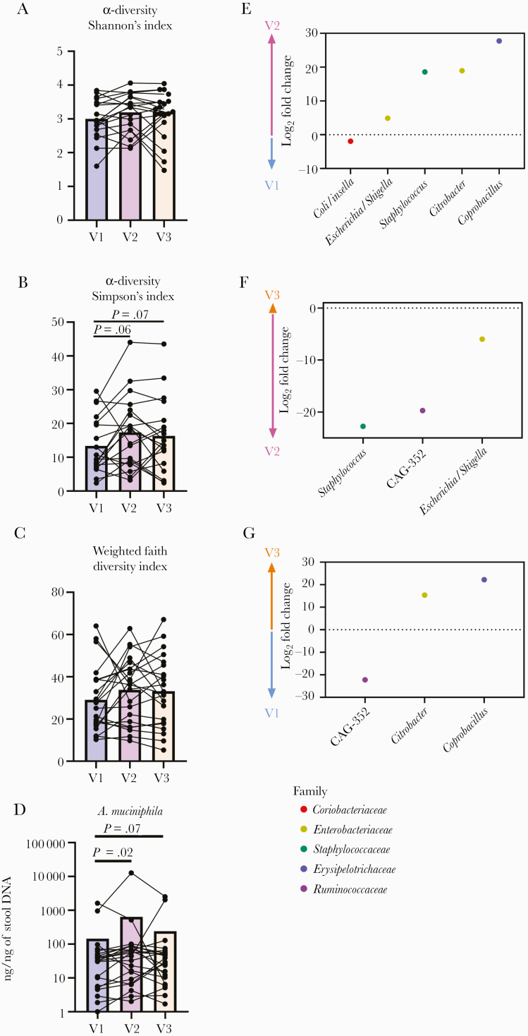
We observed no variation in α-diversity with Shannon’s index (Figure 2A) and a tendency toward an increase in diversity with the Simpson’s reciprocal index and the weighted faith diversity index (Figure 2B, C) after metformin intake and metformin discontinuation.
Figure 2.
Metformin modified the gut microbiota composition in nondiabetic antiretroviral therapy–treated people with HIV. Bacterial diversity in stools was analyzed by Shannon’s (A) and Simpson’s (B) indexes as well as the weighted faith diversity index (C). Abundance of Akkermansia muciniphila was analyzed by quantitative polymerase chain reaction in stool DNA (D) (Friedman’s test). Figure 2, E–G, depicts significant variations of the abundance of detected bacteria in stools at each visit as analyzed by DESeq2. The x-axis indicates the genus of the detected bacteria. Abbreviations: V1, baseline (blue); V2, after 12 weeks of metformin treatment (purple); V3, 12 weeks after metformin discontinuation (orange).
Abundance of A. muciniphila was significantly increased after metformin treatment (P = .02) and tended to remain elevated after metformin discontinuation (P = .07) (Figure 2D).
As depicted in Supplementary Figure 3, fecal bacterial profiles were fairly similar between visits. After metformin treatment, DESeq2 analyses showed a decrease of Collinsella and an increase of Escherichia/Shigella, Staphylococcus, Citrobacter, and Coprobacillus abundances (Figure 2E). Abundances of Staphylococcus, Clostridium CAG-352, and Escherichia/Shigella were decreased between V2 and V3 (Figure 2F). CAG352 abundance was lower at baseline compared with after discontinuation, while abundances of Citrobacter and Coprobacillus were higher after discontinuation than at baseline (Figure 2G). Moreover, LEfSe analysis showed an increase in Lachnoclostridium after metformin intake (Supplementary Figure 4). Abundance of Lachnospiraceae_NK4A136 was increased only after metformin discontinuation as compared with the baseline value (Supplementary Figure 4). We hypothesized here that metformin treatment modified stool microbiota composition in nondiabetic ART-treated PWH.
Metformin Use Was Associated With Increased Blood Levels of SCFA
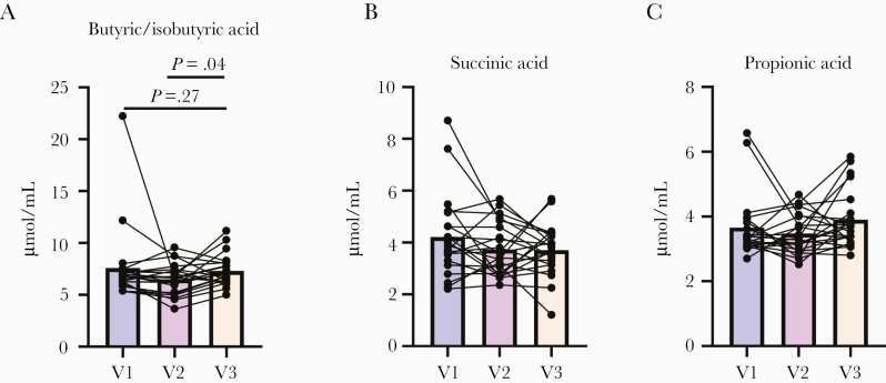
Increased abundance of A. muciniphila was linked with increased colonization of SCFA- producing bacteria, notably butyric acid [54–56]. Serum SCFA levels were compared, and butyric/isobutyric acid levels were found to be higher after metformin discontinuation as compared with metformin treatment (P = .02) (Figure 3A). Levels of succinic and propionic acids were stable during the study (Figure 3B, C).
Figure 3.
Variation of serum levels of short-chain fatty acids in nondiabetic people with HIV after metformin treatment. Butyric/isobutyric acid (A), succinic acid (B), and propionic acid (C) were quantified in serum (Friedman’s tests). Abbreviations: V1, baseline; V2, after 12 weeks of metformin treatment; V3, 12 weeks after metformin discontinuation.
Gut Damage and Microbial Translocation Were Not Changed With Metformin Treatment, but Inflammatory Response Was Decreased After Metformin Discontinuation
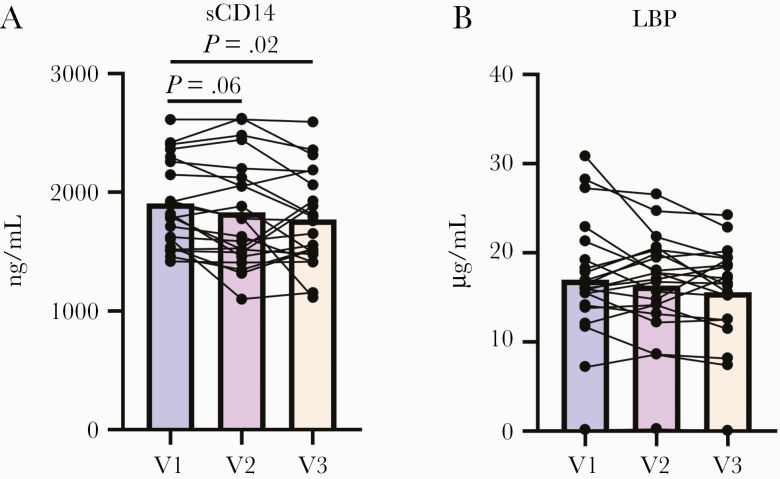
Levels of the gut damage marker REG3α, as well as and LPS and BDG, were stable across the 3 study visits (Supplementary Figure 5). Circulating levels of sCD14 tended to decrease after metformin treatment (P = .06) and further decreased after metformin discontinuation (P = .02) compared with baseline levels (Figure 4A). LBP levels tended to decrease after metformin discontinuation as compared with baseline (P = .13) (Figure 4B). Plasma levels of IL-6 and TNF-α were not affected by metformin treatment or its discontinuation (data not shown).
Figure 4.
Variation of plasma levels of sCD14 and LPS binding protein (LBP) after metformin treatment. Plasma levels of soluble CD14 (A) decreased at V3. Plasma levels of LBP (B) tended to decrease after metformin discontinuation (Friedman’s tests). Abbreviations: V1, baseline; V2, after 12 weeks of metformin treatment; V3, 12 weeks after metformin discontinuation.
Modification of the Gut Microbiota was Associated With Weight Loss and Decreased sCD14 Levels
As indicated in Table 1, abundances of Lachnospiraceae_g and other species were associated with weight loss after metformin treatment, while abundances of Prevotella_7 and other bacteria were associated with weight gain after metformin.
Table 1.
Association Between Gut Microbiota Composition and Weight, Plasma GDF-15, and sCD14 Variation After Metformin Treatment
| Stool Microbiota Composition at V2 | Fold Change at V2 | ||||||
|---|---|---|---|---|---|---|---|
| Class | Order | Family | Genus | Species | Weight | GDF-15 | sCD14 |
| Bacteroidia | Bacteroidales | Bacteroidaceae | Bacteroides | Fragilis | –0.24 | 0.47 * | 0.62 ** |
| Bacteroides | Thetaiotaomicron | –0.38 | 0.53 * | 0.30 | |||
| Muribaculaceae | Muribaculaceae_g | Muribaculaceae_g_s | 0.11 | –0.43 * | –0.13 | ||
| Prevotellaceae | Prevotella_7 | Prevotella_7_s | 0.42 * | –0.40 | –0.28 | ||
| Prevotellaceae_g | Prevotellaceae_g_s | 0.14 | –0.48 * | –0.37 | |||
| Rikenellaceae | Alistipes | Alistipes_s | –0.50 * | 0.27 | 0.44 * | ||
| Alistipes | Putredinis | –0.23 | 0.43 * | –0.20 | |||
| Rikenellaceae_RC9_gut_group | Rikenellaceae_RC9_gut_group_s | 0.22 | –0.20 | –0.49 * | |||
| Clostridia | Clostridiales | Lachnospiraceae | Blautia | Obeum | 0.44 * | 0.01 | 0.05 |
| CAG-56 | CAG-56_s | 0.19 | 0.55 ** | 0.32 | |||
| Hungatella | Hathewayi | –0.45 * | 0.21 | 0.28 | |||
| Lachnospiraceae_g | Lachnospiraceae_g_s | –0.43 * | 0.01 | 0.35 | |||
| Lachnospiraceae_ND3007_group | Lachnospiraceae_ND3007_group_s | 0.05 | –0.43 * | 0.11 | |||
| Lachnospira | Lachnospira_s | 0.02 | –0.54 ** | –0.04 | |||
| Ruminococcaceae | Faecalibacterium | CM04-06 | 0.10 | –0.42 * | –0.12 | ||
| Oscillibacter | Oscillibacter_s | –0.52 * | 0.09 | 0.23 | |||
| Ruminiclostridium_5 | Ruminiclostridium_5_s | 0.25 | –0.65 ** | –0.07 | |||
| Negativicutes | Selenomonadales | Acidaminococcaceae | Phascolarctobacterium | Faecium | –0.44 * | 0.00 | –0.03 |
| Alphaproteobacteria | Rhodospirillales | Rhodospirillales_f | Rhodospirillales_f_g | Rhodospirillales_f_g_s | –0.18 | 0.51 * | 0.12 |
| Deltaproteobacteria | Desulfovibrionales | Desulfovibrionaceae | Bilophila | Wadsworthia | 0.44 * | 0.00 | 0.07 |
| Gamma proteobacteria | Betaproteobacteriales | Burkholderiaceae | Parasutterella | Excrementihominis | –0.47 * | 0.01 | 0.25 |
| Betaproteobacteriales | Burkholderiaceae | Sutterella | Sutterella_s | 0.04 | –0 .45 * | –0.22 | |
Abbreviation: V2, after metformin treatment.
Significant values are indicated in bold. *P < .05; **P < .01; ***P < .001. Spearman’s test.
Abundances of Bacteroides fragilis, Thetaiotaomicron, and Lachnospiraceae CAG-56 were associated with increased levels of GDF-15 in plasma after metformin treatment, while abundances of Prevotellaceae_g, Lachnospira, and others were negatively associated with GDF-15 level fold changes after metformin treatment (Table 1). Finally, abundances of Bacteroides fragilis and Alistipes also correlated positively with plasma sCD14 fold change. (Table 1). These results suggest that a complex network of bacteria, including Bacteroidales and Lachnospiraceae, might influence weight and inflammation in ART-treated PWH.
DISCUSSION
In this pilot study, we investigated for the first time the influence of metformin on weight, inflammation, and gut microbiota composition in nondiabetic ART-treated PWH. We found that metformin decreased weight and modified gut microbiota composition without modifying glucose levels, CD4 and CD8 T-cell counts, or CD4/CD8 ratio.
Weight gain and obesity are escalating global health concerns contributing to morbidity via increased risk of cardiovascular disease, diabetes, nonalcoholic steatohepatitis, and cancer. In ART-treated PWH, similar comorbidities have been observed, and weight gain associated with ART is under investigation [28, 57–64]. Integrase inhibitor–containing regimens have been shown to increase weight, and several cofactors have been identified such as low CD4 nadir, being of African origin, and being female. Repurposing metformin to prevent weight gain in nondiabetic ART-treated PWH might reduce risks of non-AIDS comorbidities.
Metformin decreased participants’ weight with no diet or physical activity modification. Weight is regulated in part through a neuroendocrine axis with Ghrelin, a hunger hormone, counterbalanced by GDF-15, a growth factor that promotes satiety. Weight loss was associated with increased levels of GDF-15, but not Ghrelin. In line with our findings, Coll et al. showed that metformin-induced weight loss was dependent on increased production of GDF-15 in the colon and liver, independent of a glucose-lowering effect, in mice. Moreover, metformin has been shown to decrease weight in diabetic and/or obese people in association with increased GDF-15 [65, 66]. Interestingly, in our study, we found that plasma levels of GDF-15 returned to baseline levels 12 weeks after metformin discontinuation, concomitant with participants’ weight. Thus, the dynamics of GDF-15 before, during, and after metformin discontinuation favor a direct role of metformin on weight loss in nondiabetic people.
Variations in weight were also associated with gut microbiota composition. The Prevotellaceae family was associated with weight gain and GDF-15 decrease after metformin treatment. The Prevotellaceae and Desulfovibrionaceae families have been shown to be associated with inflammation and disease progression in men who have sex with men [26, 29–31, 33, 36, 67]. The SCFA-producing Lachnospiraceae family appears to have a role in both weight and GDF-15 modulation. In obese mice, butyrate production was shown to decrease weight through a gut–brain axis [68]. The influence of the gut microbiota, SCFA, and GDF-15 on weight change should be further studied.
Gut microbiota composition has been shown to differ in ART-treated PWH compared with uninfected controls [30]. Notably, abundance of butyrate-producing bacteria, mostly from the Lachnospiraceae family, has been shown to be decreased in PWH [29, 30]. Herein, we have shown that metformin was associated with increased levels of butyrate in the blood after metformin discontinuation. Decreased abundance of A. muciniphila has been associated with epithelial gut damage, inflammation, and metabolic disorders in diabetic people [16, 69]. Herein, we showed that metformin increased abundance of A. muciniphila in ART-treated PWH [11, 70]. Metformin increasing A. muciniphila abundance might support other species, including SCFA-producing bacteria [71, 72]. Metformin also increased abundance of Escherichia/Shigella in ART-treated PWH, which has been previously observed in diabetic people and healthy men [14, 17, 73]. Abundance of Escherichia/Shigella was associated with gastrointestinal symptoms in healthy men taking metformin, a contribution that requires further assessment in PWH. Overall, metformin appears to induce moderate but potentially beneficial shifts in nondiabetic ART-treated PWH by increasing abundance of SCFA-producing bacteria and A. muciniphila.
Increased levels of SCFA were observed after metformin discontinuation, along with a decrease of the pro-inflammatory marker sCD14, secreted by antigen-presenting cells upon detection of bacterial LPS. Shikuma et al. showed in 6 nondiabetic PWH that metformin decreased CD4 T-cell expression of the marker of cell exhaustion programmed cell death–1 (PD-1) but not T-cell activation markers CD38 and HLA-DR [74]. Butyrate has been shown to decrease the LPS-induced pro-inflammatory response of antigen-presenting cells [75]. In agreement with those results, we observed that Lachnospiraceae abundance correlated negatively with sCD14 levels, while circulating levels of LPS were not modified by metformin. Conversely, Bacteroides fragilis abundance was associated with increased levels of sCD14 after metformin treatment. Bacteroides fragilis was hypothesized to restore CD4 T-cell expansion and promote anti-inflammatory responses [76]. Moreover, increased abundance of Bacteroides fragilis and bacteria from the Erysipelotrichaceae family were associated with immune responses against colon cancer, whose risks are higher in PWH than in uninfected people [77–79]. Hence, the specific role of bacterial species in the gut microbiota must be further studied in PWH.
We acknowledge several limitations to our analysis, as our pilot study included 1 arm only. Future studies should include a placebo arm, accounting for diet and physical activity. As weight gain was predominantly observed in women of African descent taking ART, a comprehensive view of metformin’s effect on this population will be required in large randomized controlled trials. In addition, animal models would help in deciphering the mechanisms of metformin-induced microbiota modification and its influence on weight and inflammation.
CONCLUSIONS
Our study showed that metformin may be beneficial in nondiabetic ART-treated PWH by decreasing people’s weight and possibly favoring a more anti-inflammatory microbiota composition. Further, weight modulation by metformin was associated with increased GDF-15, an effect that was reversible after metformin discontinuation.
Metformin appears to mimic fasting/caloric restriction without reducing glucose levels in PWH [8]. Such a mechanism has been shown to be beneficial partially through modification of the gut microbiota [80, 81]. Hence, long-term use of metformin might decrease inflammation and thereby decrease the risk of developing non-AIDS comorbidities [42].
The extended-release form of metformin appeared to reduce weight and inflammation, with fewer side effects than the immediate-release metformin in diabetic people [82], representing a promising avenue in nondiabetic PWH. The duration and optimal metformin dosage will have to be defined before implementing a large randomized controlled trial. Our findings pave the way toward repurposing metformin to decrease non-AIDS comorbidities in ART-treated PWH.
Supplementary Data
Supplementary materials are available at Open Forum Infectious Diseases online. Consisting of data provided by the authors to benefit the reader, the posted materials are not copyedited and are the sole responsibility of the authors, so questions or comments should be addressed to the corresponding author.
Acknowledgments
The authors are grateful to the study participants for their contribution. We thank Angie Massicotte, Josée Girouard, Rosalie Ponte, Cynthia Dion, and Beatrice Choi for study coordination and support.
Financial support. This work was supported by the Vaccines & Immunotherapies core of the HIV clinical trial network from the Canadian Institute for Health Research (CIHR CTN PT07), a CIHR grant (MOP 103230 and PTJ 166049), a CanCURE 2.0 CIHR grant (HB2-164064). Dr Stephane Isnard is supported by a Fond de Recherche Quebec Sante fellowship and a CIHR/CTN Postdoctoral Fellowship Award.
Potential conflicts of interest. Y.Z. and M.F. are employees of Associates of Cape Cod, Inc., the manufacturers of Fungitell, the (1→3)-β-d-glucan in vitro diagnostic kit. All other authors: no reported conflicts. All authors have submitted the ICMJE Form for Disclosure of Potential Conflicts of Interest. Conflicts that the editors consider relevant to the content of the manuscript have been disclosed.
Author contributions. S.I. performed the experiments, analyzed the data, wrote the first draft, and revised the final draft of the manuscript. J.L., B.F., J.O., T.V.V., C.R., A.M., R.R., D.P., L.R.-M., M.M., C.P.V.d.L., I.K., D.S.A., Y.Z., M.F., B.R., and P.A. contributed to the experiments, data analysis, and critical review of the first and final drafts of the manuscript. T.V.V., C.R., A.M., R.R., M.M., and B.R. were involved in microbiota composition analysis. J.A. recruited and followed participants in Ottawa, Canada. J.P.R. designed the study, contributed to data analysis, and critically reviewed the first and final drafts of the manuscript. All authors have read and approved the content of this manuscript.
Prior presentation. Data were partially presented at the HIV Microbiome Workshop 2019 in Washington, DC (oral presentation), CROI 2020 in Boston, Massachusetts (poster), and AIDS2020 (virtual; poster).
Trial registration. NCT02659306.
References
- 1. Harper W, Clement M, Goldenberg R, et al. . Policies, guidelines and consensus statements: pharmacologic management of type 2 diabetes-2015 interim update. Can J Diabetes 2015; 39:250–2. [DOI] [PubMed] [Google Scholar]
- 2. Pollak M. The effects of metformin on gut microbiota and the immune system as research frontiers. Diabetologia 2017; 60:1662–7. [DOI] [PubMed] [Google Scholar]
- 3. Kim SA, Choi HC. Metformin inhibits inflammatory response via AMPK-PTEN pathway in vascular smooth muscle cells. Biochem Biophys Res Commun 2012; 425:866–72. [DOI] [PubMed] [Google Scholar]
- 4. Hirsch HA, Iliopoulos D, Struhl K. Metformin inhibits the inflammatory response associated with cellular transformation and cancer stem cell growth. Proc Natl Acad Sci U S A 2013; 110:972–7. [DOI] [PMC free article] [PubMed] [Google Scholar]
- 5. Li SN, Wang X, Zeng QT, et al. . Metformin inhibits nuclear factor kappaB activation and decreases serum high-sensitivity C-reactive protein level in experimental atherogenesis of rabbits. Heart Vessels 2009; 24:446–53. [DOI] [PubMed] [Google Scholar]
- 6. Victor VM, Rovira-Llopis S, Bañuls C, et al. . Metformin modulates human leukocyte/endothelial cell interactions and proinflammatory cytokines in polycystic ovary syndrome patients. Atherosclerosis 2015; 242:167–73. [DOI] [PubMed] [Google Scholar]
- 7. Arrieta O, Barron F, Padilla MS, et al. . Effect of metformin plus tyrosine kinase inhibitors compared with tyrosine kinase inhibitors alone in patients with epidermal growth factor receptor-mutated lung adenocarcinoma: a phase 2 randomized clinical trial. JAMA Oncol 2019; 5:e192553. [DOI] [PMC free article] [PubMed] [Google Scholar]
- 8. Onken B, Driscoll M. Metformin induces a dietary restriction-like state and the oxidative stress response to extend C. elegans healthspan via AMPK, LKB1, and SKN-1. PLoS One 2010; 5:e8758. [DOI] [PMC free article] [PubMed] [Google Scholar]
- 9. Martin-Montalvo A, Mercken EM, Mitchell SJ, et al. . Metformin improves healthspan and lifespan in mice. Nat Commun 2013; 4:2192. [DOI] [PMC free article] [PubMed] [Google Scholar]
- 10. Sum CF, Webster JM, Johnson AB, et al. . The effect of intravenous metformin on glucose metabolism during hyperglycaemia in type 2 diabetes. Diabet Med 1992; 9:61–5. [DOI] [PubMed] [Google Scholar]
- 11. Shin NR, Lee JC, Lee HY, et al. . An increase in the Akkermansia spp. population induced by metformin treatment improves glucose homeostasis in diet-induced obese mice. Gut 2014; 63:727–35. [DOI] [PubMed] [Google Scholar]
- 12. Cani PD, Bibiloni R, Knauf C, et al. . Changes in gut microbiota control metabolic endotoxemia-induced inflammation in high-fat diet-induced obesity and diabetes in mice. Diabetes 2008; 57:1470–81. [DOI] [PubMed] [Google Scholar]
- 13. Pilon G, Dallaire P, Marette A. Inhibition of inducible nitric-oxide synthase by activators of AMP-activated protein kinase: a new mechanism of action of insulin-sensitizing drugs. J Biol Chem 2004; 279:20767–74. [DOI] [PubMed] [Google Scholar]
- 14. Forslund K, Hildebrand F, Nielsen T, et al. ; MetaHIT Consortium Disentangling type 2 diabetes and metformin treatment signatures in the human gut microbiota. Nature 2015; 528:262–6. [DOI] [PMC free article] [PubMed] [Google Scholar]
- 15. Wu H, Esteve E, Tremaroli V, et al. . Metformin alters the gut microbiome of individuals with treatment-naive type 2 diabetes, contributing to the therapeutic effects of the drug. Nat Med 2017; 23:850–8. [DOI] [PubMed] [Google Scholar]
- 16. Depommier C, Everard A, Druart C, et al. . Supplementation with Akkermansia muciniphila in overweight and obese human volunteers: a proof-of-concept exploratory study. Nat Med 2019; 25:1096–103. [DOI] [PMC free article] [PubMed] [Google Scholar]
- 17. Bryrup T, Thomsen CW, Kern T, et al. . Metformin-induced changes of the gut microbiota in healthy young men: results of a non-blinded, one-armed intervention study. Diabetologia 2019; 62:1024–35. [DOI] [PMC free article] [PubMed] [Google Scholar]
- 18. Ramendra R, Isnard S, Mehraj V, et al. . Circulating LPS and (1→3)-β-D-glucan: a folie à deux contributing to HIV-associated immune activation. Front Immunol 2019; 10:465. [DOI] [PMC free article] [PubMed] [Google Scholar]
- 19. Brenchley JM, Price DA, Schacker TW, et al. . Microbial translocation is a cause of systemic immune activation in chronic HIV infection. Nat Med 2006; 12:1365–71. [DOI] [PubMed] [Google Scholar]
- 20. Marchetti G, Tincati C, Silvestri G. Microbial translocation in the pathogenesis of HIV infection and AIDS. Clin Microbiol Rev 2013; 26:2–18. [DOI] [PMC free article] [PubMed] [Google Scholar]
- 21. Coretti L, Natale A, Cuomo M, et al. . The interplay between defensins and microbiota in Crohn’s disease. Mediators Inflamm 2017; 2017:8392523. [DOI] [PMC free article] [PubMed] [Google Scholar]
- 22. De Smet K, Contreras R. Human antimicrobial peptides: defensins, cathelicidins and histatins. Biotechnol Lett 2005; 27:1337–47. [DOI] [PubMed] [Google Scholar]
- 23. Meade KG, O’Farrelly C. β-defensins: farming the microbiome for homeostasis and health. Front Immunol 2018; 9:3072. [DOI] [PMC free article] [PubMed] [Google Scholar]
- 24. Alzahrani J, Hussain T, Simar D, et al. . Inflammatory and immunometabolic consequences of gut dysfunction in HIV: parallels with IBD and implications for reservoir persistence and non-AIDS comorbidities. EBioMedicine 2019; 46:522–31. [DOI] [PMC free article] [PubMed] [Google Scholar]
- 25. Hsu DC, Sereti I. Serious non-AIDS events: therapeutic targets of immune activation and chronic inflammation in HIV infection. Drugs 2016; 76:533–49. [DOI] [PMC free article] [PubMed] [Google Scholar]
- 26. Vujkovic-Cvijin I, Sortino O, Verheij E, et al. . HIV-associated gut dysbiosis is independent of sexual practice and correlates with noncommunicable diseases. Nat Commun 2020; 11:2448. [DOI] [PMC free article] [PubMed] [Google Scholar]
- 27. Hill A, McCann KM, Pilkington V, et al. . Risks of metabolic syndrome, diabetes and cardiovascular disease in advance trial [a bstract 00081 ]. Paper presented at: Conference on Retroviruses and Opportunistic Infection; March 8–11, 2020; Boston, MA. [Google Scholar]
- 28. Sax PE, Erlandson KM, Lake JE, et al. . Weight gain following initiation of antiretroviral therapy: risk factors in randomized comparative clinical trials. Clin Infect Dis. In press. [DOI] [PMC free article] [PubMed] [Google Scholar]
- 29. Vujkovic-Cvijin I, Dunham RM, Iwai S, et al. . Dysbiosis of the gut microbiota is associated with HIV disease progression and tryptophan catabolism. Sci Transl Med 2013; 5:193ra91. [DOI] [PMC free article] [PubMed] [Google Scholar]
- 30. Vujkovic-Cvijin I, Somsouk M. HIV and the gut microbiota: composition, consequences, and avenues for amelioration. Curr HIV/AIDS Rep 2019; 16:204–13. [DOI] [PMC free article] [PubMed] [Google Scholar]
- 31. Lu W, Feng Y, Jing F, et al. . Association between gut microbiota and CD4 recovery in HIV-1 infected patients. Front Microbiol 2018; 9:1451. [DOI] [PMC free article] [PubMed] [Google Scholar]
- 32. Neff CP, Krueger O, Xiong K, et al. . Fecal microbiota composition drives immune activation in HIV-infected individuals. EBioMedicine 2018; 30:192–202. [DOI] [PMC free article] [PubMed] [Google Scholar]
- 33. Nowak P, Troseid M, Avershina E, et al. . Gut microbiota diversity predicts immune status in HIV-1 infection. AIDS 2015; 29:2409–18. [DOI] [PubMed] [Google Scholar]
- 34. Mutlu EA, Keshavarzian A, Losurdo J, et al. . A compositional look at the human gastrointestinal microbiome and immune activation parameters in HIV infected subjects. PLoS Pathog 2014; 10:e1003829. [DOI] [PMC free article] [PubMed] [Google Scholar]
- 35. Rocafort M, Noguera-Julian M, Rivera J, et al. . Evolution of the gut microbiome following acute HIV-1 infection. Microbiome 2019; 7:73. [DOI] [PMC free article] [PubMed] [Google Scholar]
- 36. Vázquez-Castellanos JF, Serrano-Villar S, Jiménez-Hernández N, et al. . Interplay between gut microbiota metabolism and inflammation in HIV infection. ISME J 2018; 12:1964–76. [DOI] [PMC free article] [PubMed] [Google Scholar]
- 37. Vázquez-Castellanos JF, Serrano-Villar S, Latorre A, et al. . Altered metabolism of gut microbiota contributes to chronic immune activation in HIV-infected individuals. Mucosal Immunol 2015; 8:760–72. [DOI] [PubMed] [Google Scholar]
- 38. Vesterbacka J, Rivera J, Noyan K, et al. . Richer gut microbiota with distinct metabolic profile in HIV infected elite controllers. Sci Rep 2017; 7:6269. [DOI] [PMC free article] [PubMed] [Google Scholar]
- 39. Gelpi M, Vestad B, Hansen SH, et al. . Impact of HIV-related gut microbiota alterations on metabolic comorbidities. Clin Infect Dis. In press. [DOI] [PubMed] [Google Scholar]
- 40. Sessa L, Reddel S, Manno E, et al. . Distinct gut microbiota profile in antiretroviral therapy-treated perinatally HIV-infected patients associated with cardiac and inflammatory biomarkers. AIDS 2019; 33:1001–11. [DOI] [PubMed] [Google Scholar]
- 41. Routy JP, Isnard S, Mehraj V, et al. ; Lilac Study Group Effect of metformin on the size of the HIV reservoir in non-diabetic ART-treated individuals: single-arm non-randomised Lilac pilot study protocol. BMJ Open 2019; 9:e028444. [DOI] [PMC free article] [PubMed] [Google Scholar]
- 42. Ouyang J, Isnard S, Lin J, et al. . Metformin effect on gut microbiota: insights for HIV-related inflammation. AIDS Res Ther 2020; 17:10. [DOI] [PMC free article] [PubMed] [Google Scholar]
- 43. Serrano-Villar S, Sainz T, Lee SA, et al. . HIV-infected individuals with low CD4/CD8 ratio despite effective antiretroviral therapy exhibit altered T cell subsets, heightened CD8+ T cell activation, and increased risk of non-AIDS morbidity and mortality. PLoS Pathog 2014; 10:e1004078. [DOI] [PMC free article] [PubMed] [Google Scholar]
- 44. Zong J, Borland J, Jerva F, et al. . The effect of dolutegravir on the pharmacokinetics of metformin in healthy subjects. J Int AIDS Soc 2014; 17:p400–7. [DOI] [PMC free article] [PubMed] [Google Scholar]
- 45. Anhê FF, Nachbar RT, Varin TV, et al. . Treatment with camu camu (Myrciaria dubia) prevents obesity by altering the gut microbiota and increasing energy expenditure in diet-induced obese mice. Gut 2019; 68:453–64. [DOI] [PubMed] [Google Scholar]
- 46. Kechin A, Boyarskikh U, Kel A, Filipenko M. cutPrimers: a new tool for accurate cutting of primers from reads of targeted next generation sequencing. J Comput Biol 2017; 24:1138–43. [DOI] [PubMed] [Google Scholar]
- 47. Callahan BJ, McMurdie PJ, Rosen MJ, et al. . DADA2: high-resolution sample inference from Illumina amplicon data. Nat Methods 2016; 13:581–3. [DOI] [PMC free article] [PubMed] [Google Scholar]
- 48. Quast C, Pruesse E, Yilmaz P, et al. . The SILVA ribosomal RNA gene database project: improved data processing and Web-based tools. Nucleic Acids Res 2013; 41:D590–6. [DOI] [PMC free article] [PubMed] [Google Scholar]
- 49. Wang Q, Garrity GM, Tiedje JM, Cole JR. Naive Bayesian classifier for rapid assignment of rRNA sequences into the new bacterial taxonomy. Appl Environ Microbiol 2007; 73:5261–7. [DOI] [PMC free article] [PubMed] [Google Scholar]
- 50. Pranger IG, Joustra ML, Corpeleijn E, et al. . Fatty acids as biomarkers of total dairy and dairy fat intakes: a systematic review and meta-analysis. Nutr Rev 2019; 77:46–63. [DOI] [PubMed] [Google Scholar]
- 51. McMurdie PJ, Holmes S. phyloseq: an R package for reproducible interactive analysis and graphics of microbiome census data. PLoS One 2013; 8:e61217. [DOI] [PMC free article] [PubMed] [Google Scholar]
- 52. Love MI, Huber W, Anders S. Moderated estimation of fold change and dispersion for RNA-seq data with DESeq2. Genome Biol 2014; 15:550. [DOI] [PMC free article] [PubMed] [Google Scholar]
- 53. Segata N, Izard J, Waldron L, et al. . Metagenomic biomarker discovery and explanation. Genome Biol 2011; 12:R60. [DOI] [PMC free article] [PubMed] [Google Scholar]
- 54. Liu H, Wang J, He T, et al. . Butyrate: a double-edged sword for health? Adv Nutr 2018; 9:21–9. [DOI] [PMC free article] [PubMed] [Google Scholar]
- 55. Ouyang J, Lin J, Isnard S, et al. . The bacterium Akkermansia muciniphila: a sentinel for gut permeability and its relevance to HIV-related inflammation. Front Immunol 2020; 11:645. [DOI] [PMC free article] [PubMed] [Google Scholar]
- 56. Belzer C, Chia LW, Aalvink S, et al. . Microbial metabolic networks at the mucus layer lead to diet-independent butyrate and vitamin B12 production by intestinal symbionts. mBio 2017; 8: e00770-17. [DOI] [PMC free article] [PubMed] [Google Scholar]
- 57. Eckard AR, McComsey GA. Weight gain and integrase inhibitors. Curr Opin Infect Dis 2020; 33:10–9. [DOI] [PMC free article] [PubMed] [Google Scholar]
- 58. Gomez M, Seybold U, Roider J, Harter G, Bogner JR. A retrospective analysis of weight changes in HIV-positive patients switching from a tenofovir disoproxil fumarate (TDF)- to a tenofovir alafenamide fumarate (TAF)-containing treatment regimen in one German university hospital in 2015–2017. Infection 2019; 47(1):95–102. [DOI] [PMC free article] [PubMed] [Google Scholar]
- 59. Herrin M, Tate JP, Akgün KM, et al. . Weight gain and incident diabetes among HIV-infected veterans initiating antiretroviral therapy compared with uninfected individuals. J Acquir Immune Defic Syndr 2016; 73:228–36. [DOI] [PMC free article] [PubMed] [Google Scholar]
- 60. Norwood J, Turner M, Bofill C, et al. . Brief report: weight gain in persons with HIV switched from efavirenz-based to integrase strand transfer inhibitor-based regimens. J Acquir Immune Defic Syndr 2017; 76:527–31. [DOI] [PMC free article] [PubMed] [Google Scholar]
- 61. Morse CG, Kovacs JA. Metabolic and skeletal complications of HIV infection: the price of success. JAMA 2006; 296:844–54. [DOI] [PubMed] [Google Scholar]
- 62. Achhra AC, Mocroft A, Reiss P, et al. ; D:A:D Study Group Short-term weight gain after antiretroviral therapy initiation and subsequent risk of cardiovascular disease and diabetes: the D:A:D study. HIV Med 2016; 17:255–68. [DOI] [PubMed] [Google Scholar]
- 63. Sharma A, Hoover DR, Shi Q, et al. . Relationship between body mass index and mortality in HIV-infected HAART users in the Women’s Interagency HIV Study. PLoS One 2015; 10:e0143740. [DOI] [PMC free article] [PubMed] [Google Scholar]
- 64. Vuille-Lessard É, Lebouché B, Lennox L, et al. . Nonalcoholic fatty liver disease diagnosed by transient elastography with controlled attenuation parameter in unselected HIV monoinfected patients. AIDS 2016; 30:2635–43. [DOI] [PubMed] [Google Scholar]
- 65. Coll AP, Chen M, Taskar P, et al. . GDF15 mediates the effects of metformin on body weight and energy balance. Nature 2020; 578:444–8. [DOI] [PMC free article] [PubMed] [Google Scholar]
- 66. Day EA, Ford RJ, Smith BK, et al. . Metformin-induced increases in GDF15 are important for suppressing appetite and promoting weight loss. Nat Metab 2019; 1:1202–8. [DOI] [PubMed] [Google Scholar]
- 67. Dillon SM, Lee EJ, Kotter CV, et al. . An altered intestinal mucosal microbiome in HIV-1 infection is associated with mucosal and systemic immune activation and endotoxemia. Mucosal Immunol 2014; 7:983–94. [DOI] [PMC free article] [PubMed] [Google Scholar]
- 68. Li Z, Yi CX, Katiraei S, et al. . Butyrate reduces appetite and activates brown adipose tissue via the gut-brain neural circuit. Gut 2018; 67:1269–79. [DOI] [PubMed] [Google Scholar]
- 69. Everard A, Belzer C, Geurts L, et al. . Cross-talk between Akkermansia muciniphila and intestinal epithelium controls diet-induced obesity. Proc Natl Acad Sci U S A 2013; 110:9066–71. [DOI] [PMC free article] [PubMed] [Google Scholar]
- 70. Rosario D, Benfeitas R, Bidkhori G, et al. . Understanding the representative gut microbiota dysbiosis in metformin-treated type 2 diabetes patients using genome-scale metabolic modeling. Front Physiol 2018; 9:775. [DOI] [PMC free article] [PubMed] [Google Scholar]
- 71. Reunanen J, Kainulainen V, Huuskonen L, et al. . Akkermansia muciniphila adheres to enterocytes and strengthens the integrity of the epithelial cell layer. Appl Environ Microbiol 2015; 81:3655–62. [DOI] [PMC free article] [PubMed] [Google Scholar]
- 72. Chia LW, Hornung BVH, Aalvink S, et al. . Deciphering the trophic interaction between Akkermansia muciniphila and the butyrogenic gut commensal Anaerostipes caccae using a metatranscriptomic approach. Antonie Van Leeuwenhoek 2018; 111:859–73. [DOI] [PMC free article] [PubMed] [Google Scholar]
- 73. Elbere I, Kalnina I, Silamikelis I, et al. . Association of metformin administration with gut microbiome dysbiosis in healthy volunteers. PLoS One 2018; 13:e0204317. [DOI] [PMC free article] [PubMed] [Google Scholar]
- 74. Shikuma C, Chew GM, Kohorn L, et al. . Metformin reduces CD4 T-cell exhaustion in HIV-infected adults on suppressive antiretroviral therapy. AIDS Res Hum Retroviruses 2020; 36(4):303–5. [DOI] [PMC free article] [PubMed] [Google Scholar]
- 75. Chang PV, Hao L, Offermanns S, Medzhitov R. The microbial metabolite butyrate regulates intestinal macrophage function via histone deacetylase inhibition. Proc Natl Acad Sci U S A 2014; 111:2247–52. [DOI] [PMC free article] [PubMed] [Google Scholar]
- 76. Lozupone CA, Li M, Campbell TB, et al. . Alterations in the gut microbiota associated with HIV-1 infection. Cell Host Microbe 2013; 14:329–39. [DOI] [PMC free article] [PubMed] [Google Scholar]
- 77. Roberti MP, Yonekura S, Duong CPM, et al. . Chemotherapy-induced ileal crypt apoptosis and the ileal microbiome shape immunosurveillance and prognosis of proximal colon cancer. Nat Med 2020; 26:919–31. [DOI] [PubMed] [Google Scholar]
- 78. OʼNeill TJ, Nguemo JD, Tynan AM, et al. . Risk of colorectal cancer and associated mortality in HIV: a systematic review and meta-analysis. J Acquir Immune Defic Syndr 2017; 75:439–47. [DOI] [PMC free article] [PubMed] [Google Scholar]
- 79. Mehraj V, Ghali P, Ramendra R, et al. . The evaluation of risk-benefit ratio for gut tissue sampling in HIV cure research. J Virus Erad 2017; 3:212–7. [PMC free article] [PubMed] [Google Scholar]
- 80. Di Francesco A, Di Germanio C, Bernier M, de Cabo R. A time to fast. Science 2018; 362:770–5. [DOI] [PMC free article] [PubMed] [Google Scholar]
- 81. Stekovic S, Hofer SJ, Tripolt N, et al. . Alternate day fasting improves physiological and molecular markers of aging in healthy, non-obese humans. Cell Metab 2019; 30:462–76 e5. [DOI] [PubMed] [Google Scholar]
- 82. Derosa G, D’Angelo A, Romano D, Maffioli P. Effects of metformin extended release compared to immediate release formula on glycemic control and glycemic variability in patients with type 2 diabetes. Drug Des Devel Ther 2017; 11:1481–8. [DOI] [PMC free article] [PubMed] [Google Scholar]
Associated Data
This section collects any data citations, data availability statements, or supplementary materials included in this article.


