Abstract
Background
In this study, we aimed to evaluate the efficacy and safety of primary laparoscopic repair of Morgagni hernia.
Methods
In this retrospective study, a total of 12 patients (4 males, 8 females; mean age 56.5±14.9 years; range, 32 to 80 years) who underwent primary laparoscopic repair for Morgagni hernia between January 2014 and December 2019 were included. In all cases, the hernia sac was excised and the defect was repaired primarily with non-absorbable sutures.
Results
All patients had excellent outcomes and were uneventfully discharged from the hospital after a mean length of hospital stay of 4.6±1.3 days (range, 3 to 7 days). No mortality, morbidity or recurrence were observed in any of the patients.
Conclusion
The primary laparoscopic repair is an effective and safe approach to surgical repair for Morgagni hernia in experienced hands.
Keywords: Diaphragm, laparoscopic approach, Morgagni hernia
Introduction
Diaphragmatic herniation is defined as the passage of abdominal organs from an abnormal opening into the thoracic cavity.[1] Congenital diaphragmatic hernias are developmental defects caused by non-association of embryonic components which appear less than every 3,600 live births.[1] Posterolateral diaphragmatic defects (Bochdalek hernias) dominate this group with a ratio of 95%, while retrosternal/parasternal hernias (Morgagni- Larrey) constitute a group of 5%.[1,2] On the other hand, Morgagni hernias account for about 2% of all diaphragmatic hernias.[2] It is localized immediately adjacent to the xiphoid protrusion of the sternum. It appears more often in females and is usually seen after the fifth decade of life.[3] Congenital heart anomalies and neurological problems may accompany up to 50% of cases with Morgagni hernias.[2,4] Morgagni hernia may also present with chromosomal abnormalities such as Down syndrome (Trisomy 21), Turner syndrome (Monosomy X), Edward syndrome (Trisomy 18), and Patau"s syndrome (Trisomy 13).[2,4] Coexistence with omphalocele has been also reported in 15% of cases.[5]
Children with Morgagni hernia younger than one year old are usually symptomatic, whereas adult cases are reported to be asymptomatic.[4] However, considering that Morgagni hernia is mostly detected in adulthood, it can be speculated that most of the cases are asymptomatic in childhood. The most common symptoms in adulthood are pulmonary complaints, gastrointestinal symptoms, and pain. In the neonatal period, symptomatic cases may present with cyanosis or dyspnea. The mortality rate ranges from 40 to 60%.[2] Decreased respiratory sounds or bowel sounds are important findings in the diagnosis of thoracic examination and can be diagnosed by routine antenatal ultrasound screening. On chest X-ray, abdominal structures extending into the thoracic cavity may be helpful to confirm the diagnosis.
Surgery is indicated, once the diagnosis is made for both the symptomatic and asymptomatic patients to avoid severe complications.[3] During surgery, after the reduction of hernia organs, the defect is repaired by primary closure or using prosthetic materials. The approach can be performed via laparotomy, thoracotomy, thoracoscopy, or laparoscopy.[2,3] With the advent of minimally invasive techniques, the frequency of application of open surgical approaches has decreased, as their results of recovery and discharge periods are relatively advantageous compared to open surgery. The defect can be closed by using primary non-absorbable sutures or placing prosthetic materials which can be used, if necessary.
In the present study, we aimed to evaluate the efficacy and safety of primary laparoscopic repair of Morgagni hernia.
Patients and Methods
In this retrospective study, a total of 12 patients (4 males, 8 females; mean age 56.5±14.9 years; range, 32 to 80 years) who underwent laparoscopic surgery for Morgagni hernia between January 2014 and December 2019 were included. The chest X-ray and computed tomography (CT) scan were used in all cases to confirm the diagnosis. Patients who did not undergo laparoscopy and did not want to undergo surgery were excluded. Data including sex, age, symptoms, diagnostic studies, laterality, hernia sac contents, surgical technique, duration of hospital stay, complications, and recurrence were recorded. A written informed consent was obtained from each patient. The study protocol was approved by the Atatürk University Faculty of Medicine Clinical Research Ethics Committee. The study was conducted in accordance with the principles of the Declaration of Helsinki.
Surgical technique
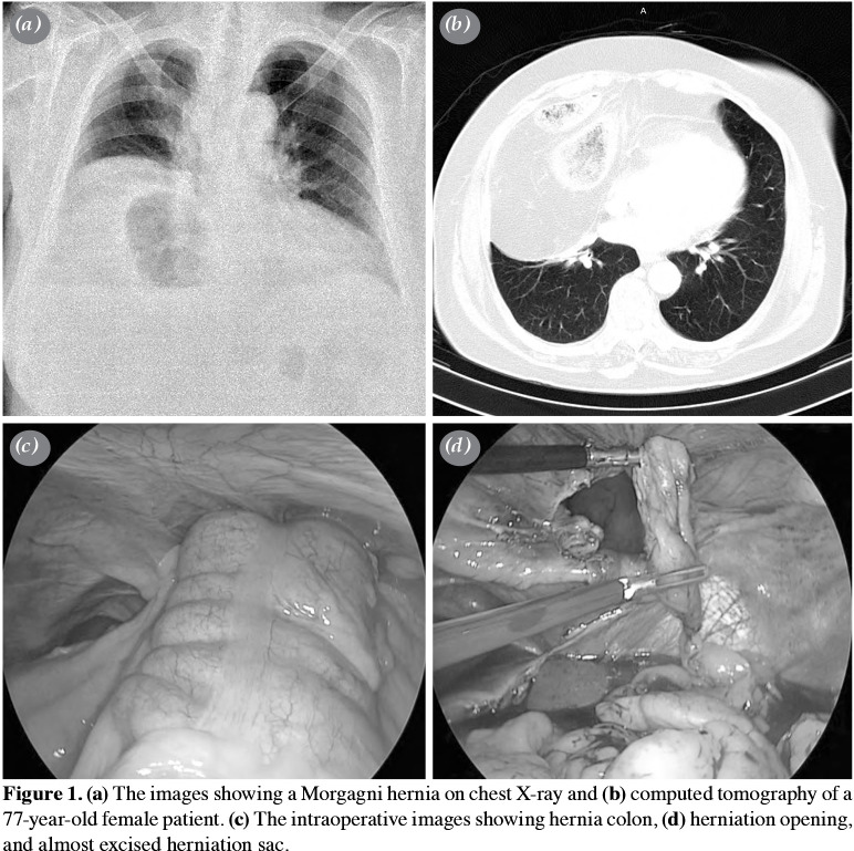
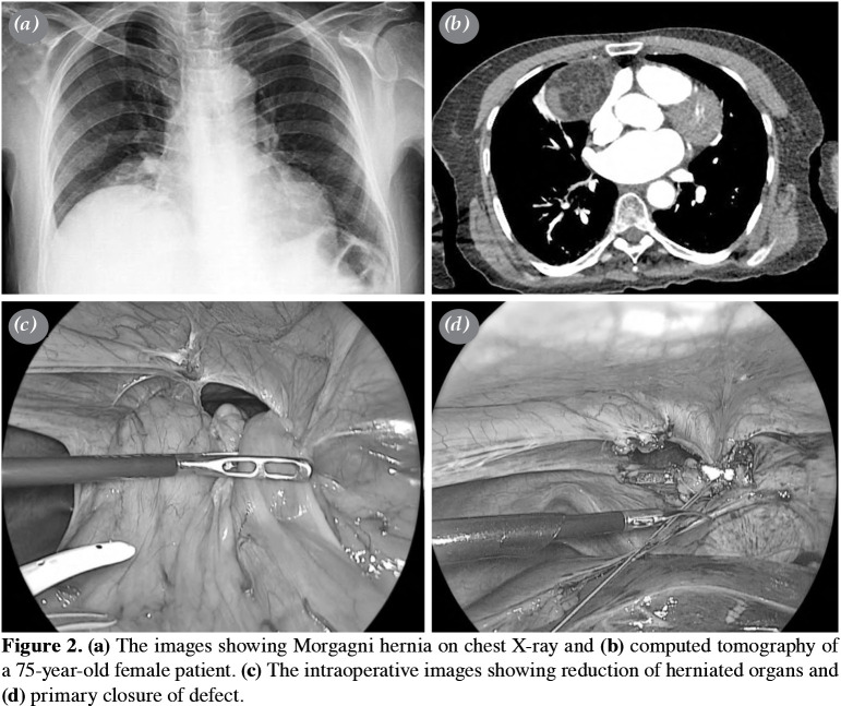
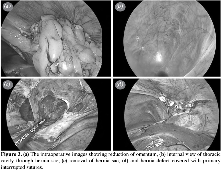
All patients underwent laparoscopic repair. They were placed in the supine position and were, then, given a reverse Trendelenburg position. The surgeon performed the operations on the right side of the patients. A total of three trocar ports were used for the operation: one (10 mm) for the camera (30° angled optic), just above the umbilicus and the others (5 mm) for the right and left upper quadrants for instruments. A pneumoperitoneum was created by inflating the abdomen with carbon dioxide gas. The defect was defined (Figure 1). The reduction of hernia organs to the abdomen was done. The hernia sac was resected in all patients (Figure 2). A chest drain was inserted into the right hemithorax and the diaphragmatic defect was primarily repaired using non-absorbable sutures (usually 0 Ethibond) placed in an interrupted fashion (Figure 3). No intra- or postoperative complications were observed. All patients were admitted to the thoracic surgery intensive care unit in the first postoperative day. The patients were discharged after a mean hospital stay of 4.6±1.3 days (range, 3 to 7 days).
Figure 1. (a) The images showing a Morgagni hernia on chest X-ray and (b) computed tomography of a 77-year-old female patient. (c) The intraoperative images showing hernia colon, (d) herniation opening, and almost excised herniation sac.
Figure 2. (a) The images showing Morgagni hernia on chest X-ray and (b) computed tomography of a 75-year-old female patient. (c) The intraoperative images showing reduction of herniated organs and (d) primary closure of defect.
Figure 3. (a) The intraoperative images showing reduction of omentum, (b) internal view of thoracic cavity through hernia sac, (c) removal of hernia sac, (d) and hernia defect covered with primary interrupted sutures.
Statistical analysis
Statistical analysis was performed using the IBM SPSS version 20.0 software (IBM Corp., Armonk, NY, USA). In the descriptive statistics, the numerical data were presented as mean and standard deviation and the categorical data were presented as numbers and percentages.
Results
The main initial symptoms were shortness of breath in seven (58.3%), chest pain in four (33.3%), abdominal pain in four (33.3%), and cough in one (8.3%) patient. The diagnosis was confirmed by CT in addition to chest X-ray. The hernia was located on the right side in all patients. The herniated organs were omentum in 12 (100%), transverse colon in seven (58.3%), and small intestine in three (25%) patients. The hernia sac was removed in all patients. All cases were laparoscopically repaired using primary sutures and no prosthetic material was used. The mean postoperative hospital stay was 4.6±1.3 days (range, 3 to 7 days). There was no complication or recurrence in the mean follow-up of 2.4 years (1 to 5 years) (Table 1).
Table 1. Characteristics of the patients.
| No | Age/Sex | The organs in the | Excision of | Prosthetic | Complication | Mortality | Postoperative | Recurrence |
| hernia sac | hernia sac | material use | hospital stay (Day) | |||||
| 1 | 67/F | Omentum, transverse | + | - | - | - | 5 | - |
| colon | ||||||||
| 2 | 59/F | Omentum, transverse | + | - | - | - | 5 | - |
| colon, small intestine | ||||||||
| 3 | 32/F | Omentum | + | - | - | - | 4 | - |
| 4 | 42/F | Omentum | + | - | - | - | 3 | - |
| 5 | 36/F | Omentum, transverse | + | - | - | - | 4 | - |
| colon | ||||||||
| 6 | 71/M | Omentum, transverse | + | - | - | - | 7 | - |
| colon | ||||||||
| 7 | 80/M | Omentum | + | - | - | - | 5 | - |
| 8 | 61/F | Omentum, transverse | + | - | - | - | 5 | - |
| colon, small intestine | ||||||||
| 9 | 70/F | Omentum | + | - | - | - | 3 | - |
| 10 | 77/F | Omentum | + | - | - | - | 7 | - |
| 11 | 60/M | Omentum, transverse | + | - | - | - | 4 | - |
| colon, small intestine | ||||||||
| 12 | 65/M | Omentum, transverse | + | - | - | - | 4 | - |
| colon |
Discussion
Morgagni hernia is thought to result from the absence of anterior costal elements joining the sternal components.[4] It was first described in 1761 by Giovanni Battista Morgagni,[6] the founder of pathological anatomy, and originates from the sternocostal trigone.[7] The Morgagni hernia is also called retrosternal, parasternal, substernal, or subcostosternal hernia. Conventionally, a right-side hernia is called Morgagni hernia, while a left-side hernia is called Larrey hernia. The ligamentum teres indicates the medial line of the hernia on both sides.[8] Although Morgagni hernias are congenital, the non-pathological appearance of previous radiographs of some patients indicates that herniations may subsequently develop from these congenital diaphragmatic defects.[9] Trauma, obesity, pregnancy, chronic constipation, and chronic cough are shown to be predisposing factors for Morgagni hernia in 41% of cases.[2-4] In addition, Morgagni hernia has been reported to be more common in males in childhood, but more common in females in adulthood.[10] Horton et al.[3] found the incidence to be 62% in women in their large series of 258 patients. In their study, the mean age was 53 years (58 for women and 50 for men). Similarly, in our study, there were eight females and four males with a mean age of 56.5±14.9 years (55.5 for women and 69 for men), indicating a higher mean age among males than females.
More than half of the patients are asymptomatic and are diagnosed incidentally.[11] Patients with Morgagni hernia can be noticed late due to the liver's ability to protect the diaphragm. They may present with non-specific respiratory or gastrointestinal symptoms. Dyspnea, chest pain, and abdominal pain are common non-specific symptoms in adults. A few of the patients present with acute abdominal symptoms caused by obstruction and strangulation.[12] In our series, acute pathologies such as obstruction, volvulus, strangulation and incarceration requiring an emergency intervention were not observed. All of the patients had nonspecific symptoms once the diagnosis was confirmed. The rate of the patients with pulmonary symptoms (n=7 dyspnea; n=1 cough) and pain (n=4 chest pain; n=4 abdominal pain) was 50%.
Patients with Morgagni hernia are usually detected incidentally by a chest X-ray or CT scanning of which the use has increased in recent years. Computed tomography is usually used to confirm the diagnosis of hernia and to measure the size of the defect. It is also the most accurate imaging method in the diagnosis and evaluation of hernia contents.[13] Lateral films show the anterior pericardiophrenic area. Barium X-rays are among the options in the inspection stage. A barium enema may show colon herniation and bowel obstruction. Magnetic resonance imaging is another useful method to differentiate Morgagni hernia from other mediastinal masses. Ultrasonographic examination is also a useful method in case of parenchymal organ hernias to the thoracic cavity.[13]
Anterior mediastinal lesions such as pericardial fat cushion, atelectasis, pneumonia, lipoma, liposarcoma, pleuro-pericardial cysts, mesothelioma, thymoma, lymphoma, teratoma, mesothelioma, and various tumors should be considered in the differential diagnosis.[3,14] in our patients, hernia was detected during laparoscopic procedures. None of the patients were misdiagnosed.
Approximately 90% of Morgagni hernias are located on the right side and 2% on the left side, while bilateral localization can be seen in 8% of cases.[4,15] Horton et al.[3] reported the anatomic distribution of Morgagni hernias as 91% on the right, 5% on the left, and 4% bilaterally in their series. In all our patients, the hernia was located on the right side. Furthermore, Morgagni hernias usually contain only the omentum during infancy and childhood. However, with the effect of negative pressure in the thoracic cavity over time, other abdominal organs may be also herniated and the defect enlarges. In our study, the organs in the hernia sac were omentum in 12 (100%), transverse colon in seven (58.3%), and small intestine in three (25%) patients.
Even in asymptomatic cases, early surgical intervention is recommended, due to rare, but lifethreatening acute complications such as intestinal obstruction and strangulation.[16,17] The main goals of surgery are the excision of the hernia sac, replacement of hernia organs back to the abdomen, and closure of the hernia defect. In their study comparing transthoracic and transabdominal approaches, Aydin et al.[2] reported similar and satisfactory results in both approaches.
In recent years, the laparoscopic approach has been also popular in the practice of thoracic surgery.[18] The first laparoscopic approach was performed in 1992 by Kuster et al.[19] In general, the thoracoscopic approach is used by surgeons due to the advantages of excellent visualization of the hernia content and the ability to easily reduction the hernia content. Better visualization of the hernia sac allows more secure dissection of possible pleural and pericardial adhesions.[2,4,17] Cases who are scheduled for the thoracoscopic approach should be evaluated with a gastrointestinal barium imaging to exclude malrotation. The thoracoscopic approach is contraindicated in the presence of malrotation. Therefore, it is more convenient to use any of the transabdominal approaches, if there is a preliminary diagnosis of ischemia or incarceration.[20] Currently, the most popular approach in the treatment of hernia is laparoscopic repair.[2,4,17,20]
In 95% of cases, there is a hernia sac. The excision of the hernia sac is still a matter of debate. Some authors oppose the excision of the hernia sac due to the risk of cardiorespiratory complications, injury to the mediastinal structures, and pneumomediastinum.[2] Akbiyik et al.[21] reported eight pediatric cases in which they everted the sacs of hernias to the peritoneal space before closing the defects. They reported that there was no recurrence and, thus, they were free of complications such as cardiac arrhythmia and pleural-pericardial damage. In our study, all of the patients had a hernia sac. We believe that the hernia sac should be resected in accordance with the classical surgical principles and, therefore, we excised the hernia sac in all patients to avoid recurrence and residual cavity. No excision-related complication was observed in any of the patients.
All the approaches including laparoscopy, laparotomy, thoracoscopy, and thoracotomy have low recurrence rates and excellent results.[2,4] Horton et al.[3] reported the complication rates of the laparoscopic approach as 5% and the mean postoperative hospital stay as 3 days. In a recent comparative study including 43 patients, Young et al.[22] found lower c omplication rates, shorter hospital stays, and similar recurrence rates in the laparoscopic group against open surgery group. In addition, similar studies comparing open surgery versus minimally invasive surgery were unable to find a significant difference in the recurrence rates.[17,20] In our opinion, routine follow-up should be done by chest X-ray. No recurrence was observed during the follow-up.
Furthermore, Morgagni hernia in association with a hiatal hernia is a very rare condition.[2] In their report, Eroglu et al.[23] presented such a case who underwent primary repair and anti-reflux surgery using the transabdominal approach and recovered without any complication. Recent studies have also demonstrated that robot-assisted laparoscopic Morgagni hernia repair is a feasible and safe method; however, the set-up time is prolonged and operation time is longer, compared to laparoscopic repair.[3]
Our study has a few limitations. These include the small study population and retrospective nature of the study.
In conclusion, Morgagni hernias, which are more frequently detected with an increased incidence of computed tomography, should be repaired as soon as they are diagnosed, and laparoscopy should be the first-line surgical approach due to the advantages such as a lower complication risk and shorter hospital stay which have been proved against open surgery. Primary repair without the use of any prosthetic material is a safe and effective treatment, except for large defects which can cause tension to the diaphragmatic ends, when closed. Removal of the hernia sac can be done in experienced hands to avoid complications.
Footnotes
Conflict of Interest: The authors declared no conflicts of interest with respect to the authorship and/or publication of this article.
Financial Disclosure: The authors received no financial support for the research and/or authorship of this article.
References
- 1.McGivern MR, Best KE, Rankin J, Wellesley D, Greenlees R, Addor MC, et al. Epidemiology of congenital diaphragmatic hernia in Europe: a register-based study. F137-44Arch Dis Child Fetal Neonatal Ed. 2015;100 doi: 10.1136/archdischild-2014-306174. [DOI] [PubMed] [Google Scholar]
- 2.Aydin Y, Altuntas B, Ulas AB, Daharli C, Eroglu A. Morgagni hernia: transabdominal or transthoracic approach. Acta Chir Belg. 2014;114:131–135. [PubMed] [Google Scholar]
- 3.Horton JD, Hofmann LJ, Hetz SP. Presentation and management of Morgagni hernias in adults: a review of 298 cases. Surg Endosc. 2008;22:1413–1420. doi: 10.1007/s00464-008-9754-x. [DOI] [PubMed] [Google Scholar]
- 4.Schumacher L, Gilbert S. Congenital diaphragmatic hernia in the adult. Thorac Surg Clin. 2009;19:469–472. doi: 10.1016/j.thorsurg.2009.08.004. [DOI] [PubMed] [Google Scholar]
- 5.Herman TE, Siegel MJ. Bilateral congenital Morgagni hernias. J Perinatol. 2001;21:343–344. doi: 10.1038/sj.jp.7200077. [DOI] [PubMed] [Google Scholar]
- 6.Morgagni GB. De sedibus, et causis morborum per anatomen indagatis libri quinque. Venetiis, Italy: Typog Remondiniana; 1761. [Google Scholar]
- 7.Kiliç D, Nadir A, Döner E, Kavukçu S, Akal M, Ozdemir N, et al. Transthoracic approach in surgical management of Morgagni hernia. Eur J Cardiothorac Surg. 2001;20:1016–1019. doi: 10.1016/s1010-7940(01)00934-4. [DOI] [PubMed] [Google Scholar]
- 8.LoCicero J. Shields" General Thoracic Surgery. Philadelphia: Lippincott Williams & Wilkins; 2018. [Google Scholar]
- 9.Kashiwagi H, Kumagai K, Nozue M, Terada Y. Morgagni hernia treated by reduced port surgery. Int J Surg Case Rep. 2014;5:1222–1224. doi: 10.1016/j.ijscr.2014.11.047. [DOI] [PMC free article] [PubMed] [Google Scholar]
- 10.McBride CA, Beasley S. Re: Laparoscopic repair of diaphragmatic Morgagni hernia in children: review of 3 cases. J Pediatr Surg. 2011;46:1470–1470. doi: 10.1016/j.jpedsurg.2011.03.073. [DOI] [PubMed] [Google Scholar]
- 11.Loong TP, Kocher HM. Clinical presentation and operative repair of hernia of Morgagni. Postgrad Med J. 2005;81:41–44. doi: 10.1136/pgmj.2004.022996. [DOI] [PMC free article] [PubMed] [Google Scholar]
- 12.Chang TH. Laparoscopic treatment of Morgagni-Larrey hernia. W V Med J. 2004;100:14–17. [PubMed] [Google Scholar]
- 13.Chaturvedi A, Rajiah P, Croake A, Saboo S, Chaturvedi A. Imaging of thoracic hernias: types and complications. Insights Imaging. 2018;9:989–1005. doi: 10.1007/s13244-018-0670-x. [DOI] [PMC free article] [PubMed] [Google Scholar]
- 14.Colakoğlu O, Haciyanli M, Soytürk M, Colakoğlu G, Simşek I. Morgagni hernia in an adult: atypical presentation and diagnostic difficulties. Turk J Gastroenterol. 2005;16:114–116. [PubMed] [Google Scholar]
- 15.Kurkcuoglu IC, Eroglu A, Karaoglanoglu N, Polat P, Balik AA, Tekinbas C. Diagnosis and surgical treatment of Morgagni hernia: report of three cases. Surg Today. 2003;33:525–528. doi: 10.1007/s10595-002-2522-z. [DOI] [PubMed] [Google Scholar]
- 16.Tarim A, Nursal TZ, Yildirim S, Ezer A, Caliskan K, Törer N. Laparoscopic repair of bilateral morgagni hernia. Surg Laparosc Endosc Percutan Tech. 2004;14:96–97. doi: 10.1097/00129689-200404000-00011. [DOI] [PubMed] [Google Scholar]
- 17.Sanford Z, Weltz AS, Brown J, Shockcor N, Wu N, Park AE. Morgagni Hernia Repair: A Review. Surg Innov. 2018;25:389–399. doi: 10.1177/1553350618777053. [DOI] [PubMed] [Google Scholar]
- 18.Park A, Doyle C. Laparoscopic Morgagni hernia repair: how I do it. J Gastrointest Surg. 2014;18:1858–1862. doi: 10.1007/s11605-014-2552-y. [DOI] [PubMed] [Google Scholar]
- 19.Kuster GG, Kline LE, Garzo G. Diaphragmatic hernia through the foramen of Morgagni: laparoscopic repair case report. J Laparoendosc Surg. 1992;2:93–100. doi: 10.1089/lps.1992.2.93. [DOI] [PubMed] [Google Scholar]
- 20.Arikan S, Dogan MB, Kocakusak A, Ersoz F, Sari S, Duzkoylu Y, et al. Morgagni's Hernia: Analysis of 21 Patients with Our Clinical Experience in Diagnosis and Treatment. Indian J Surg. 2018;80:239–244. doi: 10.1007/s12262-016-1580-0. [DOI] [PMC free article] [PubMed] [Google Scholar]
- 21.Akbiyik F, Tiryaki TH, Senel E, Mambet E, Livanelioğlu Z, Atayurt H. Is hernial sac removal necessary. Retrospective evaluation of eight patients with Morgagni hernia in 5 years. Pediatr Surg Int. 2006;22:825–827. doi: 10.1007/s00383-006-1750-4. [DOI] [PubMed] [Google Scholar]
- 22.Young MC, Saddoughi SA, Aho JM, Harmsen WS, Allen MS, Blackmon SH, et al. Comparison of Laparoscopic Versus Open Surgical Management of Morgagni Hernia. Ann Thorac Surg. 2019;107:257–261. doi: 10.1016/j.athoracsur.2018.08.021. [DOI] [PubMed] [Google Scholar]
- 23.Eroğlu A, Kürkçüoğlu IC, Karaoğlanoğlu N, Yilmaz O. Combination of paraesophageal hernia and Morgagni hernia in an old patient. Dis Esophagus. 2003;16:151–153. doi: 10.1046/j.1442-2050.2003.00315.x. [DOI] [PubMed] [Google Scholar]


