PLOS Neglected Tropical Diseases logoLink to PLOS Neglected Tropical Diseases
. 2020 Aug 31;14(8):e0007857. doi: 10.1371/journal.pntd.0007857

Telacebec (Q203)-containing intermittent oral regimens sterilized mice infected with Mycobacterium ulcerans after only 16 doses

Aurélie Chauffour 1,*, Jérôme Robert 1,2, Nicolas Veziris 1,2,3, Alexandra Aubry 1,2, Kevin Pethe 4,5, Vincent Jarlier 1,2
Editor: Abdallah M Samy6
PMCID: PMC7494103  PMID: 32866170

Abstract

Buruli ulcer (BU), caused by Mycobacterium ulcerans, is currently treated with a daily combination of rifampin and either injectable streptomycin or oral clarithromycin. An intermittent oral regimen would facilitate treatment supervision. We first evaluated the bactericidal activity of newer antimicrobials against M. ulcerans using a BU animal model. The imidazopyridine amine telacebec (Q203) exhibited high bactericidal activity whereas tedizolid (an oxazolidinone closely related to linezolid), selamectin and ivermectin (two avermectine compounds) and the benzothiazinone PBTZ169 were not active. Consequently, telacebec was evaluated for its bactericidal and sterilizing activities in combined intermittent regimens. Telacebec given twice a week in combination with a long-half-life compound, either rifapentine or bedaquiline, sterilized mouse footpads in 8 weeks, i.e. after a total of only 16 doses, and prevented relapse during a period of 20 weeks after the end of treatment. These results are very promising for future intermittent oral regimens which would greatly simplify BU treatment in the field.

Author summary

The current treatment for Buruli ulcer (BU), an infection caused by Mycobacterium ulcerans, is based on a daily antibiotic combination of rifampin associated with streptomycin or clarithromycin. A shorter or intermittent treatment without an injectable drug would clearly simplify the management in the field. We evaluated the bactericidal activity of several new antimicrobial drugs in a mouse model of BU and found that telacebec (Q203) exhibited the greatest bactericidal effect. We subsequently identified new antibiotic combinations containing telacebec with high sterilizing activity when administered twice a week for 8 weeks, i.e. at a total of only 16 doses.

Introduction

Buruli ulcer (BU), caused by Mycobacterium ulcerans, was only treated by surgery until 2004 [1]. The first medical treatment recommended by the World Health Organization (WHO) was an eight-week daily treatment based on an association of two antibiotics, rifampin (RIF), an oral ansamycin, and streptomycin (STR), an injectable aminoglycoside [1]. Currently a promising fully oral regimen combining RIF and clarithromycin (CLR), a macrolide compound [2,3], is tested clinically on a large scale (NCT01659437, clinicaltrials.gov).

The oral RIF-CLR combination was promoted to eliminate the toxic effects and injections of aminoglycosides, resulting in greater patient adherence and safety. Nevertheless, this combination is given daily for eight weeks. Shorter or intermittent treatment would facilitate adherence as well as supervision by healthcare workers. For instance, many Buruli ulcer patients with small-to-moderate size wounds are on ambulatory care and visit healthcare centres twice or three times per week for dressing changes, a rhythm that could allow receiving supervised intermittent antibiotic administration [4].

Our main objective was to identify alternative oral regimens active against BU by using a validated BU animal model. As a first step, we screened several new drug candidates for their in vivo bactericidal activity against M. ulcerans. Based on available data on their activity against M. tuberculosis or M. ulcerans, the following compounds, with either short or long half-life, were selected as potentially interesting: selamectin (SEL) and ivermectin (IVE), two drugs from the avermectin family with antiparasitic properties [57]; tedizolid (TDZ) [8], a new oxazolidinone active against M. tuberculosis that has the same mechanism of action as linezolid (LZD), a drug active against M. tuberculosis and M. ulcerans [9] with high bioavailability and a long half-life [10]; the 2-piperazino-benzothiazinone 169 (PBTZ), shown to be highly active against M. tuberculosis [11,12]; the imidazopyridine amine telacebec (Q203) that targets the respiratory terminal oxidase cytochrome bc1:aa3 in M. tuberculosis and that was recently shown to prevent mortality and reduce CFU counts in the footpads of mice infected with M. ulcerans [13] and to exhibit sterilizing activity when administrated in combination with other drugs [14].

Because the first-step screening demonstrated that Q203 was the compound with the highest bactericidal activity, and because of the long half-life of this compound [15], we measured, in a second experiment, the bactericidal and sterilizing activity of intermittent regimens containing Q203 combined with rifapentine or bedaquiline, other antibiotics with a long half-life and known to be active against M. ulcerans [16,17].

Methods

Infection of mice with M. ulcerans

In the first and the second experiment, respectively, 190 and 390 4-week-old female BALB/c/j mice were used (Janvier Labs, Le Genest Saint-Isle, France). Mice were inoculated, according to Shepard [18] in the left hind footpad with 0.03 ml of a bacterial suspension containing 5 log10 bacilli of the M. ulcerans strain Cu001. The number of live bacilli in the bacterial suspension was determined to be 5.02 and 4.6 log10 in the first and second experiment, respectively, by culturing the inoculum on Lowenstein-Jensen (LJ) medium. This strain, isolated in 1996 from a BU patient in Adzopé, Ivory Coast [19], was kindly provided by the local laboratory, blinded to patient identity. The strain is susceptible to all drugs used in BU treatment and was maintained in our laboratory through regular mouse footpad passage.

Treatment of mice

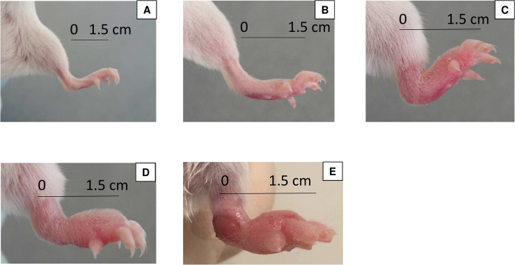
Treatment was initiated when the infection was well established, i.e. at a footpad swelling index of “2” (inflammatory swelling limited to the inoculated footpad) to “3” (inflammatory swelling involving the entire inoculated footpad) on a 4-grade scale [19] (Fig 1). This stage of infection was reached six weeks after the inoculation. Mice were randomly allocated into eight groups (1st experiment) and ten groups (2nd experiment) using a randomization table generated by the web site Randomization.com (https://www.randomization.com).

Fig 1.

Fig 1

Images representative of the lesion indexes used (A) index 0; (B) index 1; (C) index 2; (D) index 3; (E) index 4. Measure of the index was taken at the middle of the footpad (see the black bracket). Measures of indexes were as follows: 2mm for index 0; 3mm for index 1; 4.5mm for index 2; 5.5mm for index 3; and 6.5mm for index 4.

The groups were treated (with respect to drug, dosage, as well as number of doses/number of weeks, respectively) as follows:

In the first experiment, we used 8 groups including one untreated control group of 30 mice and seven antibiotic-treated groups of 20 mice, each treated with either TDZ, 10 mg/kg, 5/7; LZD, 100 mg/kg, 5/7; SEL, 12 mg/kg, 1/7; IVE, 1 mg/kg, 5/7; Q203, 5 mg/kg, 5/7 or PBTZ, 25 mg/kg, 5/7 and, as treatment control, RIF, 10 mg/kg, 5/7 alone or combined with STR, 150 mg/kg.

In the second experiment, out of 10 groups in total, we used 6 groups including one untreated control group of 27 mice and five antibiotic-treated groups of 27 mice, each receiving monotherapy with RIF, 10 mg/kg, 5/7; rifapentine (RPT), 20 mg/kg, 2/7; bedaquiline (BDQ), 25 mg/kg, 2/7; or Q203, 5 mg/kg, 5/7 or 2/7 while we used 4 groups of 57 mice each treated with drug combinations (dosages as above), i.e. Q203-RIF, 5/7; Q203-RPT, 2/7; Q203-BDQ, 2/7 or RIF-CLR, 100 mg/kg, 5/7 as control.

LZD was purchased from Pfizer, France; SEL and IVE from Merck, France; RIF from Sandoz, France; STR from Panpharma, France; and CLR from Abbott, France. TDZ was kindly provided by MSD-MERCK group, BDQ by Janssen Pharmaceutica, Belgium, and PBTZ by Stewart Cole (Global Health Institute, Ecole Polytechnique Fédérale de Lausanne, Lausanne, Switzerland; Institut Pasteur, rue du Docteur Roux, France). Q203 was custom-synthetized at GVKBio (GVK Biosciences Private Limited, Hyderabad, India) and contributed by Kevin Pethe.

Antibiotics were suspended in 0.05% agar-containing distilled water, except for STR, which was dissolved in buffered saline, Q203 in 1% DMSO-20% TPGS (D-α-Tocopherol polyethylene glycol 1000 succinate) and PBTZ in a solution of 1% carboxyl-methylcellulose-1% Tween 80. BDQ was provided by Janssen Pharmaceutica in a 20% hydropropyl-β-cyclodextrin formulation.

Treatments were administered for 4 or 8 weeks in both experiments and all drugs were orally administered by gavage in a final volume of 0.2 ml, except STR, which, in the same volume, was injected subcutaneously.

Assessment of M. ulcerans infection and effectiveness of treatment

Two methods were used for assessing the development of M. ulcerans infection and the effectiveness of treatments: (i) a clinical method consisting in weekly evaluation of the lesion index as previously described [19] and (ii) a bacteriological method consisting in the cultivation of M. ulcerans from the mouse footpads. When footpad swelling totally regresses under treatment, some redness often remains, indicative of scar tissue. We chose to assign index “1” to this stage, as index “0” is assigned to a normal footpad. For CFU enumeration in footpads, mice were sacrificed by cervical dislocation as recommended by the European directive 2010/63 and the French decree n°2013–118. Mouse footpads were removed aseptically and ground in 2 ml of Hank’s balanced salt solution in a tissue grinder (Octo Dissociator GentleMACS, Miltenyi Biotec, France). Suspensions were then plated onto LJ slants containing vancomycin (10 μg/ml), colistin (40 μg/ml) and amphotericin B (10 μg/ml) to limit contamination. In the first experiment, for the untreated group and the groups treated with RIF, TDZ, LZD, SEL, IVE or PBTZ, suspensions were serially 10-fold diluted to 10−4, and 0.1 ml of the dilutions was plated in duplicate onto LJ media. For the Q203 and the RIF-STR-treated groups, the entire volume of the footpad suspensions was plated onto LJ-media, at 0.2 ml per slant. In the second experiment, suspensions were serially diluted 10-fold to 10−4 for the untreated group, to 10−3 for the RIF and RPT groups at 2, 4 and 8 weeks and for the BDQ group at 2 weeks; to 10−2 for the Q203 2/7, Q203 5/7 and RIF-CLR groups at 2 weeks, and for the BDQ group at 4 weeks. A volume of 0.1 ml of each of these serial dilutions was plated in duplicate onto LJ media. For all the remaining time points, including those for all Q203 combinations, the entire volume of the footpad suspensions was plated onto LJ media, at 0.2 ml per slant. All tubes were incubated at 30°C for 90 days.

In the first experiment, the footpad lesion indexes were determined weekly during the 8-week period of treatment and CFUs were counted at weeks 4 and 8. In the 2nd experiment, footpad lesion indexes were determined weekly during the 8-week period of treatment and CFUs were counted at weeks 2, 4 and 8. Moreover, in the latter experiment, 30 mice that had been treated for 8 weeks with combination therapies were held without treatment during an additional period of 20 weeks to monitor relapses of M. ulcerans infection. Lesion indexes were determined weekly during this period and CFUs were counted at the end of the observation period.

MIC determination

In order to assess a possible acquisition of resistance to the antibiotics used during treatment, MICs of the antibiotics used for the treatment were determined for the bacilli isolated from relapsing mice during the observation period and for the initial strain Cu001. The isolates were suspended in distilled water to match the turbidity of a Mc Farland 3 standard. RIF and CLR were tested on Middlebrook 7H11 + 10% OADC (Oleicacid-Albumin-Dextrose-Catalase) medium (pH 7.4) and CLR was also tested on Mueller Hinton medium (pH 6.6). RIF was dissolved in dimethylformamide and CLR in distilled water, and both were twofold diluted in their respective solvent and incorporated into the culture media to obtain final concentrations ranging from 4 to 0.12 μg/ml. Suspensions (0.1 ml) of two distinct bacillus isolates (pure and diluted 10−2) were plated onto drug-containing and drug-free media used for growth control. All media were incubated at 30°C and examined after 60 and 90 days. The minimum inhibitory concentration (MIC) was defined as the lowest concentration of the drug preventing ≥99% of the growth on drug-free medium [20].

Statistical analysis

The Mann–Whitney U test was used to analyze the results (BiostaTGV). Relapse rates were also compared using Fisher's exact test. A p-value <0.05 was considered to be statistically significant. A significant decrease in the mean CFU value per footpad of treated as compared to untreated mice was used to assess the bactericidal effect of a given drug regimen.

Ethics statement

The experimental project was favorably evaluated by the ethics committee n°005 Charles Darwin localized at the Pitié-Salpêtrière Hospital and clearance was given by the French Ministry of Education and Research under the number APAFIS#9576–20170301171176185 v2. Our animal facility received the authorization to carry out animal experiments (license number C-75-13-01) on April 27, 2017. The persons who carried out the animal experiments had followed a specific training recognized by the French Ministry of Education and Research.

Results

First experiment

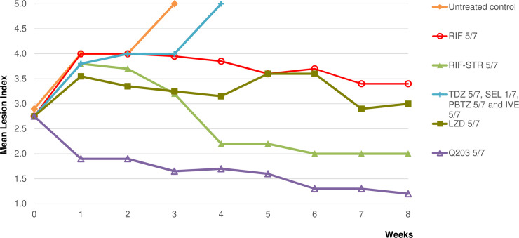
The mean lesion index (MLI) was 2.8±0.66 at the start of treatment (Fig 2). Untreated control mice with swollen footpads and MLIs that increased from 2.8 to 4 after 2 weeks, and mice with extensive lesions (inflammatory swelling of the whole limb, accompanied by a deteriorating general condition) either died or had to be sacrificed at week 4. MLIs in RIF-treated control groups increased to 3.8 after one week and then decreased to remain stable at 3.4, while decreasing to 2 in the RIF-STR group. MLIs in the SEL-, IVE-, TDZ- and PBTZ-treated groups continued to increase and mice either died or had to be sacrificed at week 4 due to advanced lesions comparable to those observed in untreated mice. In contrast, the MLI in the Q203-treated group decreased to 1.9 after 1 week of treatment and to 1.2 after 8 weeks.

Fig 2. First experiment: Mean lesion index of M. ulcerans-infected mouse footpads during treatment for 8 weeks.

Fig 2

Doses were as follow: rifampin (RIF), 10 mg/kg; streptomycin (STR), 150 mg/kg; tedizolid (TDZ), 10 mg/kg; linezolid (LZD), 100 mg/kg; selamectin (SEL), 12 mg/kg; ivermectin (IVE), 1 mg/kg; telacebec (Q203), 5 mg/kg; PBTZ169 (PBTZ), 25 mg/kg. The number of doses/number of weeks for RIF alone were 5/7; for RIF-STR, 5/7; for TDZ, 5/7; for LZD, 5/7; for SEL, 1/7; for IVE, 5/7; for Q203, 5/7 and for PBTZ, 5/7.

All untreated mice had culture-positive footpads at the beginning of treatment, with a mean of 6.93 ± 0.20 log10 CFUs, a count that remained stable at week 4 (Table 1). All mice remained culture-positive after 4 weeks of treatment in the RIF control groups but the mean CFU counts were significantly lower (p <0.05) than those in the untreated mouse group (4.58 ± 1.29 log10 for the RIF-(p = 0.0007) and 2.15 ± 1.20 log10 for the RIF-STR (p = 0.000006) treated groups, respectively). After 8 weeks of treatment, only 5 out of 8 mice in the RIF groups remained culture-positive with a low mean CFU count (<1 log10). After 4 weeks of treatment, all mice were culture-positive in the TDZ, SEL, IVE and PBTZ groups, with CFU counts not statistically different from those in the untreated control group. Due to advanced footpad lesion and mortality of mice, the evaluation point initially planned at week 8 was cancelled for these groups. After 4 weeks of treatment, 3 out of 10 mice were culture-negative in the Q203-treated group with a low mean CFU count (1.14 ± 1.30 log10), i.e. different from those in the RIF-treated group (p = 0.00002). After 8 weeks of treatment, all mice were culture-negative in the Q203 group.

Table 1. First experiment: Results of footpad cultures during the treatment of mice infected with M. ulcerans.


Regimena
(n doses/n weeks)
Results during treatment
Day 0 Week 4 Week 8
Culture positivity rate Mean (±SD) CFU per group Culture positivity rate Mean (±SD) CFU per group Culture positivity rate Mean (±SD) CFU per group
Untreated control 10/10 6.93±0.20 13/13b 6.81±0.36
RIF 5/7 9/9c 4.58±1.29 5/8c 0.47±1.16
RIF-STR 5/7 8/8d 2.15±1.20 3/9d 0.57±0.90
TDZ 5/7 7/7e 6.35±0.59 f
LZD 5/7 10/10 2.83±0.62 9/10 1.87±1.11
SEL 1/7 4/4g 6.24±0.82 f
IVE 5/7 8/8h 6.30±0.82 f
Q203 5/7 7/10 1.14±1.30 0/10
PBTZ 5/7 7/7i 7.08±0.64 f

a: treatment began 6 weeks after inoculation with 5.02 log10 bacilli per footpad when the infected swollen footpads had reached a lesion index of 2.8.

Drugs were administered 5-times a week, except for selamectin (SEL) which was administered once a week. Dosages were as follows: rifampin (RIF), 10 mg/kg; streptomycin (STR), 150 mg/kg; tedizolid (TDZ), 10 mg/kg; linezolid (LZD), 100 mg/kg; SEL, 12 mg/kg; ivermectin (IVE), 1 mg/kg; telacebec (Q203), 5 mg/kg; PBTZ169 (PBTZ), 25 mg/kg.

b: due to advanced lesions, all mice from the untreated control group were in fact sacrificed at week 3; nevertheless, footpad cultures were contaminated in 7 out of 20 mice.

c: 3 mice died in the RIF groups due to an accident during gavage.

d: 3 mice died in the RIF-STR groups due to an accident during gavage.

e: footpad cultures were contaminated due to advanced lesions in 3 mice.

f: cultures at week 8 were not performed because of advanced necrotized footpad lesions.

g: 5 mice died from M.ulcerans infection and footpad cultures were contaminated due to advanced lesion in 1 mouse.

h: 1 mouse died from M.ulcerans infection and footpad cultures were contaminated due to advanced lesion in 1 mouse.

i: footpad cultures were contaminated due to advanced lesions in 3 mice.

Second experiment

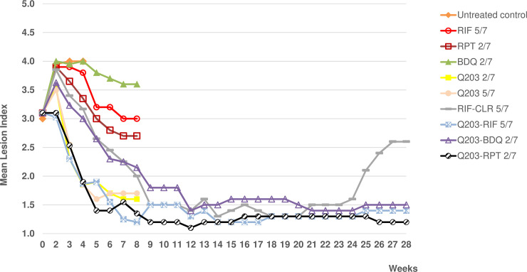
The MLI was 3±0.79 at the start of the treatment (Fig 3). The footpad MLIs of untreated control mice increased from 3 to 4 after 2 weeks and the mice had to be sacrificed at week 4 due to extensive BU lesions. The MLIs in the RIF-, RPT- and BDQ-treated groups increased to 3.8–4 after one week of treatment and then decreased, by week 8, to 3.6 (BDQ), 3 (RIF) and 2.7 (RPT). In the Q203-treated groups (n of doses/n of weeks, 5/7 or 2/7), MLIs increased slightly to 3.5 after one week of treatment and then decreased rapidly to 1.6–1.7 at week 8. The MLI in the group treated with RIF-CLR increased to 4 after 1 week of treatment, then decreased to 1.4 at week 12 and remained stable until week 20, but increased again to 2.6 at week 28. The MLI in the group treated with Q203-BDQ increased slightly to 3.6 after one week of treatment and decreased thereafter to 1.4 at week 12, a level that remained stable until week 28.The MLIs in the groups treated with Q203 combined with RIF or RPT decreased rapidly to 1.2–1.3 at week 8 and remained at that level until week 28.

Fig 3. Second experiment: Mean lesion index of M. ulcerans-infected mouse footpads during and after treatment for 8 weeks.

Fig 3

Dosages were as follows: rifampin (RIF), 10 mg/kg; rifapentine (RPT), 20 mg/kg; bedaquiline (BDQ), 25 mg/kg; Q203, 5 mg/kg; clarithromycin (CLR), 100 mg/kg. RIF was given (n of doses/n of weeks) 5/7; RPT, 2/7; BDQ, 2/7; Q203, 2/7 or 5/7; RIF-CLR, 5/7; Q203-RIF, 5/7; Q203-BDQ, 2/7 and Q203-RPT, 2/7. 1.2–1.3 at week 8 and remained at that level until week 28.

All untreated mice had culture-positive footpads at the beginning of treatment, with a mean of 6.87 ± 0.10 log10 CFUs, a count that remained stable at weeks 2 and 4 (Table 2). All treated mice remained culture-positive after 2 weeks of treatment but the CFU counts were significantly lower in all treated groups compared to those in the untreated group. Moreover, CFU counts were lower in the RPT (compared to RIF p = 0.008 and p = 0.0003 compared to BDQ), Q203 (for Q203 2/7 compared to RIF p = 0.008; compared to BDQ p = 0.001; for Q203 5/7 compared to RIF p = 0.01 and p = 0.0001 compared to BDQ) and the 4 combined-treatment groups than in the RIF and BDQ groups (p-values for combined treatment groups vs RIF: CR p = 0.003; QR p = 0.002; QB p = 0.006; QP p = 0.002; all p-values for combined treatment groups vs BDQ were 0.0002). After 4 weeks of treatment, some of the mice became culture-negative, especially in the groups treated with Q203-RPT and Q203-BDQ in which the CFU counts were very low (~0.2 log10). CFU counts at 4 weeks remained lower in the RPT and Q203 than in the RIF (p = 0.006, p = 0.04 and p = 0.002 for RPT, Q203 2/7 and Q203 5/7 respectively) and BDQ groups (p = 0.007, p = 0.001 and p = 0.001 for RPT, Q203 2/7 and Q203 5/7 respectively) and they were lower in the Q203-RPT (p = 0.04 and p = 0.03) and Q203-BDQ (p = 0.03 and p = 0.01) groups than in the Q203-RIF and RIF-CLR groups. After 8 weeks of treatment, all mice treated with Q203 alone or combined with RIF (5/7) or RPT (2/7) or BDQ (2/7) became culture-negative. Few mice remained culture-positive in the RIF, RPT and RIF-CLR groups with very low mean CFU counts (<0.5 log10) but all remained culture-positive in the BDQ group (1.28 ± 0.93 log10).

Table 2. Second experiment: Results of footpad cultures during the treatment of mice infected with M. ulcerans (for 2, 4 or 8 weeks) and relapse rate after treatment completion.


Regimena
(n doses/n weeks)
Results during treatment Results after observation period
Day 0 Week 2 Week 4 Week 8 Week 28
Culture positivity rate Mean (±SD) CFU per group Culture positivity rate Mean (±SD) CFU per group Culture positivity rate Mean (±SD) CFU per group Culture positivity rate Mean (±SD) CFU per group Culture positivity rate Mean (±SD) CFU per group
Untreated control 9/9 6.87±0.10 8/8b 7.00±0.16 6/6b 6.84±0.35
RIF 5/7 9/9 5.71±0.65 6/6c 2.78±0.81 2/8c 0.44±0.81
RPT 2/7 8/9 4.18±1.71 3/9 0.63±1.03 1/9 0.20±0.59
BDQ 2/7 7/7d 6.12±0.31 7/7d 5.48±0.14 5/5d 1.28±0.93
Q203 2/7 9/9 4.88±0.42 5/9 1.42±1.21 0/9
Q203 5/7 9/9 4.90±0.29 7/9 0.74±0.79 0/9
RIF-CLR 5/7 9/9 4.42±0.55 6/7e 1.15±0.84 1/8e 0.22±0.63 8/26e 0.87±1.36
Q203-RIF 5/7 9/9 4.52±0.36 9/9 1.20±1.04 0/8f 0/30
Q203-BDQ 2/7 9/9 4.65±0.75 2/9 0.17±0.51 0/9 0/30
Q203-RPT 2/7 9/9 4.40±0.78 2/9 0.25±0.49 0/9 0/30

a: treatments were begun 6 weeks after inoculation of 4.6 log10 CFUs per footpad when the infected swelling footpads reached a lesion index of 3. Drugs were administered two or five times a week and dosages were as follows: rifampin (RIF), 10 mg/kg; rifapentine (RPT), 20 mg/kg; bedaquiline (BDQ), 25mg/kg; telacebec (Q203), 5 mg/kg and clarithromycin (CLR), 100 mg/kg.

b: footpad cultures were contaminated due to advanced lesion in 1 mouse in the untreated Week 2 control group, and in 1 mouse in the Week 4 group.

c: footpad cultures were contaminated due to advanced lesions in 3 mice in the Week 4 RIF group, and in 1 mouse in the Week 8 group.

d: footpad cultures were contaminated due to advanced lesions in 1 mouse in the Week 2 BDQ group, in 2 mice in the Week 4 group and in 2 mice in the Week 8 group; 2 mice died due to an accident during gavage in the Week 8 group.

e: footpad cultures were contaminated due to advanced lesion in 2 mice in the Week 4 RIF-CLR group and in 3 mice in the relapse observation group; 1 mouse each died due to an accident during gavage in the Week 8 group and in the relapse observation group.

f: 1 mouse died due to an accident during gavage in the Week 8 Q203-RIF group.

During the 20-week observation period after stopping the treatment, no clinical or bacteriological relapse was observed in the 3 groups treated with Q203 combinations whereas 8 out of 26 mice treated with RIF-CLR relapsed with a mean CFU count of 0.87 ± 0.93 log10. This proportion was significantly higher than those in other groups (p = 0.001).

MICs for M. ulcerans bacilli recovered from relapsing mice

The MICs for bacilli isolated from the 8 relapsing mice in the RIF-CLR-treated group remained stable when compared to the initial MICs of RIF (0.5–1 μg/ml) and CLR (0.5 μg/ml) found for M. ulcerans Cu001 (the MICs of the latter were found to be the same on the 7H11 and MH media).

Discussion

Although nearly all Buruli ulcer lesions can be successfully treated by a two-month antibiotic combination regimen administered daily [21,22], new shorter and/or intermittent regimens would greatly simplify the treatment procedure in the field. Indeed, intermittent treatment would allow for organizing direct supervision of therapy and for combining drug administration and change of dressings in remote places where BU is prevalent.

The key finding of the present work was that regimens combining telacebec with rifapentine or bedaquiline, two long lasting drugs, sterilized infected mouse footpads after twice weekly administration during for 8 weeks, i.e. after a total of only 16 doses.

Our first in vivo screening experiment aimed at identifying newer bactericidal drugs. Indeed, available data on the activity of several new drugs against M. tuberculosis or M. ulcerans justified a systematic evaluation in a BU mouse model that has been successfully used for many years for this purpose [9]. IVE, SEL and TDZ were not bactericidal after 4 weeks of treatment and failed to prevent mortality during 8 weeks. The doses used in our experiment were taken from available pharmacokinetic data. IVE, a long-lasting drug in humans (half-life, 15-19h) and in mice (9h), when used in mice at a dose of 0.2 mg/kg, was shown to yield serum concentrations lower than those found in humans at standard therapeutic dosage [7]. We therefore used a higher dose (1 mg/kg), which also failed to control infection in our model. A similarly unfavorable result was obtained with SEL used at 12 mg/kg as proposed in a previous publication [23]. However, it has been suggested that these two avermectin compounds might be used safely at even higher doses [7], which could be evaluated in future studies. The dose of TDZ used in the present study, i.e. 10 mg/kg, was shown to yield pharmacokinetic profiles close to those observed in humans at the therapeutic dose of 200 mg [2426]. Contrasting with the disappointing results obtained with TDZ, and as reported in a previous study [9], a marked bactericidal activity was obtained with LZD, an oxazolidinone included in the experiment as a positive control for TDZ. Surprisingly, PBTZ was not bactericidal in our BU model at 25 mg/kg, a dose shown to be active against M. tuberculosis in mice [11]. Yet in M. ulcerans, as well as in M. tuberculosis, DprE1, the target of benzothiazinones, carries a cysteine at position 387 in lieu of a serine or an alanine, which have been shown to confer natural resistance to PBTZ in M. avium or M. aurum, respectively [27]. Thus, the reason for the disappointing result obtained with PBTZ in our BU model is unclear.

RIF and RIF-STR were highly bactericidal as observed in all our preceding studies [2,28]. Q203 drastically reduced the lesion index and CFU counts after 4 weeks of treatment and all mice became culture-negative after 8 weeks. These results obtained with Q203 were clearly superior to those obtained with the usual positive control with RIF alone, and even with the reference combination regimen RIF-STR.

Drugs with low bactericidal activity, when given as monotherapy, might be of interest when used in combination with other drugs. However, since our goal was to obtain the most effective combination regimen, we selected, for the second experiment, combinations of drugs shown to be highly active separately. The results of this experiment demonstrated that regimens combining Q203 with RIF or RPT or BDQ were not only bactericidal, making all footpads culture-negative after 8 weeks of treatment, but also sterilized them and prevented relapse during an observation period of 20 weeks after stopping the treatment. These promising results were obtained after administering twice weekly for 8 weeks, i.e. after a total of only 16 doses, the combinations of Q203 with either RPT or BDQ. These three drugs are long-lasting, with serum half-lives in mice after a single dose being Q203 23 h [15]; RPT 25 h [16] and BDQ 53 h [17]. Recently, regimens combining RPT with CLR or BDQ, administered twice weekly for 8 weeks, were found to be as bactericidal and as sterilizing as a daily RPT-CLR regimen [28].

We recently showed that Q203 was very active in the footpads of mice infected with M. ulcerans [13]. Lately, a study by Converse et al confirmed this finding and furthermore showed that triple combinations of Q203 administered at the higher dose of 10 mg/kg, either with RPT and clofazimine, RPT and BDQ or BDQ and clofazimine, as well as a quadruple combination of these four drugs, were sterilizing after 2 weeks of daily treatment in a BU animal model [14], leading to the conclusion that targeting the M. ulcerans respiratory chain with several drugs is an effective strategy for designing new shortened treatments of BU. Thus, Q203 appears clearly an important candidate for future treatment of BU, either in daily or intermittent regimens.

The fact that, in the present work, few bacilli were still found by culture in 1 out of 8 mice after 8 weeks of treatment with RIF-CLR 5/7, and that 8 out of 26 mice relapsed with low CFU counts within 20 weeks after the end of this regimen, was surprising since bactericidal and sterilizing activity of RIF-CLR was shown in our previous studies. Other authors have observed, 20 weeks after stopping treatment with RIF-CLR 5/7, 25% of relapses based on recurrent swelling or ectopic lesions [29]. The susceptibility to RIF and to CLR of the bacilli isolated from relapsing mice in our study was unchanged, ruling out the selection of resistant mutants during treatment. Relapses could be explained by unusually high bacterial loads reached in the present work when compared to those in our previous studies, i.e. 3–4 times higher at the start of treatment and 4–10 times higher after 2–4 weeks in untreated mice. Nevertheless, this fact strengthens further the good results obtained with the Q203-containing combination regimens.

New drugs are rare for the treatment of BU. The last active new marketed drug was BDQ, initially assessed with success for tuberculosis and found later to be very active against M. ulcerans in a BU animal model [28]. Therefore, the excellent in vivo activity of Q203 against M. ulcerans constitutes a step forward. In a recent study, the basis of this promising result was found to be reductive evolution in most strains of this species that led to hyper susceptibility to Q203 by elimination due to the absence of alternate terminal electron acceptors, thereby making the target, respiratory cytochrome bc1:aa3, crucial for survival [13].

Whereas, triple or quadruple combinations of Q203 at 10mg/kg, administered with RPT, clofazimine and BDQ were found to sterilize infected mice after 2 weeks of daily treatment, we demonstrated in the present study that double combinations of Q203 at 5 mg/kg with either RPT or BDQ, were also sterilizing after 16 doses of a twice weekly treatment, due to the long half-life and good bioavailability of these three drugs. The latter intermittent scheme of oral treatment with two drugs would greatly simplify the treatment of BU on ambulatory care in the field, when patients living in remote areas visit healthcare centers twice or three times per week for dressing changes, a rhythm that could allow receiving supervised intermittent antibiotic administration.

Data Availability

All relevant data are within the manuscript and its Supporting Information files.

Funding Statement

This work was funded by the Raoul Follereau Foundation. The funders had no role in study design, data collection and analysis, decision to publish, or preparation of the manuscript.

References

  • 1.WHO. Treatment of Mycobacterium ulcerans disease (Buruli ulcer) Guidance for health workers. 2012. Available: https://apps.who.int/iris/bitstream/handle/10665/77771/9789241503402_eng.pdf;jsessionid=AECF9887A2910FB44C46C2212817A90F?sequence=1 [Google Scholar]
  • 2.Ji B, Chauffour A, Robert J, Jarlier V. Bactericidal and sterilizing activities of several orally administered combined regimens against Mycobacterium ulcerans in mice. Antimicrob Agents Chemother. 2008;52: 1912–1916. 10.1128/AAC.00193-08 [DOI] [PMC free article] [PubMed] [Google Scholar]
  • 3.Chauty A, Ardant M-F, Marsollier L, Pluschke G, Landier J, Adeye A, et al. Oral treatment for Mycobacterium ulcerans infection: results from a pilot study in Benin. Clin Infect Dis. 2011;52: 94–96. 10.1093/cid/ciq072 [DOI] [PubMed] [Google Scholar]
  • 4.Phillips RO, Robert J, Abass KM, Thompson W, Sarfo FS, Wilson T, et al. Rifampicin and clarithromycin (extended release) versus rifampicin and streptomycin for limited Buruli ulcer lesions: a randomised, open-label, non-inferiority phase 3 trial. Lancet Lond Engl. 2020;395: 1259–1267. 10.1016/S0140-6736(20)30047-7 [DOI] [PMC free article] [PubMed] [Google Scholar]
  • 5.Lim LE, Vilcheze C, Ng C, Jacobs WR, Ramon-Garcia S, Thompson CJ. Anthelmintic Avermectins Kill Mycobacterium tuberculosis, Including Multidrug-Resistant Clinical Strains. Antimicrob Agents Chemother. 2013;57: 1040–1046. 10.1128/AAC.01696-12 [DOI] [PMC free article] [PubMed] [Google Scholar]
  • 6.Omansen TF, Porter JL, Johnson PDR, van der Werf TS, Stienstra Y, Stinear TP. In-vitro Activity of Avermectins against Mycobacterium ulcerans. Johnson C, editor. PLoS Negl Trop Dis. 2015;9: e0003549 10.1371/journal.pntd.0003549 [DOI] [PMC free article] [PubMed] [Google Scholar]
  • 7.Scherr N, Pluschke G, Thompson CJ, Ramón-García S. Selamectin Is the Avermectin with the Best Potential for Buruli Ulcer Treatment. Johnson C, editor. PLoS Negl Trop Dis. 2015;9: e0003996 10.1371/journal.pntd.0003996 [DOI] [PMC free article] [PubMed] [Google Scholar]
  • 8.Molina-Torres CA, Barba-Marines A, Valles-Guerra O, Ocampo-Candiani J, Cavazos-Rocha N, Pucci MJ, et al. Intracellular activity of tedizolid phosphate and ACH-702 versus Mycobacterium tuberculosis infected macrophages. Ann Clin Microbiol Antimicrob. 2014;13: 13 10.1186/1476-0711-13-13 [DOI] [PMC free article] [PubMed] [Google Scholar]
  • 9.Ji B, Lefrancois S, Robert J, Chauffour A, Truffot C, Jarlier V. In Vitro and In Vivo Activities of Rifampin, Streptomycin, Amikacin, Moxifloxacin, R207910, Linezolid, and PA-824 against Mycobacterium ulcerans. Antimicrob Agents Chemother. 2006;50: 1921–1926. 10.1128/AAC.00052-06 [DOI] [PMC free article] [PubMed] [Google Scholar]
  • 10.Burdette SD, Trotman R. Tedizolid: The First Once-Daily Oxazolidinone Class Antibiotic. Saravolatz LD, editor. Clin Infect Dis. 2015;61: 1315–1321. 10.1093/cid/civ501 [DOI] [PubMed] [Google Scholar]
  • 11.Makarov V, Lechartier B, Zhang M, Neres J, van der Sar AM, Raadsen SA, et al. Towards a new combination therapy for tuberculosis with next generation benzothiazinones. EMBO Mol Med. 2014;6: 372–383. 10.1002/emmm.201303575 [DOI] [PMC free article] [PubMed] [Google Scholar]
  • 12.Makarov V, Manina G, Mikusova K, Mollmann U, Ryabova O, Saint-Joanis B, et al. Benzothiazinones Kill Mycobacterium tuberculosis by Blocking Arabinan Synthesis. Science. 2009;324: 801–804. 10.1126/science.1171583 [DOI] [PMC free article] [PubMed] [Google Scholar]
  • 13.Scherr N, Bieri R, Thomas SS, Chauffour A, Kalia NP, Schneide P, et al. Targeting the Mycobacterium ulcerans cytochrome bc1:aa3 for the treatment of Buruli ulcer. Nat Commun. 2018;9: 5370 10.1038/s41467-018-07804-8 [DOI] [PMC free article] [PubMed] [Google Scholar]
  • 14.Converse PJ, Almeida DV, Tyagi S, Xu J, Nuermberger EL. Shortening Buruli Ulcer Treatment with Combination Therapy Targeting the Respiratory Chain and Exploiting Mycobacterium ulcerans Gene Decay. Antimicrob Agents Chemother. 2019;63: e00426–19, /aac/63/7/AAC.00426-19.atom. 10.1128/AAC.00426-19 [DOI] [PMC free article] [PubMed] [Google Scholar]
  • 15.Pethe K, Bifani P, Jang J, Kang S, Park S, Ahn S, et al. Discovery of Q203, a potent clinical candidate for the treatment of tuberculosis. Nat Med. 2013;19: 1157–1160. 10.1038/nm.3262 [DOI] [PubMed] [Google Scholar]
  • 16.Rosenthal IM, Williams K, Tyagi S, Peloquin CA, Vernon AA, Bishai WR, et al. Potent Twice-Weekly Rifapentine-containing Regimens in Murine Tuberculosis. Am J Respir Crit Care Med. 2006;174: 94–101. 10.1164/rccm.200602-280OC [DOI] [PMC free article] [PubMed] [Google Scholar]
  • 17.Rouan M-C, Lounis N, Gevers T, Dillen L, Gilissen R, Raoof A, et al. Pharmacokinetics and Pharmacodynamics of TMC207 and Its N-Desmethyl Metabolite in a Murine Model of Tuberculosis. Antimicrob Agents Chemother. 2012;56: 1444–1451. 10.1128/AAC.00720-11 [DOI] [PMC free article] [PubMed] [Google Scholar]
  • 18.Shepard CC. The experimental disease that follows the injection of human leprosy bacilli into foot-pads of mice. J Exp Med. 1960;112: 445–454. 10.1084/jem.112.3.445 [DOI] [PMC free article] [PubMed] [Google Scholar]
  • 19.Dega H, Bentoucha A, Robert J, Jarlier V, Grosset J. Bactericidal Activity of Rifampin-Amikacin against Mycobacterium ulcerans in Mice. Antimicrob Agents Chemother. 2002;46: 3193–3196. 10.1128/aac.46.10.3193-3196.2002 [DOI] [PMC free article] [PubMed] [Google Scholar]
  • 20.Inderlied CB, Nash KA. Antimicrobial agents: in vitro susceptibility testing, spectra of activity, mechanisms of action and resistance, and assays for activity in biologic fluids 4th ed. Antibiotics in laboratory medicine. 4th ed. The Williams & Wilkins Co., Baltimore. Md: M. D. V. Lorian; 1996. pp. 127–175. [Google Scholar]
  • 21.Wadagni AC, Steinhorst J, Barogui YT, Catraye PM, Gnimavo R, Abass KM, et al. Buruli ulcer treatment: Rate of surgical intervention differs highly between treatment centers in West Africa. PLoS Negl Trop Dis. 2019;13: e0007866 10.1371/journal.pntd.0007866 [DOI] [PMC free article] [PubMed] [Google Scholar]
  • 22.Wadagni AC, Barogui YT, Johnson RC, Sopoh GE, Affolabi D, van der Werf TS, et al. Delayed versus standard assessment for excision surgery in patients with Buruli ulcer in Benin: a randomised controlled trial. Lancet Infect Dis. 2018;18: 650–656. 10.1016/S1473-3099(18)30160-9 [DOI] [PubMed] [Google Scholar]
  • 23.Geyer J, Gavrilova O, Petzinger E. Brain penetration of ivermectin and selamectin in mdr1a,b P-glycoprotein- and bcrp—deficient knockout mice. J Vet Pharmacol Ther. 2009;32: 87–96. 10.1111/j.1365-2885.2008.01007.x [DOI] [PubMed] [Google Scholar]
  • 24.Lepak AJ, Marchillo K, Pichereau S, Craig WA, Andes DR. Comparative pharmacodynamics of the new oxazolidinone tedizolid phosphate and linezolid in a neutropenic murine Staphylococcus aureus pneumonia model. Antimicrob Agents Chemother. 2012;56: 5916–5922. 10.1128/AAC.01303-12 [DOI] [PMC free article] [PubMed] [Google Scholar]
  • 25.Kanafani ZA, Corey GR. Tedizolid (TR-701): a new oxazolidinone with enhanced potency. Expert Opin Investig Drugs. 2012;21: 515–522. 10.1517/13543784.2012.660250 [DOI] [PubMed] [Google Scholar]
  • 26.Flanagan S, Fang E, Muñoz KA, Minassian SL, Prokocimer PG. Single- and Multiple-Dose Pharmacokinetics and Absolute Bioavailability of Tedizolid. Pharmacother J Hum Pharmacol Drug Ther. 2014;34: 891–900. 10.1002/phar.1458 [DOI] [PMC free article] [PubMed] [Google Scholar]
  • 27.Incandela ML, Perrin E, Fondi M, de Jesus Lopes Ribeiro AL, Mori G, Moiana A, et al. DprE1, a new taxonomic marker in mycobacteria. FEMS Microbiol Lett. 2013;348: 66–73. 10.1111/1574-6968.12246 [DOI] [PubMed] [Google Scholar]
  • 28.Chauffour A, Robert J, Veziris N, Aubry A, Jarlier V. Sterilizing Activity of Fully Oral Intermittent Regimens against Mycobacterium Ulcerans Infection in Mice. Small PLC, editor. PLoS Negl Trop Dis. 2016;10: e0005066 10.1371/journal.pntd.0005066 [DOI] [PMC free article] [PubMed] [Google Scholar]
  • 29.Converse PJ, Tyagi S, Xing Y, Li S-Y, Kishi Y, Adamson J, et al. Efficacy of Rifampin Plus Clofazimine in a Murine Model of Mycobacterium ulcerans Disease. Phillips RO, editor. PLoS Negl Trop Dis. 2015;9: e0003823 10.1371/journal.pntd.0003823 [DOI] [PMC free article] [PubMed] [Google Scholar]
PLoS Negl Trop Dis. doi: 10.1371/journal.pntd.0007857.r001

Decision Letter 0

Richard Odame Phillips, Abdallah M Samy

20 Dec 2019

Dear Mrs Chauffour:

Thank you very much for submitting your manuscript "Q203 containing fully intermittent oral regimens exhibited high sterilizing activity against Mycobacterium ulcerans in mice" (#PNTD-D-19-01766) for review by PLOS Neglected Tropical Diseases. Your manuscript was fully evaluated at the editorial level and by independent peer reviewers. The reviewers appreciated the attention to an important problem, but raised some substantial concerns about the manuscript as it currently stands. These issues must be addressed before we would be willing to consider a revised version of your study. We cannot, of course, promise publication at that time.

We therefore ask you to modify the manuscript according to the review recommendations before we can consider your manuscript for acceptance. Your revisions should address the specific points made by each reviewer.

When you are ready to resubmit, please be prepared to upload the following:

(1) A letter containing a detailed list of your responses to the review comments and a description of the changes you have made in the manuscript.

(2) Two versions of the manuscript: one with either highlights or tracked changes denoting where the text has been changed (uploaded as a "Revised Article with Changes Highlighted" file); the other a clean version (uploaded as the article file).

(3) If available, a striking still image (a new image if one is available or an existing one from within your manuscript). If your manuscript is accepted for publication, this image may be featured on our website. Images should ideally be high resolution, eye-catching, single panel images; where one is available, please use 'add file' at the time of resubmission and select 'striking image' as the file type.

Please provide a short caption, including credits, uploaded as a separate "Other" file. If your image is from someone other than yourself, please ensure that the artist has read and agreed to the terms and conditions of the Creative Commons Attribution License at http://journals.plos.org/plosntds/s/content-license (NOTE: we cannot publish copyrighted images).

(4) If applicable, we encourage you to add a list of accession numbers/ID numbers for genes and proteins mentioned in the text (these should be listed as a paragraph at the end of the manuscript). You can supply accession numbers for any database, so long as the database is publicly accessible and stable. Examples include LocusLink and SwissProt.

(5) To enhance the reproducibility of your results, we recommend that you deposit your laboratory protocols in protocols.io, where a protocol can be assigned its own identifier (DOI) such that it can be cited independently in the future. For instructions see http://journals.plos.org/plosntds/s/submission-guidelines#loc-methods

Please note while forming your response, if your article is accepted, you may have the opportunity to make the peer review history publicly available. The record will include editor decision letters (with reviews) and your responses to reviewer comments. If eligible, we will contact you to opt in or out.

While revising your submission, please upload your figure files to the Preflight Analysis and Conversion Engine (PACE) digital diagnostic tool, https://pacev2.apexcovantage.com/ PACE helps ensure that figures meet PLOS requirements. To use PACE, you must first register as a user. Then, login and navigate to the UPLOAD tab, where you will find detailed instructions on how to use the tool. If you encounter any issues or have any questions when using PACE, please email us at figures@plos.org.

We hope to receive your revised manuscript by Feb 18 2020 11:59PM. If you anticipate any delay in its return, we ask that you let us know the expected resubmission date by replying to this email.

To submit a revision, go to https://www.editorialmanager.com/pntd/ and log in as an Author. You will see a menu item call Submission Needing Revision. You will find your submission record there.

Sincerely,

Abdallah M. Samy, PhD

Guest Editor

PLOS Neglected Tropical Diseases

Richard Phillips

Deputy Editor

PLOS Neglected Tropical Diseases

***********************

Editor comments to authors: I invited and received three reviews of your manuscript. All reviews raised substantial concerns as it currently stands. I read the manuscript myself and i must say that I completely agree with the points raised by the reviewers; all points are solid and detailed. So, I would recommend major revision for your manuscript to give you an opportunity to address all the points raised by the reviewers before considering a decision on your manuscript. We cannot, of course, promise publication at that time.

Reviewer's Responses to Questions

Key Review Criteria Required for Acceptance?

As you describe the new analyses required for acceptance, please consider the following:

Methods

-Are the objectives of the study clearly articulated with a clear testable hypothesis stated?

-Is the study design appropriate to address the stated objectives?

-Is the population clearly described and appropriate for the hypothesis being tested?

-Is the sample size sufficient to ensure adequate power to address the hypothesis being tested?

-Were correct statistical analysis used to support conclusions?

-Are there concerns about ethical or regulatory requirements being met?

Reviewer #1: These studies use a well established mouse model of M. ulcerans infection which has been a useful guide to antibiotic treatment of human infection. The methods are well applied to this study of new antibiotic combinations by authors who are familiar with the mouse model. Well designed study likely to yield clear answers. Ethical issues have been addressed.

Reviewer #2: 1) They did not show how they calculated "the M. ulcerans strain Cu001 (5.02 and 4.6 log10 in the 1st and 2nd experiment, respectively)." The right method should be calculating the CFUs from the mouse footpads on the day after infection.

2) Line 105, "one untreated control group of 30 mice and seven treated groups" should be "eight treated groups"!

3) Line 139-141: For the untreated groups, the entire volume of the footpad suspension was plated onto 10 LJ-media with 0.2 ml each. How did the authors count the CFUs and got 5 to 7 log10CFU/footpad in some treated groups? As far as I know, it is impossible to count.

4) What is the definition of their MIC? How did the authors calculate the MICs from the CFU results? Line 160 to 162, this method to test "MIC" was wrong.

5) “A regimen was considered to be bactericidal if its......”. This was totally wrong!

6) When the authors treat the mice in the RIF containing groups, did they separate RIF from other drugs? How long is the duration in between giving RIF and the other drug? In addition, maybe something wrong as CFUs in the RIF-STR group should be much lower than that from RIF group alone group in table 1. Can these 2 groups mixed?

7) Many (10) mice died of accident gavage and many (about 30) samples were contaminated, which indicated the perfermance/skill was not OK. Especially those in the week4 groups in table1, the samples should be diluted and so the contamination should be rare.

8) In table 1, in the SEL group, 5 died and 1 contaminated at week4? Is it possible because of the death due to toxicity? At that time point, how about the other 10 mice for the time point week8? If they were still alive, why not kill some of them for the CFU counts at week 4 in stead of week 8?Or they died but not indicated?

9) Why use very low dose of Q230 only at 5 mg/kg?

10) Why not test some inactive drugs in vitro first or showing the in vitro data?

Reviewer #3: See comments below

--------------------

Results

-Does the analysis presented match the analysis plan?

-Are the results clearly and completely presented?

-Are the figures (Tables, Images) of sufficient quality for clarity?

Reviewer #1: Yes and the results, figures and tables are easily understandable.

Reviewer #2: 1)The CFU results are not so clear as some mice died or many samples were contaminated.

2)Some results are confusing. For eg, the higher infection doses caused lower MLI. Oone possible reason is the MLI is to subjective. And CFUs in the STR-RIF group were even more than the RIF alone group. It is very abnormal.

3)In table 2, no any mouse died in the Q203 containing groups but 3 contaminated in RIF-CLR group? For negative CFUs, all undiluted tissue should be plated, and so they can be contaminated even easier. BTW, the relapse rates were evaluated at 20 weeks after treatment stopped. The duration is a little bit shorter.

Reviewer #3: See comments below

--------------------

Conclusions

-Are the conclusions supported by the data presented?

-Are the limitations of analysis clearly described?

-Do the authors discuss how these data can be helpful to advance our understanding of the topic under study?

-Is public health relevance addressed?

Reviewer #1: Yes to all questions.

Reviewer #2: The conclusions are OK. However, the findings that the fully intermittent oral regimens containing Q203 can cure BU in 8 weeks are not so valuable and not as good as the fully oral regimens containing Q203 which may cure BU in two weeks. And there are no anythings new except for some other drugs tested showed no activity in vivo.

Reviewer #3: See comments below

--------------------

Editorial and Data Presentation Modifications?

Use this section for editorial suggestions as well as relatively minor modifications of existing data that would enhance clarity. If the only modifications needed are minor and/or editorial, you may wish to recommend “Minor Revision” or “Accept”.

Reviewer #1: A minor point is that, although the paper is easy to understand, the English is awkward in places and could do with some editing

Reviewer #2: (No Response)

Reviewer #3: See comments below

--------------------

Summary and General Comments

Use this section to provide overall comments, discuss strengths/weaknesses of the study, novelty, significance, general execution and scholarship. You may also include additional comments for the author, including concerns about dual publication, research ethics, or publication ethics. If requesting major revision, please articulate the new experiments that are needed.

Reviewer #1: These are new and important results from testing combinations of antibiotics that could well be relevant to treatment of Buruli ulcer in humans. There was a surprising improvement in time required to kill all the bacteria, particularly after Q203 containing combinations. The poor results with rifampicin plus clarithromycin (current standard of care treatment in humans) differ from previously published studies but this is discussed by the authors and their explanation is plausible. This does not detract from the value of the paper whichrepresents a valuable contribution to the literature.

Reviewer #2: The authors test several drugs including Q203 targeting QcrB, the component in the ETC. And furthermore, they found that Q203 containing fully intermittent oral regimens can cure M. ulcerans infection effectively. However, the findings are not new to the society of Buruli ulver treatment field, as cited by this manuscript, (Converse P et al, 2019 doi:10.1128/AAC.00426-19 ), it has been demonstrated that Q203 containing regimen can cure Buruli ulcer in 2 weeks. The total doses are 10 and here at least 18 doses are needed for eight weeks. So the results and conclusion in this paper is not very meaningful. In addition, not only one papers showed the cytochrome bc1-aa3 respiratory terminal oxidase inhibitors were very effective against M. ulcerans infection published in Nature Communications in 2019.

The M. ulcerans study is very difficult and lengthy as M. ulcerans grows so slowly. So unfortunately, similar and even better results have been published when this study was going on.

In addition, many obvious mistakes both in the methods and in experimental skills lead to the much less value of this paper.

1)It is better if the authors could provide some pictures of the swelling footpads.

2) “0.03 ml of a bacterial suspension containing around 5 log10 Colony Forming Unit (CFU) of the M. ulcerans strain Cu001 (5.02 and 4.6 log10 in the 1st and 2nd experiment, respectively.” “The mean lesion index (MLI) was 2.8 at the start of the treatment in 1 st experiment”. However, MLI was 3 at the start of the treatment in 2nd experiment. According to my experience, the higher infection dose can cause higher MLI. There should be some explanation about why a little bit lower inoculation in the 2nd animal experiment cause a little bit higher the swelling degrees of mice than that in the 1st experiment. In addition, the CFUs at the time of treatment start with higher CFUs in the 2nd experiment.

There are some other obvious mistakes.

3)Line 55 “.” after (WHO) should be deleted.

4)Line 76 LNZ=LZD?

5)Line 106, abbreviation should be used after it appeared in the context at the first time.

6)Line 204, P< should be italic and there should be a space in between P and <. Similar mistakes in other places.

7)Line 212, Streptomycine should be “Streptomycin”.

8) “Buruli ulcer can be successfully treated by a two-month antibiotic combination regimen administered daily” is somewhat right. However, for serious patients, the antibiotic regimens in clinical use are only as adjuvant therapies.

Reviewer #3: PNTD 19-01766

The authors first tested five drug candidates as monotherapy in the mouse footpad model of M. ulcerans infection. The most promising candidate, in terms of bactericidal activity, was Q203. This drug has a long half-life and was tested in a second experiment administered in combination with other long half-life drugs, rifapentine or bedaquiline, in an intermittent, rather than daily, dosing regimen.

The results are very interesting and potentially of great interest for the field. However, a number of major and minor concerns are noted and should be addressed. These are enumerated below.

1. How was the half-life of Q203 determined? Is there a reference for this and also for BDQ and RPT?

2. Is the MIC for PBTZ169 against M. ulcerans known? Although the sequence of DprE1 is conserved, it is not 100% identical. There could be conformational differences that would account for the significantly reduced activity. Are the MICs known for the avermectins?

3. It is an interesting question as to whether an intermittent regimen facilitates or impedes treatment completion. If dressing changes are less frequent, remembering to take the drugs twice a week could be more problematic.

4. There are a number of missing (#20-22) and also misaligned references. Please verify the connections throughout the paper. Remove editor name in PNTD refs.

5. There are disagreements between the text and the tables. For example, the text states that BDQ monotherapy was given 5/7 whereas the table states the more likely 2/7 rhythm.

6. Which MLI scale was used? Dega et al (0-4) or Lefrancois et al (0-5)?

7. It is surprising to the reviewers that the swelling values did not go below 1 in either experiment. Please use the same scale for the two figures.

8. It is suggested that for clarity the authors use color in the graphs since PNTD is an online publication.

9. Title: why “fully”? exhibit not exhibited. Thus, “Q203-containing intermittent oral regimens exhibit high sterilizing activity against Mycobacterium ulcerans in mice”.

10. Short title: remove “and” invert Q203 and intermittent. Thus, “Intermittent Q203 oral treatment against Mycobacterium ulcerans in mice”

11. Was a software program used for Mann-Whitney analysis? Please cite. Previous publications by the authors used student T-test. Why did they switch from parametric to non-parametric analysis? Adjustment for multiple comparisons is recommended. Please do chi-square or Fisher’s exact test to compare relapse proportions.

12. In Table 1, a “.” rather than a “,” should be used for the decimals. Table 2 is correct in this regard. What is the rationale for the Q203 dose selected?

13. Consistent use of abbreviations for drugs, not LNZ for LZD. The final e should not be used for selamectin, ivermectin, tedizolid, linezolid, and streptomycin in text and legends.

14. Were suspensions or supernatants of ground suspensions plated (lines 138 and 140)?

15. In the figure legends, it would be helpful to indicate whether the regimen was 5/7 or 2/7, etc.

16. There are grammar and usage errors in the English throughout. For example, when nouns are used as adjectives, the singular form of the noun must be used. Thus, mouse footpads would be correct and should be changed in many places, starting with line 38 in the abstract. Also, mouse model on line 48 and footpad lesions on line 201. Line 300 should be disappointing not deceiving (false friend in French and English). It is highly recommended that a native English speaker correct the manuscript throughout.

17. Remove “.” after “(WHO)” (line 55).

18. Tedizolid does not have higher solubility and bioavailability than linezolid. The authors may have misunderstood a statement in the reference cited that referred to improved solubility and bioavailability of the tedizolid phosphate prodrug over tedizolid (lines 75-6).

19. “BALB/c”, not “balb/c” (line 88).

20. Where is GVKBio located (line 119)?

21. Were the lesion indexes measured weekly (line 143)?

22. “Oleic acid-Albumin-Dextrose-Catalase” (line 157).

23. Please confirm use of dimethylformamide, not dimethylsulfoxide?

24. In Table 1, there is no footnote “c” between “b” and “d”.

25. How was it determined that mice died from M. ulcerans infection (lines 220-1)?

26. In Table 2, it states that the swelling index was between 2 and 3. However, line 224 states and the figure shows 3. Please correct.

27. Table 2 is also misleading in results at 28w were not “during treatment” as suggested by the table heading.

28. In Table 2, the CFU count for Q203 2/7 at w2 has an extra “0.”

29. Line 248: “where”, not “were”

30. Please provide references for the available PK data (lines 290-291).

31. Line 298: “tedizolid” or “TDZ”, not both

32. Line 314: delete “Although”

33. Line 334: Is implantation rather than inoculum meant here? In addition, was footpad swelling also greater than usual before the onset of treatment in this experiment?

34. Line 350: probably effective rather than efficient.

35. Did the authors see any evidence of carryover of Q203 or other long half-life drugs? It may useful to readers to include a statement about the potential for drug carryover and whether it appeared to be mitigated by use of LJ.

--------------------

PLOS authors have the option to publish the peer review history of their article (what does this mean?). If published, this will include your full peer review and any attached files.

If you choose “no”, your identity will remain anonymous but your review may still be made public.

Do you want your identity to be public for this peer review? For information about this choice, including consent withdrawal, please see our Privacy Policy.

Reviewer #1: No

Reviewer #2: No

Reviewer #3: No

PLoS Negl Trop Dis. doi: 10.1371/journal.pntd.0007857.r003

Decision Letter 1

Abdallah M Samy

11 Mar 2020

Dear Mrs Chauffour,

Thank you very much for submitting your manuscript "Q203-containing intermittent oral regimens exhibit high sterilizing activity against Mycobacterium ulcerans in mice" for consideration at PLOS Neglected Tropical Diseases. As with all papers reviewed by the journal, your manuscript was reviewed by members of the editorial board and by several independent reviewers. The reviewers appreciated the attention to an important topic. Based on the reviews, we are likely to accept this manuscript for publication, providing that you modify the manuscript according to the review recommendations.

Please prepare and submit your revised manuscript within 15 days. If you anticipate any delay, please let us know the expected resubmission date by replying to this email.  

When you are ready to resubmit, please upload the following:

[1] A letter containing a detailed list of your responses to all review comments, and a description of the changes you have made in the manuscript. 

Please note while forming your response, if your article is accepted, you may have the opportunity to make the peer review history publicly available. The record will include editor decision letters (with reviews) and your responses to reviewer comments. If eligible, we will contact you to opt in or out

[2] Two versions of the revised manuscript: one with either highlights or tracked changes denoting where the text has been changed; the other a clean version (uploaded as the manuscript file).

Important additional instructions are given below your reviewer comments.

Thank you again for your submission to our journal. We hope that our editorial process has been constructive so far, and we welcome your feedback at any time. Please don't hesitate to contact us if you have any questions or comments.

Sincerely,

Abdallah M. Samy, PhD

Deputy Editor

PLOS Neglected Tropical Diseases

Richard Phillips

Deputy Editor

PLOS Neglected Tropical Diseases

***********************

Reviewer's Responses to Questions

Key Review Criteria Required for Acceptance?

As you describe the new analyses required for acceptance, please consider the following:

Methods

-Are the objectives of the study clearly articulated with a clear testable hypothesis stated?

-Is the study design appropriate to address the stated objectives?

-Is the population clearly described and appropriate for the hypothesis being tested?

-Is the sample size sufficient to ensure adequate power to address the hypothesis being tested?

-Were correct statistical analysis used to support conclusions?

-Are there concerns about ethical or regulatory requirements being met?

Reviewer #1: Please see previous report

Reviewer #3: The methods are now acceptable for this reviewer.

Reviewer #4: This is a well-designed, carefully performed animal experiment allowing for straightforward conclusions to be drawn; the number of experimental animals have been accounted for, and is likely to be justified to be able to reach at the conclusions; the paper adds to what we know about Q203 (why not use the new name telacebec?) and many questions regarding the potential benefit of intermittent therapy have been addressed, carrying out many control experiments in the same model. All failed experiments have been accounted for, making the paper very convincingly robust.

Reviewer #5: The objectives of the study are clearly articulated with a clear testable hypothesis stated. Also, the study design was appropriate to address the stated objectives.

The population was clearly described and appropriate for the hypothesis being tested, and the sample size was sufficient to ensure adequate power to address the hypothesis being tested. Correct statistical analysis were used to support conclusions but specific p values were not presented. Concerns about ethical or regulatory requirements were met. Below are some specific statements.

1. Rewrite lines 103 – 104 to read: The mice were randomly allocated into eight groups (1st experiment) and ten groups (2nd experiment) using a randomization table generated by the web site Randomization.com (http://www.randomization.com).

2. Lines 106 and 110. Write “1st experiment” as “First experiment” and 2nd Experiment as “Second experiment” since they are beginning the sentence.

3. Line 105: Please what do you mean by: The groups were as follows (drug, dosage, number of doses/week)???

4. Lines 178 – 179: Please provide a reference for your MIC definition.

5. Line 197: Heading should be “First experiment” instead of 1st experiment

6. Why are you not providing the specific p values? You mostly quoted P values as (p <0.05) line 217 and 271, (p < 0.001) line 225, (p ≤0.01), line 267 and (p <0.002) line 282. Please try and provide the specific P values, E.g (p = 0.012).

--------------------

Results

-Does the analysis presented match the analysis plan?

-Are the results clearly and completely presented?

-Are the figures (Tables, Images) of sufficient quality for clarity?

Reviewer #1: Yes

Reviewer #3: Fig. 1: The reviewer finds (A) lesion index 1 not typical of grade 1swelling; more like, 0-1, improving after treatment. Other images for grades 2, 3, and 4 are fine.

Line 139: why not 0-1?

Table 1: Please indicate 2.8± SD at start of treatment

Line 245: 3±SD

Line 265: the CFUs were not unchanged but you could say stable

Reviewer #4: results are well presented, and graphics and tables are clear.

Reviewer #5: 1. The analysis presented matched the analysis plan. The results were clearly and completely presented but the figures (Images) are not of sufficient quality for clarity. Quality of the figures need to be improved.

2. Figure 1: Authors need to calculate and show the image sizes to help with comparison of the Lesion index grades

3. Figures 2 and 3: On the Y-axis, authors have use comma (,) instead of point (.). Please correct throughout. E.g. 1,0, 1,5, and 2,0, should be 1.0, 1.5 and 2.0.

--------------------

Conclusions

-Are the conclusions supported by the data presented?

-Are the limitations of analysis clearly described?

-Do the authors discuss how these data can be helpful to advance our understanding of the topic under study?

-Is public health relevance addressed?

Reviewer #1: Yes

Reviewer #3: Line 325: Are mice good animal models for avermectins? These drugs are very potent at low doses in humans for parasites and scabies mites but much higher doses are needed in mice and may be toxic.

Line 358: The authors might note that relapse occurred at similar proportions in other mouse studies:

Almeida et al. after 4 weeks treatment in a kinetic rather than curative model, PMID: 21245920

Converse et al. after 8 weeks treatment in a curative model, Figs 3& 4, PMID: 26042792

Converse et al. after 6 weeks treatment in a curative model, Fig. 2, PMID: 30102705

Reviewer #4: conclusions are all supported by the data presented; limitations (especially, using high inoculums resulting in persisting live bacteria after 8 wk treatment) have been addressed; the fact that only one wild-type M ulcerans isolate was used was not mentioned as a potential weakness, but this does not seem a very important limitation anyway; the authors have a clear mindset to help advance treatment of Buruli ulcer in rural Africa; and they help the reader how to understand some of the scientific questions regarding drug treatment of M ulcerans infection.

Reviewer #5: 1. Line 336: The authors indicated that RIF and RIF-STR were highly bactericidal as in all their preceding works [20]. Meanwhile they cited just one publication (Ref 20).

2. The authors tested several drugs including Q203 (now named telacebec) and thus observed that Q203-containing intermittent oral regimens can cure M. ulcerans infection effectively. However, the findings are not novel with regards to Buruli ulcer treatment. A publication by Converse et al, 2019 (doi:10.1128/AAC.00426-19) observed that footpad swelling decreased more rapidly in mice treated with Q203-containing regimens than in mice treated with RIF and STR (RIF+STR) and RPT and CFZ (RPT+CFZ). In that study nearly all footpads were culture negative after only 2 weeks of treatment with regimens containing RPT, CFZ, and Q203. No relapse was detected after only 2 weeks of treatment in mice treated with any of the Q203-containing regimens. In contrast, 15% of mice receiving RIF+STR for 4 weeks relapsed. Converse et al (2019) concluded that it may be possible to cure patients with Buruli ulcer in 14 days or less using Q203-containing regimens rather than currently recommended 56-day regimens.

In the current study under review, as mentioned earlier, the authors evaluated the bactericidal activity of several new antimicrobials drugs in a mouse model of BU and found that the Q203 exhibited the highest bactericidal effect. They also subsequently identified new antibiotic combinations containing Q203 with high sterilizing activity when administrated twice a week for 8 weeks, i.e. for a total of only 16 doses. The authors need to highlight the latter more and indicate the new combinations to bring out the novelty of this study rather than portraying the already established idea that Q203-containing intermittent oral regimens exhibit high sterilizing activity against Mycobacterium ulcerans in mice. Thus, I recommend a modification to the title and key findings should also be based more on the new Q203-containing combinations identified.

REF:

Converse, P. J., Almeida, D. V., Tyagi, S., Xu, J., & Nuermberger, E. L. (2019). Shortening Buruli Ulcer Treatment with Combination Therapy Targeting the Respiratory Chain and Exploiting Mycobacterium ulcerans Gene Decay. Antimicrobial agents and chemotherapy, 63(7), e00426-19. https://doi.org/10.1128/AAC.00426-19

3. The study by Converse et al, 2019 is very instrumental with regards to the current study under review; meanwhile, the authors cited that study just once (at the last few sentences of the discussion). There are a number of areas in the earlier sections that this reference needed to be mentioned.

4. Lines 371 -381: Authors need to give a more detailed contrast of the current study and that of Converse et al, 2019. What has been indicated there is not enough.

5. Lines 379 -380: “thanks to the long half-life and good bio availabilities of these drugs” needs to be re-written as it is not a proper way of scientific writing.

6. Generally, the conclusions based on the major findings are OK, however, I am not so sure of the novelty of major findings highlighted by the authors.

--------------------

Editorial and Data Presentation Modifications?

Use this section for editorial suggestions as well as relatively minor modifications of existing data that would enhance clarity. If the only modifications needed are minor and/or editorial, you may wish to recommend “Minor Revision” or “Accept”.

Reviewer #1: Minor revision of the use of English

Reviewer #3: Abstract

Line 31: delete “the”

Line 34: (an oxazolidinone …) … (avermectin compounds)

Author summary

Line 51: the verb form should be administered. Please change throughout manuscript. Or, administering, as appropriate.

Introduction

Line 61: patient adherence

Lines 82 and 85: Please cite Pethe et al. then Rosenthal et al and Rouan et al here, too.

Methods

Line 88: 4 week-old

Line 119: Is this the correct affiliation for Dr. Cole?

Line 129: in the same volume

Line 142: ground in …… in a final

Line 160: 8 week

Line 162 and 163: for instead of during

Line 171: McFarland

Line 187 and 188: Ethics/ethics. The experiment project

Results

Line 208: Line 258: Fig 2. First experiment: Change in mean lesion index of M. ulcerans-infected mouse footpads during treatment for 8 weeks

Line 217: mouse group

Line 222: lesions

Table 1

b,e,i: advanced lesions

c and d: due a gavage accident

line 246: swelled

Fig 3

Line 258: Fig 3. Second experiment: Change in mean lesion index of M. ulcerans-infected mouse footpads during and after treatment for 8 weeks

Line 260: Dosages were as follows:

Line 267: the untreated group.

Table 2

a: treatment began

c,e: advanced lesions

e,f: due a gavage accident

Discussion:

Line 322: The same

Line 326: humans

Line 349: administering twice weekly for 8 weeks (similar correction line 352-353)

Line 356: with low CFU counts

Line 368: The reason for this

Line 371: italicize bc and aa

Line 380: bioavailability

I suggest that the first line of the Discussion section should be changed to reflect the preponderance of current expert opinion on Buruli ulcer treatment. Therefore, it should be changed accordingly:

“Although nearly all Buruli ulcer lesions can be successfully treated by a two-month antibiotic combination regimen administered daily, …”

The authors may wish to cite Wadagni et al. PMID: 31658295 and/or PMID: 29605498 as well as Clinicaltrials.gov: NCT01659437. I believe the latter study may (soon) be in press in the Lancet.

Reviewer #4: This is good science, presented in a clear fashion; and the only thing I see as something that might be improved is the use of prepositions and the English language article 'the' but it does not interfere with the clarity of the text.

Reviewer #5: 1. The authors indicated that “Buruli ulcer (BU), caused by Mycobacterium ulcerans, was only treated by surgery until 2004”. Is this on a worldwide scale? If Yes, please indicate. Also, this statement sounds too emphatic and thus would require reference(s) to support, as done for the statement which showed the first medical treatment of Buruli ulcer recommended by the World Health Organization.

2. Lines 64 – 67: Please provide references for the stamen below:

For instance, many Buruli ulcer patients with small-to-moderate size wounds are on

ambulatory care [REF], and visit healthcare centres twice or three times per week for dressing changes [REF], a rhythm that could allow receiving supervised intermittent antibiotic administration.

3. The entire manuscript needs English Grammar editing and proofreading. E.g. Line 88. There should be a comma (,) after the word “Respectively”. Also, line 88 -89, since 1st and 2nd were appearing for the first time they need to be written in full as First (1st ) and second (2nd) experiments.

--------------------

Summary and General Comments

Use this section to provide overall comments, discuss strengths/weaknesses of the study, novelty, significance, general execution and scholarship. You may also include additional comments for the author, including concerns about dual publication, research ethics, or publication ethics. If requesting major revision, please articulate the new experiments that are needed.

Reviewer #1: See below

Reviewer #3: The manuscript is much improved but there are editorial issues remaining that should have been addressed in the first revision.

Line 306: I am not sure that successful treatment with antibiotics is restricted to small-to-moderate lesions. Even edematous lesions have been reported to be cured in the various clinical trials.

Reviewer #4: I personally like a Discussion section starting with the most striking finding from the work presented ('this study provides evidence of excellent healing of M ulcerans infection without relapse in the mouse model, using the novel agent telacebec in a twice-weekly regimen for only 8 weeks'. But, as this is the report on an animal experiment, perhaps the current Discussion is good enough for readers that are used to read papers on experimental research. PLoS NTD has however also a clinical readership; no doubt, clinicians pick up the important message from the title as well.

Reviewer #5: The current study used a well-established mouse model of M. ulcerans infection which can be considered a useful guide to treatment of human infection. Ethical issues have been adequately

addressed. However, there are several major issues which need to be addressed before this manuscript can be considered for publication. Also, I am not so sure of the novelty of major findings highlighted by the authors and significance of the study was not highlighted very much in the writing.

--------------------

PLOS authors have the option to publish the peer review history of their article (what does this mean?). If published, this will include your full peer review and any attached files.

If you choose “no”, your identity will remain anonymous but your review may still be made public.

Do you want your identity to be public for this peer review? For information about this choice, including consent withdrawal, please see our Privacy Policy.

Reviewer #1: No

Reviewer #3: No

Reviewer #4: No

Reviewer #5: No

Figure Files:

While revising your submission, please upload your figure files to the Preflight Analysis and Conversion Engine (PACE) digital diagnostic tool, https://pacev2.apexcovantage.com. PACE helps ensure that figures meet PLOS requirements. To use PACE, you must first register as a user. Then, login and navigate to the UPLOAD tab, where you will find detailed instructions on how to use the tool. If you encounter any issues or have any questions when using PACE, please email us at figures@plos.org.

Data Requirements:

Please note that, as a condition of publication, PLOS' data policy requires that you make available all data used to draw the conclusions outlined in your manuscript. Data must be deposited in an appropriate repository, included within the body of the manuscript, or uploaded as supporting information. This includes all numerical values that were used to generate graphs, histograms etc.. For an example see here: http://www.plosbiology.org/article/info%3Adoi%2F10.1371%2Fjournal.pbio.1001908#s5.

Reproducibility:

To enhance the reproducibility of your results, PLOS recommends that you deposit laboratory protocols in protocols.io, where a protocol can be assigned its own identifier (DOI) such that it can be cited independently in the future. For instructions see http://journals.plos.org/plosntds/s/submission-guidelines#loc-materials-and-methods

PLoS Negl Trop Dis. doi: 10.1371/journal.pntd.0007857.r005

Decision Letter 2

Richard Odame Phillips, Abdallah M Samy

15 Jun 2020

Dear Mrs Chauffour,

We are pleased to inform you that your manuscript 'Telacebec (Q203)-containing intermittent oral regimens sterilized mice infected with Mycobacterium ulcerans after only 16 doses' has been provisionally accepted for publication in PLOS Neglected Tropical Diseases.

Before your manuscript can be formally accepted you will need to complete some formatting changes, which you will receive in a follow up email. A member of our team will be in touch with a set of requests.

Please note that your manuscript will not be scheduled for publication until you have made the required changes, so a swift response is appreciated.

IMPORTANT: The editorial review process is now complete. PLOS will only permit corrections to spelling, formatting or significant scientific errors from this point onwards. Requests for major changes, or any which affect the scientific understanding of your work, will cause delays to the publication date of your manuscript.

Should you, your institution's press office or the journal office choose to press release your paper, you will automatically be opted out of early publication. We ask that you notify us now if you or your institution is planning to press release the article. All press must be co-ordinated with PLOS.

Thank you again for supporting Open Access publishing; we are looking forward to publishing your work in PLOS Neglected Tropical Diseases.

Best regards,

Abdallah M. Samy, PhD

Deputy Editor

PLOS Neglected Tropical Diseases

Richard Phillips

Deputy Editor

PLOS Neglected Tropical Diseases

PLoS Negl Trop Dis. doi: 10.1371/journal.pntd.0007857.r006

Acceptance letter

Richard Odame Phillips, Abdallah M Samy

28 Jul 2020

Dear Mrs Chauffour,

We are delighted to inform you that your manuscript, "Telacebec (Q203)-containing intermittent oral regimens sterilized mice infected with Mycobacterium ulcerans after only 16 doses," has been formally accepted for publication in PLOS Neglected Tropical Diseases.

We have now passed your article onto the PLOS Production Department who will complete the rest of the publication process. All authors will receive a confirmation email upon publication.

The corresponding author will soon be receiving a typeset proof for review, to ensure errors have not been introduced during production. Please review the PDF proof of your manuscript carefully, as this is the last chance to correct any scientific or type-setting errors. Please note that major changes, or those which affect the scientific understanding of the work, will likely cause delays to the publication date of your manuscript. Note: Proofs for Front Matter articles (Editorial, Viewpoint, Symposium, Review, etc...) are generated on a different schedule and may not be made available as quickly.

Soon after your final files are uploaded, the early version of your manuscript will be published online unless you opted out of this process. The date of the early version will be your article's publication date. The final article will be published to the same URL, and all versions of the paper will be accessible to readers.

Thank you again for supporting open-access publishing; we are looking forward to publishing your work in PLOS Neglected Tropical Diseases.

Best regards,

Shaden Kamhawi

co-Editor-in-Chief

PLOS Neglected Tropical Diseases

Paul Brindley

co-Editor-in-Chief

PLOS Neglected Tropical Diseases

Associated Data

    This section collects any data citations, data availability statements, or supplementary materials included in this article.

    Supplementary Materials

    Attachment

    Submitted filename: Response to reviewers_PNTD D-19-01766.docx

    Attachment

    Submitted filename: Response to reviewers_PNTD D-19-01766.docx

    Data Availability Statement

    All relevant data are within the manuscript and its Supporting Information files.


    Articles from PLoS Neglected Tropical Diseases are provided here courtesy of PLOS

    RESOURCES