PLOS ONE logoLink to PLOS ONE
. 2020 Sep 17;15(9):e0239284. doi: 10.1371/journal.pone.0239284

Lack of RAC1 in macrophages protects against atherosclerosis

Sashidar Bandaru 1,#, Chandu Ala 1,#, Matias Ekstrand 2, Murali K Akula 2,3, Matteo Pedrelli 4, Xi Liu 1, Göran Bergström 2,5, Liliana Håversen 2, Jan Borén 2, Martin O Bergo 3,6, Levent M Akyürek 1,7,*
Editor: Michael Bader8
PMCID: PMC7498073  PMID: 32941503

Abstract

The Rho GTPase RAC1 is an important regulator of cytoskeletal dynamics, but the role of macrophage-specific RAC1 has not been explored during atherogenesis. We analyzed RAC1 expression in human carotid atherosclerotic plaques using immunofluorescence and found higher macrophage RAC1 expression in advanced plaques compared with intermediate human atherosclerotic plaques. We then produced mice with Rac1-deficient macrophages by breeding conditional floxed Rac1 mice (Rac1fl/fl) with mice expressing Cre from the macrophage-specific lysosome M promoter (LC). Atherosclerosis was studied in vivo by infecting Rac1fl/fl and Rac1fl/fl/LC mice with AdPCSK9 (adenoviral vector overexpressing proprotein convertase subtilisin/kexin type 9). Rac1fl/fl/LC macrophages secreted lower levels of IL-6 and TNF-α and exhibited reduced foam cell formation and lipid uptake. The deficiency of Rac1 in macrophages reduced the size of aortic atherosclerotic plaques in AdPCSK9-infected Rac1fl/fl/LC mice. Compare with controls, intima/media ratios, the size of necrotic cores, and numbers of CD68-positive macrophages in atherosclerotic plaques were reduced in Rac1-deficient mice. Moreover, we found that RAC1 interacts with actin-binding filamin A. Macrophages expressed increased RAC1 levels in advanced human atherosclerosis. Genetic inactivation of RAC1 impaired macrophage function and reduced atherosclerosis in mice, suggesting that drugs targeting RAC1 may be useful in the treatment of atherosclerosis.

Introduction

Atherosclerosis is a slowly progressive chronic inflammatory disorder which develops in response to hyperlipidemia [1]. Although immune cells, particularly bone marrow-derived macrophages (BMMs), contribute to the development of atherosclerotic plaque, the underlying molecular mechanisms are not fully explored. Cholesterol deposition into the arterial wall triggers BMMs to engulf the lipids to form foam cells [2]. The formation of foam cells and thereby buildup of vascular plaques gradually results in either plaque rupture or narrowing of the blood vessels, subsequently blocking oxygen supply to the heart as well as other vital organs that in turn lead to severe complications such as stroke and cardiac and renal failure [3]. Thus, studying macrophage involvement in atherosclerosis with genetically engineered mouse models may give us a better understanding of macrophage function, thereby providing new insights to develop new clinical targets to stop or at least slow atherosclerotic development.

RAS-related C3 botulinum toxin substrate 1 (RAC1) is a member of the RAC family of GTPases, a subfamily of RHO proteins. Among the three isoforms, RAC1 is ubiquitously expressed and extensively studied due to the involvement in regulating cell migration, phagocytosis, inflammatory responses, actin remodeling and gene expression [4]. Missense mutations in the human RAC1 gene are accompanied with cardiac abnormalities [5]. Complete inhibition of Rac1 in mice results in embryonic lethality at a very early stage due to defects in germ layer formation, indicating that RAC1 expression is critical for organogenesis [6]. Cardiomyocyte-specific deletion of Rac1 decreases myocardial hypertrophy by reducing oxidative stress through NADPH inhibition [7]. Recent observations indicate that RAC1 could be a potential target in atherosclerosis. Firstly, RAC1 induces systemic inflammation by interacting with various proteins and inhibition of RAC1 reduces inflammatory response which could reduce the burden of atherosclerotic plaques [8]. Secondly, RAC1 induces actin reorganization during cell membrane ruffling and phagocytosis [9] and inhibition of targeting actin dynamics in BMMs could be a potential target for treating atherosclerosis [10]. Finally, actin remodeling partly regulates lipid uptake in macrophages, RAC1 targets actin-binding protein filamin A (FLNA) to remodel the cytoskeleton [11] and FLNA is involved in both of these processes [12].

Elevated RAC1 expression in intimal macrophages from human carotid endarterectomies with advanced atherosclerotic lesions prompted us to hypothesize that inhibiting RAC1 could reduce atherosclerosis in vivo. To do this, we generated mice deficient for RAC1 in macrophages using the Cre-loxP system and induced atherosclerosis to define the consequences of Rac1-deficiency on macrophage function and plaque development. We have further provided new insights into the role of RAC1 in macrophages.

Materials and methods

Ethics statement

The animal experiments were conducted in accordance with the guidelines of the Animal Care and Use Committee of the University of Gothenburg. This investigation was approved by Institutional Review Board of Swedish Board of Agriculture and conforms to the research animal Directive 2019/2649 for mouse breeding and 2016/59 for mouse model of atherosclerosis. All mice were euthanized by placing them into an induction chamber (Abbott Scandinavia AB) up to 10 min, where mice inhaled 4% isoflurane (Forene isoflurane, Abbott Scandinavia AB) and killed by cervical dislocation and surgical excision of the heart. These animals were housed two to three per cage with free access to food and water on a 12 h light/dark cycle. Every effort was made to minimize the number and suffering of animals.

Human carotid arteries

Human intermediate (type III) and advanced (type VI) atherosclerotic plaques (obtained from the same human carotid endarterectomies as previously described [13]) were stained using multiple immunofluorescence with the anti-Mac-2 antibody to detect macrophages (Cedarlane), as well as, RAC1 (Sigma-Aldrich) and SM22α antibodies (Abcam) to detect smooth muscle cells (SMCs) (n = 9 in each group). Alexa 488 anti-mouse (Jackson ImmunoResearch), Alexa 594 anti-rat (Jackson ImmunoResearch), and Alexa 647 anti-goat (Jackson ImmunoResearch) were applied as secondary antibodies. Nuclear staining was done by DAPI. the JACoP Plugin [14] using ImageJ Software [15] was used to analyze overlapping RAC1 signals with macrophages or SMCs in the intimal area of both intermediate and advanced plaques. The same signal intensity threshold was applied to all analyzed images to measure RAC1 levels in macrophages and SMCs between the different groups.

Mice

All mouse experimental procedures were followed in accordance with institutional guidelines of University of Gothenburg, Sweden. To produce mice that are deficient for RAC1 in macrophages, female C57BL/6 mice homozygously expressing the floxed Rac1 gene (Rac1fl/fl) [6] were crossbred with male mice homozygously expressing Cre under the monocyte-specific lysozyme M promoter (LC) [12]. Mice were genotyped for Rac1 deficiency as previously described [16].

Primary murine bone marrow-derived macrophages

Bone marrow-derived monocytes were extracted from both femur and tibia of the mice as described elsewhere [10]. To differentiate the monocytes into macrophages, cells were cultured with DMEM medium containing 10% FBS, 1% glutamine, 1% gentamicin, 1% HEPES, 0.01% β-mercaptoethanol, and 10% CMG14-12 cell line supernatant as a source of mouse M-CSF. One week later, BMMs were plated and assayed at different time points. BMMs were isolated from Rac1 (Rac1fl/fl) or lacking Rac1 (Rac1fl/fl/LC) mice as described elsewhere [10]. To analyze the purity of the isolated BMMs, Rac1 (F 5’-AGAGTACATCCCCACCGTCTT-3’ and R 5’- GTCTTGAGTCCTCGCTGTGT-3’), CD68 (F 5’-ACCTACATCAGAGCCCGAGT-3’ and R 5’-CGCCTAGTCCAAGGTCCAAG-3’), and SM22α [17] expression were assessed using total RNA extracted from BMMs and reverse transcription polymerase chain reaction (RT-PCR). 18S transcripts were included as internal loading controls. Purity of BMMs was shown by CD68 immunofluorescence staining as previously described [12]. BMMs were then stained by immunofluorescence using an anti-actin phalloidin antibody (Life Technologies) as well as FLNACT (Bethyl Laboratories). To measure cell elongation ratio, images of Rac1fl/fl and Rac1fl/fl/LC BMMs were captured by an Axio Imager M1 microscope (Zeiss) and cell length and width were measured using Biopix software (GU Ventures). BMMs were stimulated with 10 ng/ml LPS 15 minutes before immunoblotting and 8 hours before ELISA. Macrophage cell lysates were immunoblotted with primary antibodies directed against RAC1 (Merck Millipore), FLNA (Chemicon, Bethyl Laboratories), SR-B1 (Santa Cruz), LXRα/β, CD36, COX2, ABCG1, and ABCA1 (Novus Biologicals) [18]. The band densities were quantified by ImageQuant software (Bio-Rad) in at least triplicated experiments. For co-immunoprecipitation assay, total cell lysates were isolated from BMMs, immunoprecipitated with either FLNACT antibody (Novus Biologicals) or RAC1 and then immunoblotted against RAC1 or FLNACT according to the manufacturer´s protocol (Thermo Scientific). Secreted levels of IL-6, IL-10, IL-12 and TNF-α were detected by ELISA (eBioscience) from either mouse serum or cultured BMMs treated by LPS [10]. Using modified Boyden chambers, proliferation and migration of BMMs were assessed up to 6 days and 18 hours, respectively [12].

Mouse model of atherosclerosis by adenoviral infection with AdPCSK9

To induce atherogenesis by hypercholesterolemia, 8-10-week-old male Rac1fl/fl or Rac1fl/fl/LC mice were infected with adenoviral vector overexpressing proprotein convertase subtilisin/kexin type 9 (AdPCSK9) at a dose of 2x10¹¹ viral particles/mouse [19]. These mice were then fed a high-fat diet for 20 weeks and whole aortas were then fixed, pinned and stained with Sudan IV [20]. Paraffin-embedded mouse aortic arches were sectioned and stained using immunofluorescence with antibodies against CD68 (Abcam) and SM22α (Abcam) and secondary antibodies rat (Jackson ImmunoResearch), and Alexa 647 anti-goat (Jackson ImmunoResearch) [13]. Immunofluorescence staining intensity was quantified in the entire wall of aortic arches as reported elsewhere [12]. Pinned whole aortas and aortic arch histological images were captured with a Leica Microsystems microscope and plaques with lipid content was quantified using Biopix software (GU Ventures) [10]. In Hematoxylin & Eosin (H&E) stained cross sections of aortic arches, intima/media ratios were quantified as described earlier [12]. Sirius red stain was performed as described earlier [19]. The percentage of Sirius red positivity and necrotic core areas within intimal thickening formed in Rac1fl/fl and Rac1fl/fl/LC aortas were quantified using Biopix software (GU Ventures).

Analysis of plasma lipid lipoproteins

Lipoproteins were separated from 2.5 μL of individual plasma samples by size-exclusion chromatography using a Superose 6 PC 3.2/30 column (GE Healthcare BioSciences AB). The sephadex column separated the lipoproteins into fractions of very low-density lipoproteins (VLDL), low-density lipoproteins (LDL), and high-density lipoproteins (HDL). Triglycerides (TG) and total cholesterol concentrations were calculated after integration of the individual chromatograms [21, 22], generated by the enzymatic-colorimetric reaction (Cholesterol CHOD-PAP and TG GPO-PAP kits, Roche Diagnostics).

Foam cell formation assay

For foam cell formation, BMMs were incubated with 50 μg/mL minimally oxidized-LDL (mmLDL, Kalen Biomedical) for 24 hours and accumulated intracellular mmLDL were detected by Oil-Red-O staining. Images were captured by a Zeiss light microscope and analyzed using Biopix software (GU Ventures) [10].

Lipoprotein uptake assay

LDL was prepared as previously described [23]. Oxidized LDL (oxLDL) was prepared by incubating LDL with 5 μM CuSO4 at 37°C for 8 and 24 h. LDL and oxLDL were labeled with dil and BMMs were treated for 3 h with 10 μg/ml dil-labeled LDL or dil-oxLDL as previously described [23], with the exception that incubation was done in the absence of oleic acid.

Confocal microscopy

BMMs were cultured in 4-well chamber slides and double immunofluorescence performed with RAC1 and FLNA antibodies to determine co-localization, as previously described [13].

Statistics

To determine the difference in cell-specific RAC1 expression levels in intermediate and advanced human carotid artery plaques, Mander’s overlap coefficients were calculated for each sample as described earlier [14]. Due to the paired nature of the samples, a Wilcoxon signed rank test was used to assess statistical significance.

Comparisons between multiple groups were evaluated using ANOVA and the Tukey-Kramer modification of Tukey’s test, and comparisons between two groups were evaluated with Student’s t test using GraphPad Prism 7.02 (GraphPad Software). All results were reported as means ± SEM. A P value ≤0.05 was considered to be statistically significant.

Results

Expression of RAC1 is increased in the intimal macrophages of advanced human atherosclerotic plaques

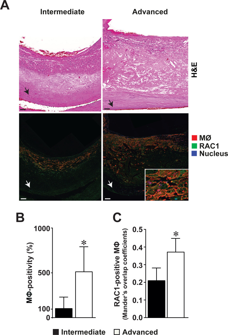
We observed that RAC1 was expressed within the intimal thickening of both intermediate and advanced atherosclerotic plaques of human carotid endarterectomies (Fig 1A). Co-localization studies indicated that RAC1 was expressed by intimal macrophages in atherosclerotic plaques (Fig 1A, right lower panel inset). An increased number of intimal macrophages were detected in advanced atherosclerotic lesions compared to intermediate plaques (100% versus 517%, p<0.05, Fig 1B). RAC1 expression was increased in intimal macrophages within the advanced atherosclerotic lesions compared to the macrophages within the intermediate lesions (0.41 versus 0.37 Mander´s overlap coefficient, p<0.05, Fig 1C). In addition to macrophages, RAC1 was expressed in vascular SMCs. Although not statistically significant, fewer RAC1-expressing SMCs were found in advanced lesions compared to intermediate lesions (0.75 versus 0.68 Mander´s overlap coefficient, p = 0.20, S1 Fig in S1 File).

Fig 1. Increased RAC1 expression in intimal macrophages of advanced human atherosclerotic carotid plaques.

Fig 1

(A) Hematoxylin & Eosin-stained sections of intermediate and advanced atherosclerotic plaques in human carotid endarterectomies (upper panels). Immunofluorescence detection of RAC1 (green), and macrophages (red, MΦ) in the intermediate and advanced atherosclerotic plaques (lower panels). Nuclear staining by DAPI (blue). Arrowhead points to the internal elastic lamina bordering the intimal thickening from the medial layer. Scale bars represent 100 μm. Co-localization of RAC1 expression in MΦ in an advanced intimal thickening shown in enlarged inset. (B) Number of macrophages in intermediate and advanced atherosclerotic plaques. (C) Number of macrophages expressing RAC1 expression within the intimal thickening of intermediate and advanced atherosclerotic plaques. Mean ± SEM values (n = 9 in each). Wilcoxon signed-rank test was used. *p<0.05.

Deficiency of RAC1 alters macrophage cell shape

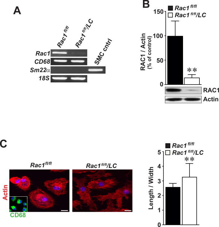
We produced mice with Rac1-deficient macrophages (Rac1fl/fl/LC) and extracted BMMs either from control Rac1fl/fl or Rac1fl/fl/LC mice. Rac1fl/fl/LC mice were fertile and did not show any pathology. Neither Rac1 mRNA (Fig 2A) nor RAC1 protein (Fig 2B) were detected in Rac1fl/fl/LC BMMs. These macrophages expressed CD68 mRNA and lacked SM22α mRNA, indicating the purity of the cultured BMMs (Fig 2A). Compared to Rac1fl/fl control BMMs, Rac1fl/fl/LC BMMs were more elongated as detected by immunofluorescence staining of actin phalloidin (Fig 2C, left panel) and quantification of cell length and width (Fig 2C, right graphs) (3.28 ± 0.06 folds versus 2.58 ± 0.04 folds, p<0.01). There was no difference in proliferation rates between Rac1fl/fl or Rac1fl/fl/LC BMMs, as assayed up to 5 days (S2A Fig in S1 File). Stimulation with LPS did not significantly alter the number of migrated Rac1fl/fl or Rac1fl/fl/LC BMMs after 4 and 16 hours (S2B Fig in S1 File).

Fig 2. Bone marrow-derived macrophages deficient for RAC1 display longer cell shape.

Fig 2

(A) RT-PCR analysis of Rac1, CD68, and Sm22α in cultured BMMs by gel electrophoresis. 18S mRNA serves as an internal loading control. (B) Immunoblot analysis of RAC1 in Rac1fl/fl and Rac1fl/fl/LC BMMs. Actin was included as an internal loading control. (C) Morphological shape of Rac1fl/fl and Rac1fl/fl/LC BMMs as detected by actin immunofluorescence staining (left images). Inset demonstrates CD68-positivity in BMMs. Scale bars represent 10 μm. Quantification of ratios of macrophage cell elongation (right graphs). Mean ± SEM values of percentage or fold changes in at least triplicated data. Student’s t-test was used. **p<0.01.

Mice lacking RAC1 in macrophages develop smaller aortic atherosclerotic plaques

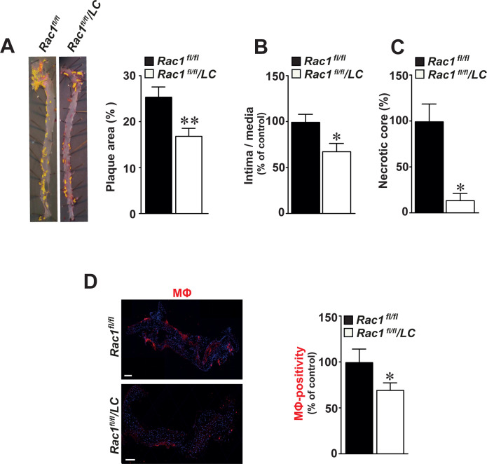
We observed a 33% reduction in the size of atherosclerotic plaques in whole aortas of Rac1fl/fl/LC mice infected with AdPCSK9 compared with Rac1fl/fl control mice (p<0.01, Fig 3A). Similarly, a 31% reduction in ratio of intima/media was observed in Rac1fl/fl/LC aortic arches (p<0.05, Fig 3B). Compared to Rac1fl/fl aortic controls, necrotic core areas formed within intimal thickening were reduced in Rac1fl/fl/LC aortas by 81% (p<0.05, Fig 3C). In these aortic arches, the number of CD68-positive macrophages was reduced by 30.2% compared with Rac1fl/fl control aortic arches (p = 0.05, Fig 3D). Once intimal thickening formed, there was no difference in collagen compositions as detected by Sirius red stain in Rac1fl/fl/LC and Rac1fl/fl aortas (p>0.05, S3 Fig in S1 File).

Fig 3. Mice lacking RAC1 in macrophages develop smaller aortic atherosclerotic plaques.

Fig 3

(A) Representative images of Sudan IV-stained atherosclerotic aortic plaques in aortas obtained from Rac1fl/fl/LC mice infected with AdPCSK9 (n = 12) as compared to Rac1fl/fl control mice infected with AdPCSK9 (n = 12) (left panels). Quantification of atherosclerotic plaque size by image analysis in Rac1fl/fl and Rac1fl/fl/LC aortas infected with AdPCSK9 (right graphs). (B) Intima/media ratios in Rac1fl/fl and Rac1fl/fl/LC aortic arches from mice infected with AdPCSK9. (C) Percentage of intimal necrotic core areas in Rac1fl/fl and Rac1fl/fl/LC aortic arches. (D) Immunofluorescent detection of macrophages (MΦ) using anti-CD68 antibodies in Rac1fl/fl and Rac1fl/fl/LC aortic arches from mice infected with AdPCSK9 (left panels). Number of MΦ in Rac1fl/fl and Rac1fl/fl/LC aortic arches (right graphs). Scale bars represent 100 μm. Mean ± SEM values. Student’s t-test was used. *p≤0.05.

Secretion of inflammatory cytokines is reduced in RAC1-deficient macrophages

Reduced secretion of IL-6 (5.2 ± 0.2 ng/ml versus 6.8 ± 0.3 ng/ml, p<0.01, Fig 4A) as well as TNF-α (0.8 ± 0.08 ng/ml versus 1.0 ± 0.04 ng/ml, p<0.05, Fig 4A) was observed in cultured Rac1fl/fl/LC BMMs compared to Rac1fl/fl controls. Similarly, serum levels of IL-6 were lower in atherogenic Rac1fl/fl/LC mice compared to Rac1fl/fl control mice as detected by ELISA (0.40 ± 0.08 ng/ml versus 0.71 ± 0.05 ng/ml, p<0.05, Fig 4B). However, serum levels of TNF-α did not reach statistical significance in Rac1fl/fl/LC mice (0.20 ± 0.09 ng/ml versus 0.31 ± 0.17 ng/ml, p>0.05, Fig 4B). IL-10 and IL-12 levels were not changed between Rac1fl/fl/LC and Rac1fl/fl BMMs and serum levels of these cytokines were not detectable in Rac1fl/fl/LC and Rac1fl/fl mice.

Fig 4. Secretion of inflammatory cytokines is reduced in bone marrow-derived macrophages deficient for RAC1.

Fig 4

(A) Secretion of IL-6 and TNF-α in primary Rac1fl/fl or Rac1fl/fl/LC BMMs as detected by ELISA (n = 6 in each) (B) Serum blood levels of secreted IL-6 and TNF-α in atherogenic Rac1fl/fl or Rac1fl/fl/LC mice (n = 6 in each). Mean ± SEM values. Student’s t-test was used. *p<0.05 and **p<0.01.

Deficiency of RAC1 in macrophages decreases lipid uptake

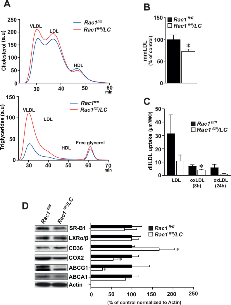
Compared to Rac1fl/fl control mice, Rac1fl/fl/LC mice produced higher serum levels of triglyceride, particularly in the VLDL/remnants and LDL-particles (Fig 5A, S4 Fig in S1 File). Compared to cultured Rac1fl/fl BMMs, Rac1fl/fl/LC BMMs displayed lower levels of mmLDL by 26% (p<0.05, Fig 5B). As compared to Rac1fl/fl BMMs, Rac1fl/fl/LC BMMs displayed reduced uptake of dil-labeled OxLDL after 8 hours (4.29 ± 0.29 versus 7.09 ± 0.92, p<0.05, Fig 5C).

Fig 5. Deficiency of RAC1 in bone marrow-derived macrophages decreases lipid uptake.

Fig 5

(A) Blood levels of cholesterol and triglyceride in fast protein liquid chromatography–fractionated plasma pooled from Rac1fl/fl mice (n = 5) that are infected with AdPCSK9 as compared to Rac1fl/fl/LC mice (n = 5) that are infected with AdPCSK9 after a high-fat diet for 24 weeks. HDL, high-density lipoprotein; IDL, intermediate-density lipoprotein; LDL, low-density lipoprotein; and VLDL, very-low-density lipoprotein. (B) Levels of minimally-modified LDL (mmLDL) in Rac1fl/fl and Rac1fl/fl/LC BMMs. Student’s t-test was used. Mean ± SEM values. (C) Uptake of total LDL and oxidized LDL (OxLDL) for 8 and 24 hours in Rac1fl/fl and Rac1fl/fl/LC BMMs. Multiple groups were statistically evaluated by ANOVA and the Tukey-Kramer modification of Tukey’s test and comparisons between two groups were evaluated by Student’s t test. (D) Representative Immunoblots and densitometry reading of SR-B1, LXRα/β, CD36, COX2, ABCG1, and ABCA1 bands in Rac1fl/fl and Rac1fl/fl/LC BMMs. Mean ± SEM values of triplicated data. Student’s t-test was used. *p<0.05.

We then assessed the expression levels of proteins that are involved in cholesterol metabolism using immunoblotting. Increased levels of CD36 (by 67%, p<0.05) and decreased levels of both COX2 (by 45%, p<0.05), and ABCG1 (by 71%, P<0.05) were observed in Rac1fl/fl/LC BMMs compared to control Rac1fl/fl BMMs (Fig 5D). However, no difference was observed in the expression of SR-B1, LXRα/β, and ABCA1 proteins.

RAC1 interacts with FLNA in macrophages

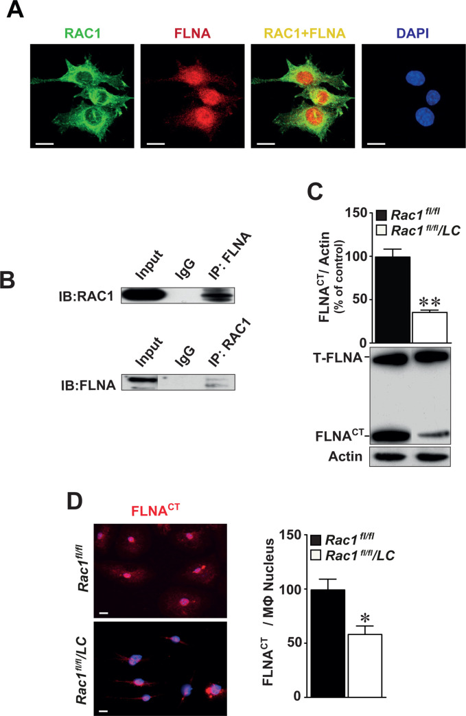
As cell polarity is regulated by the organization of cytoskeleton, partly by actin-binding proteins, and activity of RAC1 is reduced in macrophages deficient for FLNA [12], we first performed co-localization studies using immunofluorescently-labeled antibodies against RAC1 and FLNA. These data indicated that RAC1 and FLNA are co-expressed mainly in the cytoplasm of cultured BMMs (Fig 6A). Co-immunoprecipitation assay showed that RAC1 protein interacts physically with FLNA and vice versa (Fig 6B). In the absence of RAC1, we observed reduced levels of the cleaved C-terminal fragment of FLNA (FLNACT by 54%, p<0.01, Fig 6C). We also detected reduced levels of nuclear FLNACT expression in BMMs using immunofluorescence staining (Fig 6D, left panel) and quantified by image analysis (Fig 6D, right graph).

Fig 6. RAC1 interacts with FLNA in the cytoplasm and deletion of RAC1 reduces the production of cleaved C-terminal fragment of filamin A (FLNACT).

Fig 6

(A) Expression of RAC1 (green) and FLNA (red) is co-localized mainly in the cytoplasm of BMMs (yellow) as detected by immunofluorescence staining. Scale bar represents 10 μm. (B) Co-immunoprecipitation identifying FLNACT as an interacting partner of RAC1. Total proteins obtained from BMMs immunoprecipitated with FLNACT and then immunoblotted with RAC1 antibodies (upper) or vice versa (lower). IgG served as negative controls. Full-length of FLNA and FLNACT were indicated and actin served as internal loading control. (C) Immunoblotting of FLNA in Rac1fl/fl and Rac1fl/fl/LC BMMs. (D) Immunofluorescence staining of FLNACT in Rac1fl/fl and Rac1fl/fl/LC BMMs. Quantification of nuclear FLNACT expression in Rac1fl/fl or Rac1fl/fl/LC BMMs. Scale bar represents 20 μm. Mean ± SEM values of at least quadruplicated experiments. Student’s t-test was used. *p<0.05, **p<0.01.

Discussion

This study is the first to show that lack of RAC1 in macrophages in vivo inhibits aortic plaque size in an in vivo model of atherosclerosis. Macrophage functions including secretion of interleukins and foam cell formation are key events during the development of atherosclerosis. In the absence of RAC1, macrophages displayed impaired secretion of proinflammatory cytokines, reduced lipid uptake and foam cell formation. Furthermore, cytoskeletal FLNACT produced by calpain cleavage physically interacted with RAC1 and we have previously shown that lack of FLNA reduces macrophage activity and atherosclerosis [12]. These new insights not only provide an evident link between the biological role of RAC1 and atherogenesis, but also identify targets that may modify atherosclerotic plaque formation.

Studies in macrophages identified RAC1 as a regulator of the organization of actin cytoskeleton [24]. In our study, RAC1-deficiency in macrophages resulted in elongated cell shape. Besides, it is known that RAC1-deficiency reduces filopodial formation, but filopodial structures that are formed display increased extension [25]. Similarly, our images confirm this previous finding as RAC1-deficiency in macrophages leads to longer filopodial structures, but in less number. In addition to cytoskeletal functions, RAC1 regulates adhesion and migration [26], cell spreading and ruffling [25], phagocytosis [27], and transcriptional factors [28]. These pieces of evidence support a key role for RAC1 in many aspects of atherosclerotic plaque development. In this study, we observed an increased number of RAC1-expressing macrophages in advanced human carotid atherosclerotic plaques. As the knowledge on these pathophysiological processes modulated by macrophage-specific RAC1 is scanty and sometime contradictory, we initiated in vitro and in vivo experimental studies. To define whether elevated RAC1 expression in human atherosclerotic plaques may have a biological role in macrophages, we induced atherosclerosis in mice deficient for RAC1 in macrophages and observed fewer macrophages as well as smaller atherosclerotic plaque size. RAC1-deficiency in macrophages did not alter collagen composition, but reduced the size of necrotic cores within the atherosclerotic plaques.

Macrophages secrete multiple pro- and anti-inflammatory cytokines, as well as, depend on diverse stimuli. Currently, data points to interleukins and TNF-α as cytokines that, at least in some settings, are effective targets to reduce cardiovascular disease progression [29]. We observed decreased levels of the proinflammatory cytokine IL-6 and TNF-α in Rac1-deficient BMMs. It has been shown that IL-6 secretion is regulated via sphingosine-1-phosphate receptor 2 involving RHO/RHO-kinase and RAC1 signaling pathways [30]. Furthermore, studies using protein kinase inhibitors and dominant negative constructs demonstrate phosphatidylinositol 3-kinase/RAC1/PAK/JNK and phosphatidylinositol 3-kinase/RAC1/PAK/p38 pathways contribute to important roles in the late stages of TNF-α down-regulation of macrophage scavenger receptor expression [31]. We have earlier reported that both FLNA deficiency and inhibition of cleavage of FLNACT by calpeptin results in reduced macrophage cell migration and proliferation [12]. However, RAC1-deficient BMMs expressing reduced levels of FLNACT did not show differences in macrophage cell proliferation or migration in the present study.

RAC1 activation induced by free cholesterol accumulation in the plasma membrane is partly due to the activity of the membrane transporters, which modulates plasma membrane cholesterol content and lipid organization and, in the absence of an efficient extracellular acceptor, may cause cholesterol accumulation. The uptake and degradation of matrix-bound lipoproteins by macrophages require Rho family GTPases [24]. We found that Rac1-deficient BMMs display impaired lipid uptake as well as altered levels of CD36, COX2, and ABCG1. The fact that RAC1-deficient macrophages showed a decreased uptake of oxLDL despite elevated CD36 protein expression seems to be counterintuitive since CD36 has been reported to be an oxLDL receptor [32]. However, our experiments were performed in the absence of fatty acids which are important for binding of oxLDL to CD36 [23, 33]. The finding on the interaction between oxidized oxLDL and CD36 inducing loss of macrophage polarity and inhibiting macrophage locomotion [34], supports our observation on elevated CD36 expression in Rac1-deficient BMMs with impaired polarity and cell shape. Macrophage activation during motility increases COX2 protein levels [35]. Interestingly, our study showed that RAC1 deficiency in BMMs decreases COX2 levels. Furthermore, ABCG1 is robustly upregulated in macrophages taken from obese mice and regulates macrophage cholesterol levels [36]. Loss of ABCG1 from hematopoietic cells results in smaller atherosclerotic lesions populated with increased apoptotic macrophages [37]. Similarly, we found decreased levels of ABCG1 in Rac1-deficient BMMs that show decreased lipid uptake, which resulted in smaller fatty atherosclerotic plaques.

Once retention of LDL takes place in the subendothelium, the internalization and degradation of matrix-retained and aggregated LDL by macrophages may involve the actin-myosin cytoskeleton [24]. In addition, phagocytosis is the mechanism of internalization used by specialized cells such as macrophages and also requires macrophage shape changes and phenotypic polarization by actin filaments. Recently, we reported that RAC1 activity is reduced in FLNA-deficient BMMs [12]. In this study, we identified actin-binding protein FLNA as an interacting partner of RAC1 in BMMs. Interestingly, RAC1 activators and downstream effectors control the local reorganization of the actin cytoskeleton beneath bound particles during ingestion [24]. FLNA is required for differentiation of monocytes by regulating actin dynamics via Rho GTPases that control monocyte migration [24] and also may be a scaffold for the spatial organization of Rho-GTPase-mediated signaling pathways [11]. In the absence of FLNA, levels of active RAC1 are reduced [18]. Human FLNA mutations associated with valvular dystrophy alter the balance between RHOA and RAC1 GTPases activities in favor of RHOA, providing evidence for a role of the RAC1-specific GTPase activating protein FilGAP, a RAC-specific Rho-GTPase-activating protein [38]. Furthermore, we observed lower levels of cleaved FLNACT and its nuclear expression in Rac1-deficient BMMs in this study. We have recently reported that genetic inactivation of FLNA and chemical inhibition of calpain-dependent cleavage of FLNA impairs macrophage signaling and function, and reduces atherosclerosis in mice [12]. Thus, this way of chemical inhibition would also be administered to target RAC1 signaling.

Overall, our study reporting RAC1-dependent cytokine release and lipid biology in macrophages and FLNA as an interacting partner of RAC1 may indicate new potential mechanisms behind atherogenesis in vivo. The finding of increased RAC1 expression in human macrophages within advanced carotid artery plaques suggests that RAC1 expression can be a prognostic biomarker for atherogenesis. Besides, RAC1 represents an attractive therapeutic target for cardiovascular diseases; however, the clinical search for effective RAC1 inhibitors is still underway [4]. Nevertheless, characterizing these novel mechanisms provide invaluable information regarding major macrophage events that are mediated by aberrant RAC1 signaling. Importantly, our results can be utilized to further facilitate the development of effective pharmacological agents that can inhibit RAC1 signaling in macrophages in atherosclerosis-related cardiovascular disorders.

Supporting information

S1 File

(PDF)

Acknowledgments

The authors thank Kristina Skålen (The Wallenberg Laboratory, University of Gothenburg, Sweden), Shahin de Lara and Samad Parhizkar (Department of Clinical Pathology, Sahlgrenska University Hospital, Sweden) for excellent technical help and Toshima Parris (University of Gothenburg, Sweden) for editing the manuscript.

Abbreviations

AdPCSK9

adenoviral vector overexpressing proprotein convertase subtilisin/kexin type 9

BMMs

bone marrow macrophages

FLNA

filamin A

FLNACT

C-terminal fragment of FLNA

LC

lysosome M promoter

mmLDL

minimally oxidized-LDL

oxLDL

oxidized LDL

RAC1

RAS-related C3 botulinum toxin substrate 1

Rac1fl/fl

mice homozygously expressing the floxed Rac1 gene

Rac1fl/fl/LC

mice lacking Rac1 in macrophages

SMCs

smooth muscle cells

Data Availability

All relevant data are within the manuscript and its Supporting Information files.

Funding Statement

This work was supported by the Strategic Fund from the Institute of Biomedicine, University of Gothenburg, the Swedish Cancer Foundation (Contract number 17 0171), and the ALF fund (ALFGBG-495961) from the Sahlgrenska Academy Hospital in the Västra Götalandregionen to LMA.

References

  • 1.Getz GS, Reardon CA. Natural killer T cells in atherosclerosis. Nat Rev Cardiol. 2017;14(5):304–314. 10.1038/nrcardio.2017.2 . [DOI] [PubMed] [Google Scholar]
  • 2.Pourcet B, Staels B. Alternative macrophages in atherosclerosis: not always protective! J Clin Invest. 2018;128(3):910–912. 10.1172/JCI120123 . [DOI] [PMC free article] [PubMed] [Google Scholar]
  • 3.Saric M, Kronzon I. Aortic atherosclerosis and embolic events. Curr Cardiol Rep. 2012;14(3):342–349. 10.1007/s11886-012-0261-2 . [DOI] [PubMed] [Google Scholar]
  • 4.Marei H, Malliri A. Rac1 in human diseases: The therapeutic potential of targeting Rac1 signaling regulatory mechanisms. Small GTPases. 2017;8(3):139–163. 10.1080/21541248.2016.1211398 . [DOI] [PMC free article] [PubMed] [Google Scholar]
  • 5.Reijnders MRF, Ansor NM, Kousi M, Yue WW, Tan PL, Clarkson K, et al. RAC1 Missense Mutations in Developmental Disorders with Diverse Phenotypes. Am J Hum Genet. 2017;101(3):466–477. 10.1016/j.ajhg.2017.08.007 . [DOI] [PMC free article] [PubMed] [Google Scholar]
  • 6.Sugihara K, Nakatsuji N, Nakamura K, Nakao K, Hashimoto R, Otani H, et al. Rac1 is required for the formation of three germ layers during gastrulation. Oncogene. 1998;17(26):3427–3433. 10.1038/sj.onc.1202595 . [DOI] [PubMed] [Google Scholar]
  • 7.Satoh M, Ogita H, Takeshita K, Mukai Y, Kwiatkowski DJ, Liao JK. Requirement of Rac1 in the development of cardiac hypertrophy. Proc Natl Acad Sci U S A. 2006;103(19):7432–7437. 10.1073/pnas.0510444103 . [DOI] [PMC free article] [PubMed] [Google Scholar]
  • 8.Hwaiz R, Hasan Z, Rahman M, Zhang S, Palani K, Syk I, et al. Rac1 signaling regulates sepsis-induced pathologic inflammation in the lung via attenuation of Mac-1 expression and CXC chemokine formation. J Surg Res. 2013;183(2):798–807. 10.1016/j.jss.2013.02.045 . [DOI] [PubMed] [Google Scholar]
  • 9.Moldovan L, Irani K, Moldovan NI, Finkel T, Goldschmidt-Clermont PJ. The actin cytoskeleton reorganization induced by Rac1 requires the production of superoxide. Antioxid Redox Signal. 1999;1(1):29–43. 10.1089/ars.1999.1.1-29 . [DOI] [PubMed] [Google Scholar]
  • 10.Khan OM, Akula MK, Skalen K, Karlsson C, Stahlman M, Young SG, et al. Targeting GGTase-I activates RHOA, increases macrophage reverse cholesterol transport, and reduces atherosclerosis in mice. Circulation. 2013;127(7):782–790. 10.1161/CIRCULATIONAHA.112.000588 . [DOI] [PMC free article] [PubMed] [Google Scholar]
  • 11.Bellanger JM, Astier C, Sardet C, Ohta Y, Stossel TP, Debant A. The Rac1- and RhoG-specific GEF domain of Trio targets filamin to remodel cytoskeletal actin. Nat Cell Biol. 2000;2(12):888–892. 10.1038/35046533 . [DOI] [PubMed] [Google Scholar]
  • 12.Bandaru S, Ala C, Salimi R, Akula MK, Ekstrand M, Devarakonda S, et al. Targeting filamin A reduces macrophage activity and atherosclerosis. Circulation. 2019;140(1):67–79. 10.1161/CIRCULATIONAHA.119.039697 . [DOI] [PubMed] [Google Scholar]
  • 13.Ekstrand M, Widell E, Hammar A, Akyurek LM, Johansson M, Fagerberg B, et al. Depletion of ATP and glucose in advanced human atherosclerotic plaques. PLoS One. 2017;12(6):e0178877 10.1371/journal.pone.0178877 . [DOI] [PMC free article] [PubMed] [Google Scholar]
  • 14.Bolte S, Cordelieres FP. A guided tour into subcellular colocalization analysis in light microscopy. J Microsc. 2006;224(Pt 3):213–232. 10.1111/j.1365-2818.2006.01706.x . [DOI] [PubMed] [Google Scholar]
  • 15.Schneider CA, Rasband WS, Eliceiri KW. NIH Image to ImageJ: 25 years of image analysis. Nat Methods. 2012;9(7):671–675. 10.1038/nmeth.2089 . [DOI] [PMC free article] [PubMed] [Google Scholar]
  • 16.Huang Z, Kim J, Lacruz RS, Bringas P Jr., Glogauer M, Bromage TG, et al. Epithelial-specific knockout of the Rac1 gene leads to enamel defects. Eur J Oral Sci. 2011;119 Suppl 1:168–176. 10.1111/j.1600-0722.2011.00904.x . [DOI] [PMC free article] [PubMed] [Google Scholar]
  • 17.Nallapalli RK, Ibrahim MX, Zhou AX, Bandaru S, Sunkara SN, Redfors B, et al. Targeting filamin A reduces K-RAS-induced lung adenocarcinomas and endothelial response to tumor growth in mice. Mol Cancer. 2012;11:50 10.1186/1476-4598-11-50 . [DOI] [PMC free article] [PubMed] [Google Scholar]
  • 18.Bandaru S, Grönros J, Redfors B, Cil C, Pazooki D, Salimi R, et al. Deficiency of filamin A in endothelial cells impairs left ventricular remodelling after myocardial infarction. Cardiovasc Res. 2015;105(2):151–159. 10.1093/cvr/cvu226 . [DOI] [PubMed] [Google Scholar]
  • 19.Björklund MM, Hollensen AK, Hagensen MK, Dagnaes-Hansen F, Christoffersen C, Mikkelsen JG, et al. Induction of Atherosclerosis in Mice and Hamsters Without Germline Genetic Engineering. Circulation Research. 2014;114(11):1684–1689. 10.1161/CIRCRESAHA.114.302937 . [DOI] [PubMed] [Google Scholar]
  • 20.Sayin VI, Khan OM, Pehlivanoglu LE, Staffas A, Ibrahim MX, Asplund A, et al. Loss of One Copy of Zfp148 Reduces Lesional Macrophage Proliferation and Atherosclerosis in Mice by Activating p53. Circulation Research. 2014;115(9):781–791. 10.1161/CIRCRESAHA.115.304992 . [DOI] [PubMed] [Google Scholar]
  • 21.Parini P, Johansson L, Broijersen A, Angelin B, Rudling M. Lipoprotein profiles in plasma and interstitial fluid analyzed with an automated gel-filtration system. Eur J Clin Invest. 2006;36(2):98–104. 10.1111/j.1365-2362.2006.01597.x . [DOI] [PubMed] [Google Scholar]
  • 22.Pedrelli M, Davoodpour P, Degirolamo C, Gomaraschi M, Graham M, Ossoli A, et al. Hepatic ACAT2 knock down increases ABCA1 and modifies HDL metabolism in mice. PLoS One. 2014;9(4):e93552 10.1371/journal.pone.0093552 . [DOI] [PMC free article] [PubMed] [Google Scholar]
  • 23.Håversen L, Sundelin JP, Mardinoglu A, Rutberg M, Stahlman M, Wilhelmsson U, et al. Vimentin deficiency in macrophages induces increased oxidative stress and vascular inflammation but attenuates atherosclerosis in mice. Sci Rep. 2018;8(1):16973 10.1038/s41598-018-34659-2 . [DOI] [PMC free article] [PubMed] [Google Scholar]
  • 24.Sakr SW, Eddy RJ, Barth H, Wang F, Greenberg S, Maxfield FR, et al. The uptake and degradation of matrix-bound lipoproteins by macrophages require an intact actin Cytoskeleton, Rho family GTPases, and myosin ATPase activity. J Biol Chem. 2001;276(40):37649–37658. 10.1074/jbc.M105129200 . [DOI] [PubMed] [Google Scholar]
  • 25.Wells CM, Walmsley M, Ooi S, Tybulewicz V, Ridley AJ. Rac1-deficient macrophages exhibit defects in cell spreading and membrane ruffling but not migration. J Cell Sci. 2004;117(Pt 7):1259–1268. 10.1242/jcs.00997 . [DOI] [PubMed] [Google Scholar]
  • 26.Ridley AJ. Regulation of macrophage adhesion and migration by Rho GTP-binding proteins. J Microsc. 2008;231(3):518–523. 10.1111/j.1365-2818.2008.02064.x . [DOI] [PubMed] [Google Scholar]
  • 27.Castellano F, Montcourrier P, Chavrier P. Membrane recruitment of Rac1 triggers phagocytosis. J Cell Sci. 2000;113 (Pt 17):2955–2961. . [DOI] [PubMed] [Google Scholar]
  • 28.Bosco EE, Mulloy JC, Zheng Y. Rac1 GTPase: a "Rac" of all trades. Cell Mol Life Sci. 2009;66(3):370–374. 10.1007/s00018-008-8552-x . [DOI] [PMC free article] [PubMed] [Google Scholar]
  • 29.Williams JW, Huang LH, Randolph GJ. Cytokine circuits in cardiovascular disease. Immunity. 2019;50(4):941–954. 10.1016/j.immuni.2019.03.007 . [DOI] [PMC free article] [PubMed] [Google Scholar]
  • 30.Goyal P, Brunnert D, Ehrhardt J, Bredow M, Piccenini S, Zygmunt M. Cytokine IL-6 secretion by trophoblasts regulated via sphingosine-1-phosphate receptor 2 involving Rho/Rho-kinase and Rac1 signaling pathways. Mol Hum Reprod. 2013;19(8):528–538. 10.1093/molehr/gat023 . [DOI] [PubMed] [Google Scholar]
  • 31.Hsu HY, Twu YC. Tumor necrosis factor-a -mediated protein kinases in regulation of scavenger receptor and foam cell formation on macrophage. J Biol Chem. 2000;275(52):41035–41048. 10.1074/jbc.M003464200 . [DOI] [PubMed] [Google Scholar]
  • 32.Endemann G, Stanton LW, Madden KS, Bryant CM, White RT, Protter AA. CD36 is a receptor for oxidized low density lipoprotein. J Biol Chem. 1993;268(16):11811–11816. . [PubMed] [Google Scholar]
  • 33.Jay AG, Chen AN, Paz MA, Hung JP, Hamilton JA. CD36 binds oxidized low density lipoprotein (LDL) in a mechanism dependent upon fatty acid binding. J Biol Chem. 2015;290(8):4590–4603. 10.1074/jbc.M114.627026 . [DOI] [PMC free article] [PubMed] [Google Scholar]
  • 34.Park YM, Drazba JA, Vasanji A, Egelhoff T, Febbraio M, Silverstein RL. Oxidized LDL/CD36 interaction induces loss of cell polarity and inhibits macrophage locomotion. Mol Biol Cell. 2012;23(16):3057–3068. 10.1091/mbc.E11-12-1051 . [DOI] [PMC free article] [PubMed] [Google Scholar]
  • 35.Go A, Ryu YK, Lee JW, Moon EY. Cell motility is decreased in macrophages activated by cancer cell-conditioned medium. Biomol Ther (Seoul). 2013;21(6):481–486. 10.4062/biomolther.2013.076 . [DOI] [PMC free article] [PubMed] [Google Scholar]
  • 36.Wei H, Tarling EJ, McMillen TS, Tang C, LeBoeuf RC. ABCG1 regulates mouse adipose tissue macrophage cholesterol levels and ratio of M1 to M2 cells in obesity and caloric restriction. J Lipid Res. 2015;56(12):2337–2347. 10.1194/jlr.M063354 . [DOI] [PMC free article] [PubMed] [Google Scholar]
  • 37.Tarling EJ, Bojanic DD, Tangirala RK, Wang X, Lovgren-Sandblom A, Lusis AJ, et al. Impaired development of atherosclerosis in Abcg1–/–Apoe–/–mice: identification of specific oxysterols that both accumulate in Abcg1–/–Apoe–/–tissues and induce apoptosis. Arterioscler Thromb Vasc Biol. 2010;30(6):1174–1180. 10.1161/ATVBAHA.110.205617 . [DOI] [PMC free article] [PubMed] [Google Scholar]
  • 38.Duval D, Lardeux A, Le Tourneau T, Norris RA, Markwald RR, Sauzeau V, et al. Valvular dystrophy associated filamin A mutations reveal a new role of its first repeats in small-GTPase regulation. Biochim Biophys Acta. 2014;1843(2):234–244. 10.1016/j.bbamcr.2013.10.022 . [DOI] [PMC free article] [PubMed] [Google Scholar]

Decision Letter 0

Esther Lutgens

Transfer Alert

This paper was transferred from another journal. As a result, its full editorial history (including decision letters, peer reviews and author responses) may not be present.

19 Mar 2020

PONE-D-20-06077

Lack of RAC1 in macrophages protects against atherosclerosis

PLOS ONE

Dear Dr Ayürek,,

Thank you for submitting your manuscript to PLOS ONE. After careful consideration, we feel that it has merit but does not fully meet PLOS ONE’s publication criteria as it currently stands. Therefore, we invite you to submit a revised version of the manuscript that addresses the points raised during the review process.

Please answer all the questions or reviewers 1 and 2.

We would appreciate receiving your revised manuscript by July 2020. When you are ready to submit your revision, log on to https://www.editorialmanager.com/pone/ and select the 'Submissions Needing Revision' folder to locate your manuscript file.

If you would like to make changes to your financial disclosure, please include your updated statement in your cover letter.

To enhance the reproducibility of your results, we recommend that if applicable you deposit your laboratory protocols in protocols.io, where a protocol can be assigned its own identifier (DOI) such that it can be cited independently in the future. For instructions see: http://journals.plos.org/plosone/s/submission-guidelines#loc-laboratory-protocols

Please include the following items when submitting your revised manuscript:

  • A rebuttal letter that responds to each point raised by the academic editor and reviewer(s). This letter should be uploaded as separate file and labeled 'Response to Reviewers'.

  • A marked-up copy of your manuscript that highlights changes made to the original version. This file should be uploaded as separate file and labeled 'Revised Manuscript with Track Changes'.

  • An unmarked version of your revised paper without tracked changes. This file should be uploaded as separate file and labeled 'Manuscript'.

Please note while forming your response, if your article is accepted, you may have the opportunity to make the peer review history publicly available. The record will include editor decision letters (with reviews) and your responses to reviewer comments. If eligible, we will contact you to opt in or out.

We look forward to receiving your revised manuscript.

Kind regards,

Esther Lutgens

Academic Editor

PLOS ONE

Journal Requirements:

When submitting your revision, we need you to address these additional requirements.

1. Please ensure that your manuscript meets PLOS ONE's style requirements, including those for file naming. The PLOS ONE style templates can be found at

http://www.journals.plos.org/plosone/s/file?id=wjVg/PLOSOne_formatting_sample_main_body.pdf and http://www.journals.plos.org/plosone/s/file?id=ba62/PLOSOne_formatting_sample_title_authors_affiliations.pdf

2. PLOS ONE now requires that authors provide the original uncropped and unadjusted images underlying all blot or gel results reported in a submission’s figures or Supporting Information files. This policy and the journal’s other requirements for blot/gel reporting and figure preparation are described in detail at https://journals.plos.org/plosone/s/figures#loc-blot-and-gel-reporting-requirements and https://journals.plos.org/plosone/s/figures#loc-preparing-figures-from-image-files. When you submit your revised manuscript, please ensure that your figures adhere fully to these guidelines and provide the original underlying images for all blot or gel data reported in your submission. See the following link for instructions on providing the original image data: https://journals.plos.org/plosone/s/figures#loc-original-images-for-blots-and-gels.

In your cover letter, please note whether your blot/gel image data are in Supporting Information or posted at a public data repository, provide the repository URL if relevant, and provide specific details as to which raw blot/gel images, if any, are not available. Email us at plosone@plos.org if you have any questions.

[Note: HTML markup is below. Please do not edit.]

Reviewers' comments:

Reviewer's Responses to Questions

Comments to the Author

1. Is the manuscript technically sound, and do the data support the conclusions?

The manuscript must describe a technically sound piece of scientific research with data that supports the conclusions. Experiments must have been conducted rigorously, with appropriate controls, replication, and sample sizes. The conclusions must be drawn appropriately based on the data presented.

Reviewer #1: Partly

Reviewer #2: Yes

**********

2. Has the statistical analysis been performed appropriately and rigorously?

Reviewer #1: Yes

Reviewer #2: Yes

**********

3. Have the authors made all data underlying the findings in their manuscript fully available?

The PLOS Data policy requires authors to make all data underlying the findings described in their manuscript fully available without restriction, with rare exception (please refer to the Data Availability Statement in the manuscript PDF file). The data should be provided as part of the manuscript or its supporting information, or deposited to a public repository. For example, in addition to summary statistics, the data points behind means, medians and variance measures should be available. If there are restrictions on publicly sharing data—e.g. participant privacy or use of data from a third party—those must be specified.

Reviewer #1: No

Reviewer #2: Yes

**********

4. Is the manuscript presented in an intelligible fashion and written in standard English?

PLOS ONE does not copyedit accepted manuscripts, so the language in submitted articles must be clear, correct, and unambiguous. Any typographical or grammatical errors should be corrected at revision, so please note any specific errors here.

Reviewer #1: No

Reviewer #2: Yes

**********

5. Review Comments to the Author

Please use the space provided to explain your answers to the questions above. You may also include additional comments for the author, including concerns about dual publication, research ethics, or publication ethics. (Please upload your review as an attachment if it exceeds 20,000 characters)

Reviewer #1: The manuscript of Akyürek and colleagues describes the interesting finding of decreased atherogenesis upon macrophage-specific deletion of RAC1. The authors show an increased expression of RAC1 in macrophages in human advanced plaques and demonstrate that deletion of RAC1 in (mouse) macrophages affects their shape and function. Differences in shape are explained by interactions between RAC1 and FLNA to remodel the cytoskeleton for migration and lipid uptake. Mice with RAC1-deficient macrophages show decreased plaque size and decreased macrophage content in the plaques. In vitro, isolated macrophages show decreased inflammatory activation by decreased cytokine secretion and decreased lipid uptake. The data described in this manuscript are potentially of significance for clinical application and in the identification of novel therapeutic targets.

However, some key information seems to be missing:

1. First, in the in vitro experiments cultured macrophages are used to validate in vivo findings. However, the culturing and activation of these cells is not clearly described. Concerning the isolation, the material and method section refers to a paper in which only the culturing is explained. Throughout the result section it is not always clear if the authors are talking about cultured BMMs or about lesional macrophages nor what they mean with “extracted macrophages”. It should be clarified in the M&M, text, figures and legends which macrophages are being investigated and how they were stimulated (included, timing and concentrations). This crucial information is now missing. Moreover, untreated macrophages should be shown along to their LPS-treated ones.

2. The authors indicate purity of the cultured macrophages by showing a lack of SM22a mRNA in the culture. However, it would be more informative if the purity is validated by FACS staining for both CD11b and F4/80 as the absence of SM22a does not prove purity.

3. Authors state in the discussion that macrophages display impaired secretion of proinflammatory cytokines, but only show that some are impaired and some are not. Also, it is unclear how these macrophages are activated. What is the concentration and the timing of the LPS used in this study? It would be of added value to also plot the cytokine secretion of unstimulated macrophages. Where in vitro 2 out of 4 cytokines show a decrease in secreted cytokine levels, in the serum only 1 cytokine was measured. Are serum cytokine levels behaving similarly to cytokine secretion by cultured macrophages?

4. The authors show a decreased ability for lipid uptake in the RAC1 deficient macrophages, but do not show the implications of the found proteins in this pathway. In fact, the deregulation of the proteins appears in conflict to the observed changes in lipid uptake. This should at least be discussed.

5. In the final paragraph of the results, the mechanistic link between RAC1 and FLNA is shown, but the rationale of doing these experiments is missing in there. It is unclear what the importance and relevance of these results is and this becomes only somewhat apparent when reading the discussion. The results section should be improve in a way that readers understand why particular experiments were done and what the importance of the findings is.

6. In the last paragraph of the discussion, the authors state to have found a causative correlation between RAC1-dependent cytokine release in macrophages. This should be rephrased or should be functionally proven as the current layout of experiments does not allow to make such statements.

7. Figure 1 shows the positivity of macrophages in pixels. It would be of added value to plot the data per µm2 or as a ratio to the total amount of pixels.

8. Figure 3C shows a reduced macrophage-positivity in RAC1fl/fl/LC. How is this quantified? Is this per µm2 of plaque, or in the picture in general? Could smaller plaques explain reduced numbers of macrophages seen in this picture? This should be clarified.

9. Figure 6B (bottom) and C shows an immunoblot which has clearly been edited (cut and paste). are these data from different blots? This can only be allowed if the complete blots are provided as supplemental data, highlight the parts that were cut and used in the actual figures. Preferentially, a blot should be performed in which the conditions of interest are blotted next to each other. Otherwise, the protein levels cannot be directly compared.

Moreover, the text should be reviewed and edited by a native English speaker to correct difficult statements and sentences. For example: accompanied with -> accompanied by; “macrophages were more elongated compared to Rac1fl/fl control macrophages using immunofluorescence staining of actin phalloidin”; “Compared to Rac1fl/fl controls, higher serum levels of triglyceride, particularly in the VLDL/remnants and LDL-particles, were observed in Rac1fl/fl/LC mice (Fig 5A).”; “macrophages displayed lower levels of mmLDL by 26% compared with Rac1fl/fl macrophages”, etc.

Reviewer #2: Bandaru et al Pone 2020

The manuscript by Bandaru et al. describes the presence of RAC1 in macrophages and smooth muscle cells in human atherosclerotic plaques, prompting them to investigate its role in experimental atherosclerosis and macrophages using a Lysosyme-driven Cre recombinase approach.

They show that mice lacking RAC1 in macorphages develop smaller lesions, and that the macrophages are less inflammatory, and decreased lipid uptake.

The manuscript seems technically sound.

A few issues need to be addressed.

1. Fig 2C. The authors describe that the macrophages have an altered length-to-width ratio. However, the KO macrophages also seem to develop more filopodia-like structures, which remains undiscussed

2. Fig. S2B. The authors measure macrophage migration. Please provide the rationale for measuring this parameter. In addition, the methods do not seem to be described.

3. Plaques of KO mice are smaller, however what are other plaque parameters such as severity, necrosis, collagen content,...

4. Fig3B. How was intima/media ratio in mice measured?

5. Fig. 3C: was this total macrophage area or relative to plaque size?

6. The authors describe the inflammatory parameters of the KO macrophages and in KO mice. Please clearly/explicitely describe what was measeured in vitro and what was measured in vivo. Was in vitro secretion after inflammatory stimulation or in basal conditions? What were the levels of TNF in vivo? And given the recent results in the Cantos trial, IL1b measurements could strengthen the paper

7. Fig. 5A. What were total serum cholesterol and TG levels?

8. Fig5B. The authors describe KO macrophages to display lower levels of mmLDL. How was this measured? Or do the authors mean reduced uptake of mmLDL?

9. The authors nicely show FLNA interaction in macrophages. Can they also provide evidence of reduced levels in the KO mice in vivo? And localization of FLNA in the human plaques?

**********

6. PLOS authors have the option to publish the peer review history of their article (what does this mean?). If published, this will include your full peer review and any attached files.

If you choose “no”, your identity will remain anonymous but your review may still be made public.

Do you want your identity to be public for this peer review? For information about this choice, including consent withdrawal, please see our Privacy Policy.

Reviewer #1: No

Reviewer #2: Yes: Kristiaan Wouters

[NOTE: If reviewer comments were submitted as an attachment file, they will be attached to this email and accessible via the submission site. Please log into your account, locate the manuscript record, and check for the action link "View Attachments". If this link does not appear, there are no attachment files to be viewed.]

While revising your submission, please upload your figure files to the Preflight Analysis and Conversion Engine (PACE) digital diagnostic tool, https://pacev2.apexcovantage.com/. PACE helps ensure that figures meet PLOS requirements. To use PACE, you must first register as a user. Registration is free. Then, login and navigate to the UPLOAD tab, where you will find detailed instructions on how to use the tool. If you encounter any issues or have any questions when using PACE, please email us at figures@plos.org. Please note that Supporting Information files do not need this step.

PLoS One. 2020 Sep 17;15(9):e0239284. doi: 10.1371/journal.pone.0239284.r002

Author response to Decision Letter 0


31 May 2020

Reviewer #1:

1. First, in the in vitro experiments cultured macrophages are used to validate in vivo findings.However, the culturing and activation of these cells is not clearly described. Concerning the isolation, the material and method section refers to a paper in which only the culturing is explained. Throughout the result section it is not always clear if the authors are talking about cultured BMMs or about lesional macrophages nor what they mean with “extracted macrophages”. It should be clarified in the M&M, text, figures and legends which macrophages are being investigated and how they were stimulated (included, timing and concentrations). This crucial information is now missing. Moreover, untreated macrophages should be shown along to their LPS-treated ones.

Thanks for these comments and we do apologize for the confusion. In this study, we have not cultured lesional macrophages and we have presented assays using bone marrow macrophages (BMMs) exposed to LPS in all experimental groups. Thus, the wording “macrophages” within the context of our experiments has been replaced with “BMMs” throughout the manuscript including M&M, text body, figures, and figure legends as necessary. We have extracted bone marrow monocytes from both femur and tibia of the mice as described elsewhere (ref# 12). To differentiate monocytes into macrophages, bone marrow monocytes were cultured with DMEM medium containing 10% FBS, 1% glutamine, 1% gentamicin,1% HEPES, 0.01% β-mercaptoethanol, and 10% CMG14-12 cell line supernatant as a source of mouse M-CSF. One week later, BMMs were plated and assayed up to 3 days. We have now provided this information (page 7, lines 4–8). We stimulated BMMs with 10 ng/ml LPS 15 minutes before immunoblotting and 8 hours before ELISA (page 7, lines 20-21). As requested, additional experimental groups without LPS have been included in experimental setting. We tested untreated BMMs for ELISA (Reviewer Figure 1) as well as for immunoblotting of the cultured BMMs without LPS (Reviewer Figure 2), however, these assays did not result in significant differences. Please see the attached figure including these untreated BMMs along with LPS-treated BMMs to convince the Reviewer.

2. The authors indicate purity of the cultured macrophages by showing a lack of SM22a mRNA in the culture. However, it would be more informative if the purity is validated by FACS staining for both CD11b and F4/80 as the absence of SM22a does not prove purity.

In addition to experimental proof of purity by mRNA expression, we have immunofluorescently stained cultured BMMs using a macrophage-specific CD68 antibody as reported earlier (ref# 12). An inset of image demonstrating CD68-positive BMMs has been included in Figure 2C. This information has been added to M&M (page 7, line 15-16) and figure legends.

3. Authors state in the discussion that macrophages display impaired secretion of proinflammatory cytokines, but only show that some are impaired and some are not. Also, it is unclear how these macrophages are activated. What is the concentration and the timing of the LPS used in this study? It would be of added value to also plot the cytokine secretion of unstimulated macrophages. Where in vitro 2 out of 4 cytokines show a decrease in secreted cytokine levels, in the serum only 1 cytokine was measured. Are serum cytokine levels behaving similarly to cytokine secretion by cultured macrophages?

In addition to cultured BMMs, we studied mouse blood serum levels of IL-10, IL-12, and TNF-α. However, no significant differences were seen in secretion of these cytokines between the experimental mouse groups. The only difference that we observed was the level of IL-6, which has been provided in Figure 4B. Blood levels of other cytokines in Rac1-deficient mice were not decreased as compared to in vitro experiments probably due to the dilutional effects omitting local changes in vivo. It seems that BMMs secrete high levels of IL-6 which seem to be enough to detect even in vivo. However, IL-10, IL-12, and TNF-α are secreted already at low levels as detected in vitro, making it difficult to measure them in vivo. Prior to the ELISA assays, BMMs were stimulated with 10 ng/ml LPS for 8 hours. For ELISA experiments, we also included untreated BMMs for comparison with LPS-stimulated BMMs, but comparisons of these experimental groups did not result in significant differences. As mentioned above, we have now provided these data to convince you without including it in the manuscript (Reviewer Figures 1 and 2).

4. The authors show a decreased ability for lipid uptake in the RAC1 deficient macrophages, but do not show the implications of the found proteins in this pathway. In fact, the deregulation of the proteins appears in conflict to the observed changes in lipid uptake. This should at least be discussed.

This issue had been initially discussed in the fourth paragraph of the Discussion. To clear this further, we have now provided the following argument in the Discussion. “The fact that RAC1-deficient macrophages showed a decreased uptake of oxLDL despite elevated CD36 protein expression seems to be counterintuitive since CD36 has been reported to be an oxLDL receptor [32]. However, our experiments were performed in the absence of fatty acids which are important for binding of oxLDL to CD36 [23, 33]” in (page 14, lines 12–16).

5. In the final paragraph of the results, the mechanistic link between RAC1 and FLNA is shown, but the rationale of doing these experiments is missing in there. It is unclear what the importance and relevance of these results is and this becomes only somewhat apparent when reading the discussion. The results section should be improve in a way that readers understand why particular experiments were done and what the importance of the findings is.

In this study, macrophages deficient for RAC1 displayed cell shape changes (Figure 2C and 2D). Cell polarity is regulated by organization of cytoskeleton, partly by actin-binding proteins including FLNA. We have recently reported that activity of RAC1 is reduced in macrophages deficient for FLNA (ref# 12). To provide some insights into mechanisms behind the reduced atherosclerotic plaque formation as a result of RAC1 deficiency in macrophages, we determined if RAC1 physically interacts with FLNA. In addition, RAC1-deficient macrophages express lower levels of the C-terminal fragment of FLNA (FLNACT) that is produced by calpains, providing new tools to modulate these mechanisms by chemicals. These motivations have been included in the Results (page 12, lines 2-3) and Discussion (page 14, lines 3–6) as well as importance of this interaction in the Discussion (page 15, lines 14–16).

6. In the last paragraph of the discussion, the authors state to have found a causative correlation between RAC1-dependent cytokine release in macrophages. This should be rephrased or should be functionally proven as the current layout of experiments does not allow to make such statements.

We do agree with this criticism and have rephrased this sentence as suggested in the Discussion.

7. Figure 1 shows the positivity of macrophages in pixels. It would be of added value to plot the data per μm2 or as a ratio to the total amount of pixels.

In our images, one μm2 contains 13.2 pixels. Instead of converting the pixel data to μm2 in this panel (Figure 1B), we have now presented it as percentages to keep its comparative presentation consistent with all other figures.

8. Figure 3C shows a reduced macrophage-positivity in RAC1fl/fl/LC. How is this quantified? Is this per μm2 of plaque, or in the picture in general? Could smaller plaques explain reduced numbers of macrophages seen in this picture? This should be clarified.

In Figure 3C, macrophage-positivity detected by immunofluorescence staining was quantified in the entire area of the aortic vessel wall. The total number of pixels have been normalized to the control group and differences were presented by percentages. This information has been added to the M&M (page 8, lines 9-10).

9. Figure 6B (bottom) and C shows an immunoblot which has clearly been edited (cut and paste). are these data from different blots? This can only be allowed if the complete blots are provided as supplemental data, highlight the parts that were cut and used in the actual figures. Preferentially, a blot should be performed in which the conditions of interest are blotted next to each other. Otherwise, the protein levels cannot be directly compared.

As required, we have provided the original unadjusted and uncropped images for all gel data and immunoblots included in this manuscript as supporting information (S4 Fig). As requested, the protein bands that were cut and used in the actual figures have been highlighted in rectangular red frames for convenience.

Moreover, the text should be reviewed and edited by a native English speaker to correct difficult statements and sentences. For example: accompanied with -> accompanied by; “macrophages were more elongated compared to Rac1fl/fl control macrophages using immunofluorescence staining of actin phalloidin”; “Compared to Rac1fl/fl controls, higher serum levels of triglyceride, particularly in the VLDL/remnants and LDL-particles, were observed in Rac1fl/fl/LC mice (Fig 5A).”; “macrophages displayed lower levels of mmLDL by 26% compared with Rac1fl/fl macrophages”, etc.

These sentences have been rephrased as requested and the entire manuscript text has been reviewed by a native English-speaking scientist.

Reviewer #2:

1. Fig 2C. The authors describe that the macrophages have an altered length-to-width ratio. However, the KO macrophages also seem to develop more filopodia-like structures, which remains undiscussed.

In this study, ratios of macrophage length-to-width have been given without analyses of filopodia-like structures. As pointed out, it is known that RAC1-deficiency increases extension of filopodial formation; however, filopodial formation was reduced. This finding has been discussed and the reference (ref# 25) has been indicated in the Discussion (page 13, lines 12–14).

2. Fig. S2B. The authors measure macrophage migration. Please provide the rationale for measuring this parameter. In addition, the methods do not seem to be described.

We have earlier reported that both FLNA deficiency and inhibition of cleavage of FLNACT by calpeptin results in reduced macrophage cell migration (ref # 12). In this study, we observed that RAC1 deficiency reduces FLNACT levels (Figure 6C). Thus, we questioned if reduced levels of FLNACT in RAC1-deficient macrophages could also reduce macrophage cell migration. However, RAC1 deficiency in macrophages did not alter migration (S2B Fig). This information has been added to the Discussion (page 14, lines 2). BMMs were assayed for migration using modified Boyden chambers up to 18 hours as described earlier (ref# 12). This information has been added to the M&M (page 7, last two lines).

3. Plaques of KO mice are smaller, however what are other plaque parameters such as severity, necrosis, collagen content,...

We have analyzed cellular composition including macrophages and vascular smooth muscle cells within the atherosclerotic plaques and quantified lipid content as we considered it as the most relevant parameter; nevertheless, severity of plaque type, necrosis or fibrosis are also interesting features of plaques as pointed out. Hopefully, we will include these parameters in future more detailed histopathological studies.

4. Fig3B. How was intima/media ratio in mice measured?

As reported earlier (ref # 12, page 8, lines 12-13), paraffin-embedded mouse aortic arches were sectioned near the aortic valves and stained with H&E. Images of sectioned aortas were scanned using the Mirax Scanner (Zeiss, Germany). To measure intima/media ratios, surface areas of intimal and medial layers were outlined manually in captured images and quantified separately by BioPix iQ.

5. Fig. 3C: was this total macrophage area or relative to plaque size?

In Figure 3C, macrophage-positivity detected by immunofluorescence staining was quantified in the entire area of aortic vessel wall. This information has been added to the M&M (page 8, lines 9-10).

6. The authors describe the inflammatory parameters of the KO macrophages and in KO mice. Please clearly/explicitely describe what was measeured in vitro and what was measured in vivo. Was in vitro secretion after inflammatory stimulation or in basal conditions? What were the levels of TNF in vivo? And given the recent results in the Cantos trial, IL1b measurements could strengthen the paper.

In BMMs, secreted levels of IL-6, IL-10, IL-12 and TNF-α were detected (Figures 4A and 4CE); however, only secreted levels of IL-6 were presented from mouse serum (Figure 4B). Cultured BMMs were stimulated by LPS. In this manuscript, we have not included data on basal BMMs without LPS stimulation, but we have now provided data on BMMs without LPS stimulation to convince the Reviewers in this letter (Reviewer Figure 1). In this study, TNF-α levels were presented only from cultured BMMs extracted from experimental mice. We have also analyzed this cytokine in vivo; however, no difference was measured. TNF-α secretion by intimal macrophages may also be increased; nevertheless, it seems to not be enough to detect in vivo, probably due to dilutional effects. We analyzed levels of IL-1β secretion both in cultured BMMs and experimental mice; however, we did not detect this cytokine either in vitro or in vivo.

7. Fig. 5A. What were total serum cholesterol and TG levels?

As requested, we have now included levels of total serum cholesterol and TG (S3 Fig). In the absence of RAC1, levels of total cholesterol were not altered, whereas increased levels of TG were detected in Rac1-deficient blood serum.

8. Fig5B. The authors describe KO macrophages to display lower levels of mmLDL. How was this measured? Or do the authors mean reduced uptake of mmLDL?

As reported earlier (ref# 12), BMMs were incubated with 50 μg/mL mmLDL. After 24 hours, intracellular levels of mmLDL were measured. BMMs were then fixed with ethanol and stained with Oil-Red-O. Images of BMMs were captured using a Zeiss microscope and intracellular staining were measured using BioPix iQ software. This assay was used for foam cell formation, not for mmLDL uptake.

9. The authors nicely show FLNA interaction in macrophages. Can they also provide evidence of reduced levels in the KO mice in vivo? And localization of FLNA in the human plaques?

In this study, we have not studied the level of FLNA in aortic atherosclerotic plaques. However, we would like to emphasize that reduced levels of FLNA expressed by cultured macrophages have been obtained from Rac1-deficient mice (Figure 6C), not from BMMs silenced for mRNA expression of Rac1. Nevertheless, we have earlier localized expression of FLNA to intimal macrophages in human carotid artery plaques (ref# 12, Figures 1C–1H). In addition to human macrophages, human intimal vascular smooth cells were also positive for FLNA expression.

Attachment

Submitted filename: Response letter.pdf

Decision Letter 1

Michael Bader

14 Jul 2020

PONE-D-20-06077R1

Lack of RAC1 in macrophages protects against atherosclerosis

PLOS ONE

Dear Dr. Akyürek,

Thank you for submitting your manuscript to PLOS ONE. After careful consideration, we feel that it has merit but does not fully meet PLOS ONE’s publication criteria as it currently stands. Therefore, we invite you to submit a revised version of the manuscript that addresses the points still raised by both reviewers.

Please submit your revised manuscript by Aug 28 2020 11:59PM. If you will need more time than this to complete your revisions, please reply to this message or contact the journal office at plosone@plos.org. When you're ready to submit your revision, log on to https://www.editorialmanager.com/pone/ and select the 'Submissions Needing Revision' folder to locate your manuscript file.

Please include the following items when submitting your revised manuscript:

  • A rebuttal letter that responds to each point raised by the academic editor and reviewer(s). You should upload this letter as a separate file labeled 'Response to Reviewers'.

  • A marked-up copy of your manuscript that highlights changes made to the original version. You should upload this as a separate file labeled 'Revised Manuscript with Track Changes'.

  • An unmarked version of your revised paper without tracked changes. You should upload this as a separate file labeled 'Manuscript'.

If you would like to make changes to your financial disclosure, please include your updated statement in your cover letter. Guidelines for resubmitting your figure files are available below the reviewer comments at the end of this letter.

If applicable, we recommend that you deposit your laboratory protocols in protocols.io to enhance the reproducibility of your results. Protocols.io assigns your protocol its own identifier (DOI) so that it can be cited independently in the future. For instructions see: http://journals.plos.org/plosone/s/submission-guidelines#loc-laboratory-protocols

We look forward to receiving your revised manuscript.

Kind regards,

Michael Bader

Academic Editor

PLOS ONE

[Note: HTML markup is below. Please do not edit.]

Reviewers' comments:

Reviewer's Responses to Questions

Comments to the Author

1. If the authors have adequately addressed your comments raised in a previous round of review and you feel that this manuscript is now acceptable for publication, you may indicate that here to bypass the “Comments to the Author” section, enter your conflict of interest statement in the “Confidential to Editor” section, and submit your "Accept" recommendation.

Reviewer #1: (No Response)

Reviewer #2: (No Response)

**********

2. Is the manuscript technically sound, and do the data support the conclusions?

The manuscript must describe a technically sound piece of scientific research with data that supports the conclusions. Experiments must have been conducted rigorously, with appropriate controls, replication, and sample sizes. The conclusions must be drawn appropriately based on the data presented.

Reviewer #1: Yes

Reviewer #2: Partly

**********

3. Has the statistical analysis been performed appropriately and rigorously?

Reviewer #1: Yes

Reviewer #2: Yes

**********

4. Have the authors made all data underlying the findings in their manuscript fully available?

The PLOS Data policy requires authors to make all data underlying the findings described in their manuscript fully available without restriction, with rare exception (please refer to the Data Availability Statement in the manuscript PDF file). The data should be provided as part of the manuscript or its supporting information, or deposited to a public repository. For example, in addition to summary statistics, the data points behind means, medians and variance measures should be available. If there are restrictions on publicly sharing data—e.g. participant privacy or use of data from a third party—those must be specified.

Reviewer #1: Yes

Reviewer #2: Yes

**********

5. Is the manuscript presented in an intelligible fashion and written in standard English?

PLOS ONE does not copyedit accepted manuscripts, so the language in submitted articles must be clear, correct, and unambiguous. Any typographical or grammatical errors should be corrected at revision, so please note any specific errors here.

Reviewer #1: No

Reviewer #2: Yes

**********

6. Review Comments to the Author

Please use the space provided to explain your answers to the questions above. You may also include additional comments for the author, including concerns about dual publication, research ethics, or publication ethics. (Please upload your review as an attachment if it exceeds 20,000 characters)

Reviewer #1: The authors addressed most of my and the other's reviewers’ concerns and in principal the manuscript is ready for publication after some minor, but important corrections.

1/ While the response letter suggests the authors included the naïve controls, this information is not included in the figures. This should be included in the figures of the paper. Also, I did not receive the “attached figure” mentioned.

“As requested, additional experimental groups without LPS have

been included in experimental setting. We tested untreated BMMs for ELISA (Reviewer Figure

1) as well as for immunoblotting (Reviewer Figure 2), however, these assays did not result in

significant differences. Please see the attached figure including these untreated BMMs along

with LPS-treated BMMs to convince the Reviewer.”

2/ The text should be copyedited by a professional to improve readability and correct mistakes. A few have been corrected but orthers remain. E.g. stimulated by LPS -> stimulated with LPS.

Reviewer #2: The authors have attempted to improve the manuscript by clarifying some issues in the text. Moreover, they performed an additional measurement of IL1b in vitro and in vivo in addition to describing TNF protein levels in vivo.

Although these efforts have certainly improved the quality of the manuscript, some issues remain.

1. The authors state that “ it is known that RAC1-deficiency increases extension of filopodial formation; however, filopodial formation was reduced”. However, on the picture show it seems that filopodia are increased in the KO. Does this mean that the picture presented in Fig. 2C is not representative?. Moreover, the text added in the discussion does not clarify the observation to me.

2. The authors state that they hopefully will perform more detailed analysis of plaque parameters in the future. However, given that they have paraffin embedded slides and HE stainings, I see no reason why it would not be possible to do a scoring of necrotic core on these sections and one additional Sirius red staining (which is relatively simple to perform).

3. It remains unclear whether IL10 and IL12 were not measured in vivo or not presented. I would also argue to add the measurements of TNF in vivo to the manuscript and to discuss these.

**********

7. PLOS authors have the option to publish the peer review history of their article (what does this mean?). If published, this will include your full peer review and any attached files.

If you choose “no”, your identity will remain anonymous but your review may still be made public.

Do you want your identity to be public for this peer review? For information about this choice, including consent withdrawal, please see our Privacy Policy.

Reviewer #1: No

Reviewer #2: No

[NOTE: If reviewer comments were submitted as an attachment file, they will be attached to this email and accessible via the submission site. Please log into your account, locate the manuscript record, and check for the action link "View Attachments". If this link does not appear, there are no attachment files.]

While revising your submission, please upload your figure files to the Preflight Analysis and Conversion Engine (PACE) digital diagnostic tool, https://pacev2.apexcovantage.com/. PACE helps ensure that figures meet PLOS requirements. To use PACE, you must first register as a user. Registration is free. Then, login and navigate to the UPLOAD tab, where you will find detailed instructions on how to use the tool. If you encounter any issues or have any questions when using PACE, please email PLOS at figures@plos.org. Please note that Supporting Information files do not need this step.

PLoS One. 2020 Sep 17;15(9):e0239284. doi: 10.1371/journal.pone.0239284.r004

Author response to Decision Letter 1


11 Aug 2020

Reviewer #1:

1/ While the response letter suggests the authors included the naïve controls, this information is not included in the figures. This should be included in the figures of the paper. Also, I did not receive the “attached figure” mentioned.

“As requested, additional experimental groups without LPS have been included in experimental setting. We tested untreated BMMs for ELISA (Reviewer Figure 1) as well as for immunoblotting (Reviewer Figure 2), however, these assays did not result in significant differences. Please see the attached figure including these untreated BMMs along with LPS-treated BMMs to convince the Reviewer.”

We do apologize for any technical error that may have occurred during the submission procedure. We are now re-attaching these figures to the end of our Response letter for the Reviewer’s information.

ELISA analysis revealed no detectable levels of IL-6, IL-10, IL-12 or TNF-alpha in cultured BMMs without LPS (Reviewer Figure 1). Furthermore, immunoblotting showed that protein levels did not alter between the groups of BMMs (with or without RAC1) without stimulation with LPS (Reviewer Figure 2). Please notice that these figures will not be included for publication.

2/ The text should be copyedited by a professional to improve readability and correct mistakes. A few have been corrected but orthers remain. E.g. stimulated by LPS -> stimulated with LPS.

The manuscript has been linguistically reviewed by a native English-speaking scientist and her contribution has been acknowledged in the manuscript.

Reviewer #2:

1. The authors state that “it is known that RAC1-deficiency increases extension of filopodial formation; however, filopodial formation was reduced”. However, on the picture show it seems that filopodia are increased in the KO. Does this mean that the picture presented in Fig. 2C is not representative?. Moreover, the text added in the discussion does not clarify the observation to me.

It is known that RAC1-deficiency reduces filopodial formation in macrophages, but filopodial structures that are formed display increased extension (Wells CM, et al. J Cell Sci. 2004;117:1259–1268). In our study, we have not repeated these experiments; however, images represented in Figure 2C support that macrophages deficient for RAC1 display increased extension of filopodial formation. Please note that filopodial structures are longer, but fewer in number in RAC1-deficient macrophages compared to RAC1-expressing macrophages. This has been clearly stated in the Discussion (page 13, lines 13–16).

2. The authors state that they hopefully will perform more detailed analysis of plaque parameters in the future. However, given that they have paraffin embedded slides and HE stainings, I see no reason why it would not be possible to do a scoring of necrotic core on these sections and one additional Sirius red staining (which is relatively simple to perform).

We have now sectioned the remaining distal parts of aortic arches and stained them with Sirius red, as suggested. The number of red color pixels representing collagen composition, regardless of the extent of intimal thickening, have been quantified as percentages with an image analysis software. There was no difference in the percentage of collagen composition between the aortic sections from mice expressing or lacking RAC1 in macrophages. These data have been mentioned in the Results and presented as supplemental data (S3 Fig).

In Sirius red-stained aortic sections, we identified necrotic cores that we quantified as the percentage of intimal thickened areas, as suggested. In aortic sections that are lacking RAC1 in macrophages, the size of necrotic cores was significantly reduced. This data has been included as a new figure panel (Figure 3C). One should remember that the extent of intimal thickening is different between the groups (Figure 3A), but we presented data in percentages regardless of the magnitude of intimal thickening.

3. It remains unclear whether IL10 and IL12 were not measured in vivo or not presented. I would also argue to add the measurements of TNF in vivo to the manuscript and to discuss these.

As previously mentioned, we tried to measure in vivo serum levels of IL-10 and IL-12 but they were under detectable levels. These data have been mentioned in the Results (page 11, lines 20–21).

As suggested, we have included the data on the in vivo level of TNF-alpha between the experimental groups (new Figure 4B), although these levels did not reach statistical significance. Data presenting the in vitro levels of IL-10 and IL-12 between the cultured macrophages with or without RAC1 have been removed from Figure 4, as they did not yield any statistical differences. Nevertheless, these data have been mentioned in the Results (page 11, lines 18–21).

Attachment

Submitted filename: Response to Reviewers.docx

Decision Letter 2

Michael Bader

3 Sep 2020

Lack of RAC1 in macrophages protects against atherosclerosis

PONE-D-20-06077R2

Dear Dr. Akyürek,

We’re pleased to inform you that your manuscript has been judged scientifically suitable for publication and will be formally accepted for publication once it meets all outstanding technical requirements.

Within one week, you’ll receive an e-mail detailing the required amendments. When these have been addressed, you’ll receive a formal acceptance letter and your manuscript will be scheduled for publication.

An invoice for payment will follow shortly after the formal acceptance. To ensure an efficient process, please log into Editorial Manager at http://www.editorialmanager.com/pone/, click the 'Update My Information' link at the top of the page, and double check that your user information is up-to-date. If you have any billing related questions, please contact our Author Billing department directly at authorbilling@plos.org.

If your institution or institutions have a press office, please notify them about your upcoming paper to help maximize its impact. If they’ll be preparing press materials, please inform our press team as soon as possible -- no later than 48 hours after receiving the formal acceptance. Your manuscript will remain under strict press embargo until 2 pm Eastern Time on the date of publication. For more information, please contact onepress@plos.org.

Kind regards,

Michael Bader

Academic Editor

PLOS ONE

Additional Editor Comments (optional):

Reviewers' comments:

Reviewer's Responses to Questions

Comments to the Author

1. If the authors have adequately addressed your comments raised in a previous round of review and you feel that this manuscript is now acceptable for publication, you may indicate that here to bypass the “Comments to the Author” section, enter your conflict of interest statement in the “Confidential to Editor” section, and submit your "Accept" recommendation.

Reviewer #1: All comments have been addressed

Reviewer #2: All comments have been addressed

**********

2. Is the manuscript technically sound, and do the data support the conclusions?

The manuscript must describe a technically sound piece of scientific research with data that supports the conclusions. Experiments must have been conducted rigorously, with appropriate controls, replication, and sample sizes. The conclusions must be drawn appropriately based on the data presented.

Reviewer #1: Yes

Reviewer #2: Yes

**********

3. Has the statistical analysis been performed appropriately and rigorously?

Reviewer #1: Yes

Reviewer #2: Yes

**********

4. Have the authors made all data underlying the findings in their manuscript fully available?

The PLOS Data policy requires authors to make all data underlying the findings described in their manuscript fully available without restriction, with rare exception (please refer to the Data Availability Statement in the manuscript PDF file). The data should be provided as part of the manuscript or its supporting information, or deposited to a public repository. For example, in addition to summary statistics, the data points behind means, medians and variance measures should be available. If there are restrictions on publicly sharing data—e.g. participant privacy or use of data from a third party—those must be specified.

Reviewer #1: Yes

Reviewer #2: Yes

**********

5. Is the manuscript presented in an intelligible fashion and written in standard English?

PLOS ONE does not copyedit accepted manuscripts, so the language in submitted articles must be clear, correct, and unambiguous. Any typographical or grammatical errors should be corrected at revision, so please note any specific errors here.

Reviewer #1: Yes

Reviewer #2: Yes

**********

6. Review Comments to the Author

Please use the space provided to explain your answers to the questions above. You may also include additional comments for the author, including concerns about dual publication, research ethics, or publication ethics. (Please upload your review as an attachment if it exceeds 20,000 characters)

Reviewer #1: The authors addressed all points requested by me and the other reviewer and can be published in the current form

Reviewer #2: All of my remarks were addressed adequately by the authors

**********

7. PLOS authors have the option to publish the peer review history of their article (what does this mean?). If published, this will include your full peer review and any attached files.

If you choose “no”, your identity will remain anonymous but your review may still be made public.

Do you want your identity to be public for this peer review? For information about this choice, including consent withdrawal, please see our Privacy Policy.

Reviewer #1: No

Reviewer #2: No

Acceptance letter

Michael Bader

8 Sep 2020

PONE-D-20-06077R2

Lack of RAC1 in macrophages protects against atherosclerosis

Dear Dr. Akyürek:

I'm pleased to inform you that your manuscript has been deemed suitable for publication in PLOS ONE. Congratulations! Your manuscript is now with our production department.

If your institution or institutions have a press office, please let them know about your upcoming paper now to help maximize its impact. If they'll be preparing press materials, please inform our press team within the next 48 hours. Your manuscript will remain under strict press embargo until 2 pm Eastern Time on the date of publication. For more information please contact onepress@plos.org.

If we can help with anything else, please email us at plosone@plos.org.

Thank you for submitting your work to PLOS ONE and supporting open access.

Kind regards,

PLOS ONE Editorial Office Staff

on behalf of

Prof. Michael Bader

Academic Editor

PLOS ONE

Associated Data

    This section collects any data citations, data availability statements, or supplementary materials included in this article.

    Supplementary Materials

    S1 File

    (PDF)

    Attachment

    Submitted filename: Response letter.pdf

    Attachment

    Submitted filename: Response to Reviewers.docx

    Data Availability Statement

    All relevant data are within the manuscript and its Supporting Information files.


    Articles from PLoS ONE are provided here courtesy of PLOS

    RESOURCES