

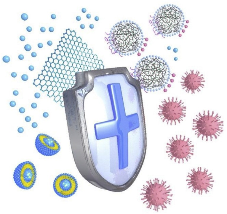
Graphical abstract
Keywords: Engineered nanomaterials, Antivirus, Nanoagents, Therapeutics, Mechanism
Abstract
The recent COVID-19 outbreak has increasingly engaged researchers in the search for effective antiviral drugs as well as therapeutic treatment options. The shortcomings of existing antiviral agents such as narrow spectrum and low bioavailability, can be overcome through the use of engineered nanomaterials, which, therefore, are considered as a significant next-generation therapeutic option. Thus, the development of novel antiviral nanoagents will certainly help address several future challenges and knowledge gaps.
Introduction
The novel coronavirus (COVID-19) outbreak has turned into a global pandemic, causing 810,492 deaths and 23,518,343 infected cases in 216 countries, areas, or territories as of August 25, 2020 [1], which is an irreparable loss. Unfortunately, despite considerable advancements in the field of antiviral drug discovery over the past years [2], there is still no approved drug or vaccine that can treat this disease [3]. Likewise, there are over 200 other infectious diseases [2]. In the last two decades, there have been a number of pathogenic viruses that have caused public health emergencies and socioeconomic losses (Fig. 1 a). For instance, the severe acute respiratory syndrome (SARS) outbreak in 2003 induced 774 deaths and 8096 infection cases [4], and its economic cost was estimated to be over $US 40 billion [5]. Similarly, approximately 37.9 million people worldwide suffered from the human immunodeficiency virus (HIV) around the end of 2018 [6], leading to an increasing healthcare burden in low-income countries.
Fig. 1.

(a) Major global virus epidemics in the last two decades.
HIV: https://www.who.int/hiv/mediacentre/events/en/.
SARS: https://www.who.int/csr/don/archive/disease/severe_acute_respiratory_syndrome/en/.
Hemagglutinin type1 neuraminidase type 1 (H1N1): https://www.who.int/csr/don/2009_12_30/en/.
Middle East respiratory syndrome (MERS): https://www.who.int/emergencies/mers-cov/en/.
Zika virus (ZIKV): https://www.who.int/en/news-room/fact-sheets/detail/zika-virus
HCV: https://www.who.int/news-room/fact-sheets/detail/hepatitis-c.
Ebola: https://www.who.int/csr/disease/ebola/en/.
COVID-19: https://www.who.int/emergencies/diseases/novel-coronavirus-2019
(b) Published papers on antibacterial, anticancer, and antiviral studies based on nanomaterials. The data were collected from the Web of Science (http://www.webofknowledge.com).
Generally, small-molecule drugs, vaccines, monoclonal antibodies, peptides, interferon, and oligonucleotide-based therapies can be used to combat viral infections such as the application of a SARS vaccine or combinatorial antiretroviral therapy (cART) in the treatment of HIV [7,8]. Nevertheless, up to 2016, only 90 antiviral drugs had been approved for the treatment of 9 types of human transmissible diseases, namely HIV, hepatitis B virus (HBV), hepatitis C virus (HCV), herpesvirus, influenza virus, human cytomegalovirus, varicella-zoster virus, respiratory syncytial virus (RSV), and human papilloma virus (HPV) [2]. Although developments of new antiviral drugs, vaccines, and antibodies are powerful prevention and treatment strategies for viral infections, most of these selectively target a single virus [9]. Moreover, viral mutation, virus-induced secondary infections, side effects from drugs and vaccines, and long development cycles bring many uncertainties and pose challenges to the search for new antiviral drugs. Hence, there is an urgent need to realize more effective therapeutic strategies to combat viral emergence.
Nanomaterials present new opportunities for antiviral drug development
Engineered nanomaterials (ENMs) offer many therapeutic advantages over traditional drugs, such as higher efficiency of drug delivery to lesions, more broad-spectrum drug activity, fewer side effects on the human body, easily tunable physicochemical properties, lower cost and higher throughput synthesis, etc. These superior properties of ENMs could be considered as an emerging solution to the problems faced during novel drug development. Hence, ENMs have been intensively investigated for their applications in next-generation therapeutics, such as anticancer and antibacterial treatments. However, antiviral applications of ENMs are still in their infancy (Fig. 1b). Reportedly (Table 1 ), antiviral nanoagents can be roughly classified into three strategies, namely virus or host cell surface receptor interactions, antiviral agent delivery, and nanovaccines (Fig. 2 ).
Table 1.
Representative antiviral nanoagents in published papers.
| ENMs | Size | Surface modification / antiviral agent | Virusa | Study | Mechanism | Ref. | |
|---|---|---|---|---|---|---|---|
| Ag NPs | 16 nm | Polyvinylpyrrolidone / bovine serum albumin | HIV-1 | In vitro | Virus or host cell surface receptor interactions | Binding to gp120 glycoprotein knobs on HIV-1 surface. | [12] |
| 50 nm | HBV | In vitro | Impeding the transcription of viral RNA by directly binding to HBV DNA. | [14] | |||
| GO | HSV-1 | In vitro | Mimicking the cell surface receptor heparan sulfate to inhibit HSV binding to host cells. | [16] | |||
| Au NPs | 2.3 nm | Ligands containing undecane sulfonic acid | HSV, HPV, RSV, DENV | In vivo, in vitro | Au NPs coated with undecane sulfonic acid mimicking the heparin sulfate proteoglycans to prevent the virus–cell interaction. | [18] | |
| PLGA NPs | 395 nm | Acyclovir | HSV | In vivo, in vitro | Antiviral agent delivery | PLGA NPs loaded with acyclovir for oral delivery. | [20] |
| 180 nm | 3D8 scFv antibody | VSV | In vivo, in vitro | Cytoplasmic delivery of a monoclonal antibody with nucleic acid-hydrolyzing activity. | [34] | ||
| Liposome | 150 nm | Suramin | NV | In vitro | Delivery the suramin-loaded liposome to inhibit NV polymerases. | [22] | |
| 124 nm | Stearylamine | BV, HSV-1 | In intro | Cationic liposomes with incorporated stearylamine inhibit viral infectivity. | [35] | ||
| Au NPs | 5, 50, 100 nm | Thiol cysteamine | HCV | In vivo, in vitro | Nanovaccines | DNA segment specific to HCV core gene conjugated on the AuNPs to simulate the immune response. | [27] |
| 2 2, 5, 8, 12, 17, 37, 50 nm | FMDV peptide | FMDV | In vivo, in vitro | C-terminus of the FMDV peptide conjugated on AuNPs to induce the magnitude of the immune response. | [36] | ||
| 20,40 nm | WNV envelope E protein | WNV | In vivo, in vitro | WNV envelope E protein coated AuNPs enhance the immune response. | [37] | ||
| 20, 40, 80 nm | ED III protein | DENV | In vivo, in vitro | Serotype 2 of DENV as dengue subunit to functionalize Au NPs to induce a high level of antibody to neutralizing DENV. | [38] | ||
DENV: dengue virus; VSV: vesicular stomatitis virus; BV: baculovirus; ED III: domain III of envelope glycoprotein derived from serotype 2 of dengue virus. RANTES: regulated upon activation, normal T expressed and secreted.
Fig. 2.

Three antiviral strategies for nanoagents: (a) virus or host cell surface receptor interactions, (b) antiviral agent delivery, and (c) nanovaccines. Red cross denotes the targets of ENMs against the virus−cell interactions.
Virus or host cell surface receptor interactions
A virus is an extremely small particle with an outer protein shell called a capsid and an inner core of nucleic acids (either RNA or DNA). For self-replication, the virus has to invade host cells through binding itself to specific “cell surface receptor molecules” (Fig. 2a), such as the angiotensin-converting enzyme 2 (ACE2) receptor for COVID-19 [10]. Antiviral nanoagents can be used to disrupt the viral replication cycle. As shown in Table 1, multifarious nanoagents have been widely investigated for their antiviral activities, such as metallic (e.g., silver), metal oxide (e.g., titanium dioxide), and carbonaceous (e.g., graphene oxide (GO)) nanomaterials. The controllable physicochemical properties (e.g., size, shape, and surface) of ENMs facilitate their direct interactions with viral particles or host cell surface receptors to inhibit virus-cell interactions [11]. For instance, silver nanoparticles (Ag NPs) can directly interact with virus envelope proteins [12,13], and nucleic acids [14]. Furthermore, our recent study revealed that AgNPs could block the primary infection of Kaposi’s sarcoma-associated herpesvirus (KSHV) by directly destroying its virion protein [15]. In addition, GO and its derivatives were reported to inhibit virus infections through competitive binding with the cell receptors [16], or inducing conformational damage of the viral protein [17]. Analogously, by mimicking heparan sulfate proteoglycans on the eukaryotic cell surface, viral attachment ligand (VAL)-modified gold nanoparticles (Au NPs) exhibit a broad-spectrum nontoxic antiviral activity [18].
Antiviral agent delivery
Many antiviral agents have been approved for clinical use or in preclinical tests based on the basis of disturbing the virus life cycle, which can be used to develop new antiviral approaches in combination with ENMs (Fig. 2b). In this way, ENMs can not only function as an ideal vehicle for these agents to improve targeting efficiency, bioavailability and blood circulation, but also reduce side effects [19]. For instance, as an approved antiviral drug, acyclovir can be used to treat herpes simplex virus (HSV) infection through disruption of the viral DNA polymerase function by acting as an analog of deoxyguanosine triphosphate to compete with 2′-deoxyaguanosine-5′-triphosphate [2]. However, its clinical application is hampered by its short half-life of less than 3 h and low bioavailability (only 10 %−20 %) through oral administration [20]. These limitations can be overcome by using poly(lactic acid-co-glycolic acid) (PLGA) as an ideal biodegradable vehicle for drug delivery, which has been approved for clinical application by U.S. Food and Drug Administration. Encapsulation of acyclovir into PLGA-based NPs for oral administration has been demonstrated as an effective way that resulted in a 2.6-fold improvement in its bioavailability [20]. Furthermore, liposomes hold promise as a kind of antiviral drug delivery agent. For instance, although suramin is an active antiretroviral agent that can inhibit HBV, HIV, ZIKV and norovirus (NV) [21], its lipophilicity and high negative charge lead to low membrane permeability and limited cell internalization [22]. However, its encapsulation into cationic liposomal NPs has been reported to result in better inhibition of murine NV infections [22].
Nanovaccines
The biological basis for developing nanovaccines is the activation of the host’s immune system against viruses through nanoagents (Fig. 2c). For this purpose, the dominant antigens of viruses can be used to construct nanovaccines for pathogenic viruses. Thus, ENMs loaded with viral antigens (viral nucleic acids or inactivated proteins) can pre-stimulate the immune system to produce specific virus antibodies that promote antiviral inflammatory responses [23,24]. For instance, Au NPs modified with West Nile virus (WNV) envelope proteins have been demonstrated to stimulate the release of proinflammatory factors (i.e., tumor necrosis factor-α (TNF-α), interleuin-6 (IL-6), interleuin-2 (IL-2) and granulocyte-macrophage colony-stimulating factor) to achieve antiviral immune responses [25]. Similarly, the loading of viral DNA or RNA onto ENMs is another effective way of preparing nanovaccines by introducing an eukaryotic expression vector to encode the viral antigen protein into a host cell to provoke the immune system into combating viral infections [26]. Moreover, ENMs can also be used as an auxiliary to overcome the obstacle of inefficient DNA vaccine intake [27]. The coadministration of Au NPs with an anti-HCV DNA vaccine was observed to generate vibrational and dipole-like oscillations, increase the DNA vaccine uptake, and ultimately improve antibody levels and immune response to the HCV infection in immunized mice [27]. Furthermore, nanovaccines based on genetic engineering, bioconjugation, infusion, or mineralization have been successfully applied to treat infections due to HIV-1, Ebola, and foot-and-mouth disease virus (FMDV) [28].
Challenges and knowledge gaps
The COVID-19 outbreak has led to increased efforts toward the development of more antiviral nanoagents as a reserve strategy in the fight against viral epidemics. Research thus far has mostly focused only on the development of new drugs and vaccines, or reusing approved antiviral drugs [3], while the absence of antiviral nanoagents during this emergency is conspicuous. Moreover, despite the promising versatility of ENMs in antiviral applications, practical therapeutics are limited in terms of the development of new strategies and improvements in efficacy and safety. The following are a few such limitations:
-
(i)
Although most reported antiviral nanoagents exhibit outstanding performance in mammalian cell lines or rodents, they may cause adverse or more complicated responses in humans. Hence, before rushing into the clinic, more preclinical tests using promising animal models (e.g., ferrets, macaques, and other primates) that better mimic human disease response must be performed to assess the health risks as well as the antiviral activity of nanoagents. More importantly, massive preclinical tests are prerequisite for the development of nanoagents with better efficacy and broad-spectrum antiviral activity than current approved antiviral drugs.
-
(ii)
Compared to bacteria, virus infections are more complicated, involving faster transmission and a variable incubation period. For instance, although both SARS and COVID-19 belong to the coronavirus family, they exhibit different infectious and pathogenic effects on patients [29]. Therefore, the design of novel antiviral nanoagents for a severe viral pandemic requires not only specific and broad-spectrum activities of antiviral nanoagents, but also a better understanding of the infection mechanisms, especially for viruses exhibiting a high mutation rate and tolerance to approved drugs or vaccine treatments [30].
-
(iii)
The envelope, capsid, and nucleic acids are the primary targets in the design of antiviral nanoagents. However, the interactions between ENMs and these targets remain unclear, which hampers a deeper understanding of the mechanisms that inhibit virus infection. Therefore, the possible interaction mechanisms, including the damage caused by the direct binding of ENMs to viruses, the disturbance of interactions between virus and host cell, or the indirect effects on the activation of innate and adaptive immunity, should be clarified.
-
(iv)
A viral infection involves the following stages: attachment to membrane receptors, penetration into cells, intracellular uncoating, nucleic acid synthesis, and the transfer of progeny virus between cells. Existing databases and bioinformatics should be better used to propose strategies for designing novel antiviral nanoagents that can inhibit the abovementioned processes. Furthermore, the prediction and screening of the interactions between ENMs and viruses during the infection process requires more powerful and high-throughput analytical platforms.
-
(v)
The antiviral nanoagent treatments may also act on the immune system. Therefore, better manipulation of the physicochemical properties (e.g., surface modification) of ENMs is necessary to avoid their side effects, in addition to the well-balanced dose administration. In addition, the reasons for secondary adverse outcomes remain unclear, which can be caused by either ENM-induced nonspecific immune response or virus-incurred inflammation during treatment. Therefore, besides virus infection, the immune activation associated with the type and dosage of the applied ENMs should be considered as well.
-
(vi)
The controllable delivery of antiviral nanoagents to lesions is an important concern during the treatment of diverse viruses. Generally, intravenous administration may be the most commonly used clinical route, especially for treating an infection due to influenza virus. However, for gastrointestinal infections caused by rotavirus or hepatitis virus, oral administration is better, while intranodal administration may be better for treating infections caused by respiratory viruses such as SARS and COVID-19. Therefore, the stability and bioavailability of antiviral nanoagents must be carefully assessed, considering the features of both diverse administration methods and different viruses.
-
(vii)
The rapid increase in the variety of ENMs provides space for healthcare innovations as well as opportunities for the development of novel antiviral agents. However, both the advantages and limitations of ENMs must be well understood to avoid additional risks before practical applications. For example, owing to their high surface-to-volume ratio and modifiable surface properties, two-dimensional ENMs such as GO have been demonstrated as useful tools for anticancer drug delivery [31,32], although their biocompatibility remains a question. Therefore, the possible use of two-dimensional ENMs for antiviral treatments warrants further exploration in the future [33].
Conclusions and perspectives
The development of novel antiviral nanoagents may be a vital and highly desirable precaution against the emergence and recurrence of viral outbreaks. Compared with state-of-the-art approaches such as interfering RNAs, ENMs offer distinct advantages for antiviral therapy, including tunable physicochemical properties, high drug-loading efficacy and bioavailability, feasible incorporation of both hydrophilic and hydrophobic antiviral drugs, and outstanding stability under physiological conditions. Moreover, although diverse ENM-based antiviral strategies have already been proposed based on lab studies, their application in practical therapeutics is still in its infancy. Thus, the current challenges and knowledge gaps in the development of novel antiviral nanoagents, such as the therapeutic efficacy and potential health risks need to be addressed. Nevertheless, future research on antiviral nanoagents and combination therapy using approved antiviral drugs and ENMs is expected to produce promising developments.
Funding
This work has been supported by grants from the National Natural Science Foundation of China (21922611, 21707161, 21637004, and 21920102007), the Youth Innovation Promotion Association CAS (2019042), the Beijing Natural Science Foundation (8191002), and the international collaboration key grant from the Chinese Academy of Sciences (121311KYSB20190010).
Author contributions
Literature review and initial drafting of the manuscript were performed by Y. C., J. M., and M. X. Critical revisions of the manuscript were performed by M. X. and S. L. Each author has read and approved the final manuscript.
Declaration of Competing Interest
The authors report no declarations of interest.
Biographies

Yongjiu Chen is currently a Ph.D. student under the supervision of Professor Sijin Liu in the State Key Laboratory of Environmental Chemistry and Ecotoxicology, the Research Center for Eco-Environmental Sciences, Chinese Academy of Sciences (CAS). He has received his B.S. degree from Shandong University in 2015. His research interests include the translational toxicology of ENMs.

Juan Ma is currently an associate professor at the State Key Laboratory of Environmental Chemistry and Ecotoxicology, Research Center for Eco-Environmental Sciences, Chinese Academy of Sciences (CAS). She has received her bachelor’s degree in 2011 from South China University of Technology and Ph.D. in 2015 from the University of Chinese Academy of Sciences. Her research focuses on the immune toxicities of pollutants and the underlying mechanisms, especially the adverse outcomes caused by nanomaterials and airborne fine particles.

Ming Xu has obtained his B.S. and Ph.D. degrees from Xiamen University in 2006 and 2011. He was a postdoctoral fellow in Prof. Ryszard Lobinski’s group at the Centre National de la Recherche Scientifique (CNRS) from 2011 to 2013. He joined the Research Center for Eco-Environmental Sciences (RCEES), Chinese Academy of Sciences (CAS), in 2014, and became an associate professor in 2016. His research interests include the health risks and toxicological mechanisms of heavy metals and nanoparticles. He was awarded the “National Science Fund for Excellent Young Scholars” by the National Natural Science Foundation of China (NSFC) in 2019. He has also been a member of the “Youth Innovation Promotion Association” of CAS since 2018.

Sijin Liu is currently a professor at the State Key Laboratory of Environmental Chemistry and Ecotoxicology, Research Center for Eco-Environmental Sciences, Chinese Academy of Sciences (CAS). His research interests include: (i) Environmental behaviors, biological effects, and health risks of engineered nanomaterials and fine air particles. (ii) The molecular mechanisms responsible for environmental pollutant-induced oncogenic effects and disordered element metabolism. Dr. Liu is a recipient of the “Outstanding Young Scientist” award from the National Natural Science Foundation of China (NSFC). He had served as the Chief Scientist for the project of Environmental Behaviors and Toxicity of Engineered Nanomaterials under the “National Basic Research Program of China (973 Program)” from 2014 to 2018.
References
- 1.Coronavirus disease (COVID-2019) situation reports, WHO, https://www.who.int/emergencies/diseases/novel-coronavirus-2019/situation-reports.
- 2.De Clercq E., Li G. Clin. Microbiol. Rev. 2016;29:695–747. doi: 10.1128/CMR.00102-15. [DOI] [PMC free article] [PubMed] [Google Scholar]
- 3.Li G., De Clercq E. Nat. Rev. Drug Discov. 2020;19:149–150. doi: 10.1038/d41573-020-00016-0. [DOI] [PubMed] [Google Scholar]
- 4.World Health Organization: Summary of probable SARS cases with onset of illness from 1 November 2002 to 31 July 2003, https://www.who.int/csr/sars/country/table2004_04_21/en/.
- 5.Knobler S. National Academies Press; Washington, DC: 2004. Learning From SARS: Preparing for the Next Disease Outbreak: Workshop Summary. [PubMed] [Google Scholar]
- 6.World Health Organization . 2018. Summary of the Global HIV Epidemic.https://www.who.int/gho/hiv/en/ [Google Scholar]
- 7.Marshall E., Enserink M. Science. 2004;303:944–946. doi: 10.1126/science.303.5660.944. [DOI] [PubMed] [Google Scholar]
- 8.Zhan P., Pannecouque C., De Clercq E., Liu X. J. Med. Chem. 2016;59:2849–2878. doi: 10.1021/acs.jmedchem.5b00497. [DOI] [PubMed] [Google Scholar]
- 9.Ianevski A., Zusinaite E., Kuivanen S., Strand M., Lysvand H., Teppor M., Kakkola L., Paavilainen H., Laajala M., Kallio-Kokko H. Antiviral Res. 2018;154:174–182. doi: 10.1016/j.antiviral.2018.04.016. [DOI] [PMC free article] [PubMed] [Google Scholar]
- 10.Patel A.B., Verma A. JAMA. 2020;323:1769–1770. doi: 10.1001/jama.2020.4812. [DOI] [PubMed] [Google Scholar]
- 11.Ye S., Shao K., Li Z., Guo N., Zuo Y., Li Q., Lu Z., Chen L., He Q., Han H. ACS Appl. Mater. Interfaces. 2015;7:21571–21579. doi: 10.1021/acsami.5b06876. [DOI] [PubMed] [Google Scholar]
- 12.Elechiguerra J.L., Burt J.L., Morones J.R., Camacho-Bragado A., Yacaman M.J. J. Nanobiotechnology. 2005;3:1–10. doi: 10.1186/1477-3155-3-6. [DOI] [PMC free article] [PubMed] [Google Scholar]
- 13.Lara H.H., Ayala-Nuñez N.V., Ixtepan-Turrent L., Rodriguez-Padilla C. J. Nanobiotechnology. 2010;8:1–10. doi: 10.1186/1477-3155-8-1. [DOI] [PMC free article] [PubMed] [Google Scholar]
- 14.Lu L., Sun R., Chen R., Hui C., Ho C., Luk J., Lau G., Che C. Antivir. Ther. 2008;13:253–262. [PubMed] [Google Scholar]
- 15.Wan C., Tai J., Zhang J., Guo Y., Zhu Q., Ling D., Gu F., Gan J., Zhu C., Wang Y., Liu S., Wei F., Cai Q. Cell Death Dis. 2019;10:392–408. doi: 10.1038/s41419-019-1624-z. [DOI] [PMC free article] [PubMed] [Google Scholar]
- 16.Sametband M., Kalt I., Gedanken A., Sarid R. ACS Appl. Mater. Interfaces. 2014;6:1228–1235. doi: 10.1021/am405040z. [DOI] [PubMed] [Google Scholar]
- 17.Zeng S., Zhou G., Guo J., Zhou F., Chen J. Scientific Reports-UK. 2016;6:1–7. doi: 10.1038/srep24906. [DOI] [PMC free article] [PubMed] [Google Scholar]
- 18.Cagno V., Andreozzi P., D’Alicarnasso M., Silva P.J., Mueller M., Galloux M., Le Goffic R., Jones S.T., Vallino M., Hodek J. Nat. Mater. 2018;17:195–203. doi: 10.1038/nmat5053. [DOI] [PubMed] [Google Scholar]
- 19.Lembo D., Donalisio M., Civra A., Argenziano M., Cavalli R. Expert Opin. Drug Deliv. 2018;15:93–114. doi: 10.1080/17425247.2017.1360863. [DOI] [PubMed] [Google Scholar]
- 20.Choudhary S. Int. J. Drug Deliv. 2013;5:331–343. [Google Scholar]
- 21.Tan C.W., Sam I.-C., Chong W.L., Lee V.S., Chan Y.F. Antiviral Res. 2017;143:186–194. doi: 10.1016/j.antiviral.2017.04.017. [DOI] [PubMed] [Google Scholar]
- 22.Mastrangelo E., Mazzitelli S., Fabbri J., Rohayem J., Ruokolainen J., Nykänen A., Milani M., Pezzullo M., Nastruzzi C., Bolognesi M. ChemMedChem. 2014;9:933–939. doi: 10.1002/cmdc.201300563. [DOI] [PubMed] [Google Scholar]
- 23.Rodríguez-Gascón A., del Pozo-Rodríguez A., Solinís M.Á. Int. J. Nanomedicine. 2014;9:1833–1843. doi: 10.2147/IJN.S39810. [DOI] [PMC free article] [PubMed] [Google Scholar]
- 24.Zholobak N.M., Mironenko A.P., Shcherbakov A.B., Shydlovska O.A., Spivak M.Y., Radchenko L.V., Marinin A.I., Ivanova O.S., Baranchikov A.E., Ivanov V.K. Antiviral Res. 2016;127:1–9. doi: 10.1016/j.antiviral.2015.12.013. [DOI] [PubMed] [Google Scholar]
- 25.Niikura K., Matsunaga T., Suzuki T., Kobayashi S., Yamaguchi H., Orba Y., Kawaguchi A., Hasegawa H., Kajino K., Ninomiya T. ACS Nano. 2013;7:3926–3938. doi: 10.1021/nn3057005. [DOI] [PubMed] [Google Scholar]
- 26.Liu J., Wu J., Wang B., Zeng S., Qi F., Lu C., Kimura Y., Liu B. J. Med. Virol. 2014;86:886–894. doi: 10.1002/jmv.23768. [DOI] [PubMed] [Google Scholar]
- 27.Draz M.S., Wang Yi., Chen F.F., Xu Y., Shafiee H. Adv. Funct. Mater. 2016;27:1604139–1604149. doi: 10.1002/adfm.201604139. [DOI] [PMC free article] [PubMed] [Google Scholar]
- 28.Wen A.M., Steinmetz N.F. Chem. Soc. Rev. 2016;45:4074–4126. doi: 10.1039/c5cs00287g. [DOI] [PMC free article] [PubMed] [Google Scholar]
- 29.Guarner J. Am. J. Clin. Pathol. 2020:1–2. doi: 10.1093/ajcp/aqaa029. [DOI] [PMC free article] [PubMed] [Google Scholar]
- 30.Coughlan L., Palese P. Cell Host Microbe. 2018;24:18–24. doi: 10.1016/j.chom.2018.06.016. [DOI] [PubMed] [Google Scholar]
- 31.Zhu J., Xu M., Gao M., Zhang Z., Xu Y., Xia T., Liu S. ACS Nano. 2017;11:2637–2651. doi: 10.1021/acsnano.6b07311. [DOI] [PubMed] [Google Scholar]
- 32.Wang S., Ren L., Qi Y., Chen Y., Wang R., Ma M., Xu M., Liu S. In: Frontiers of Nanoscience. Parak W.J., Feliu N., editors. Elsevier; 2020. Chapter 7 - Two-dimensional nanoparticles for the delivery of anticancer drugs and cancer therapy; pp. 151–199. [Google Scholar]
- 33.Palmieri V., Papi M. Nano Today. 2020;33 doi: 10.1016/j.nantod.2020.100883. [DOI] [PMC free article] [PubMed] [Google Scholar]
- 34.Joung Y.K., Son S., Jang J.Y., Kwon M.H., Park K.D. Pharm. Res. 2012;29:932–942. doi: 10.1007/s11095-011-0633-0. [DOI] [PubMed] [Google Scholar]
- 35.Tahara K., Kobayashi M., Yoshida S., Onodera R., Inoue N., Takeuchi H. Int. J. Pharm. 2018;543:311–317. doi: 10.1016/j.ijpharm.2018.04.001. [DOI] [PubMed] [Google Scholar]
- 36.Chen Y.S., Hung Y.C., Lin W.H., Huang G.S. Nanotechnology. 2010;21 doi: 10.1088/0957-4484/21/19/195101. [DOI] [PubMed] [Google Scholar]
- 37.Niikura K., Matsunaga T., Suzuki T., Kobayashi S., Yamaguchi H., Orba Y., Kawaguchi A., Hasegawa H., Kajino K., Ninomiya T., Ijiro K., Sawa H. ACS Nano. 2013;7:3926–3938. doi: 10.1021/nn3057005. [DOI] [PubMed] [Google Scholar]
- 38.Quach Q.H., Ang S.K., Chu J.H.J., Kah J.C.Y. Acta Biomater. 2018;78:224–235. doi: 10.1016/j.actbio.2018.08.011. [DOI] [PubMed] [Google Scholar]