Abstract
Background
Lifetime glycemic exposure and its relationship with age at diagnosis in type 2 diabetes (T2D) are unknown. Pharmacologic glycemic management strategies for young-onset T2D (age at diagnosis <40 years) are poorly defined. We studied how age at diagnosis affects glycemic exposure, glycemic deterioration, and responses to oral glucose-lowering drugs (OGLDs).
Methods and findings
In a population-based cohort (n = 328,199; 47.2% women; mean age 34.6 and 59.3 years, respectively, for young-onset and usual-onset [age at diagnosis ≥40 years] T2D; 2002–2016), we used linear mixed-effects models to estimate the association between age at diagnosis and A1C slope (glycemic deterioration) and tested for an interaction between age at diagnosis and responses to various combinations of OGLDs during the first decade after diagnosis. In a register-based cohort (n = 21,016; 47.1% women; mean age 43.8 and 58.9 years, respectively, for young- and usual-onset T2D; 2000–2015), we estimated the glycemic exposure from diagnosis until age 75 years.
People with young-onset T2D had a higher mean A1C (8.0% [standard deviation 0.15%]) versus usual-onset T2D (7.6% [0.03%]) throughout the life span (p < 0.001). The cumulative glycemic exposure was >3 times higher for young-onset versus usual-onset T2D (41.0 [95% confidence interval 39.1–42.8] versus 12.1 [11.8–12.3] A1C-years [1 A1C-year = 1 year with 8% average A1C]). Younger age at diagnosis was associated with faster glycemic deterioration (A1C slope over time +0.08% [0.078–0.084%] per year for age at diagnosis 20 years versus +0.02% [0.016–0.018%] per year for age at diagnosis 50 years; p-value for interaction <0.001). Age at diagnosis ≥60 years was associated with glycemic improvement (−0.004% [−0.005 to −0.004%] and −0.02% [−0.027 to −0.0244%] per year for ages 60 and 70 years at diagnosis, respectively; p-value for interaction <0.001). Responses to OGLDs differed by age at diagnosis (p-value for interaction <0.001). Those with young-onset T2D had smaller A1C decrements for metformin-based combinations versus usual-onset T2D (metformin alone: young-onset −0.15% [−0.105 to −0.080%], usual-onset −0.17% [−0.179 to −0.169%]; metformin, sulfonylurea, and dipeptidyl peptidase-4 inhibitor: young-onset −0.44% [−0.476 to −0.405%], usual-onset −0.48% [−0.498 to −0.459%]; metformin and α-glucosidase inhibitor: young-onset −0.40% [−0.660 to −0.144%], usual-onset −0.25% [−0.420 to −0.077%]) but greater responses to other combinations containing sulfonylureas (sulfonylurea alone: young-onset −0.08% [−0.099 to −0.065%], usual-onset +0.06% [+0.059 to +0.072%]; sulfonylurea and α-glucosidase inhibitor: young-onset −0.10% [−0.266 to 0.064%], usual-onset: 0.25% [+0.196% to +0.312%]). Limitations include possible residual confounding and unknown generalizability outside Hong Kong.
Conclusions
In this study, we observed excess glycemic exposure and rapid glycemic deterioration in young-onset T2D, indicating that improved treatment strategies are needed in this setting. The differential responses to OGLDs between young- and usual-onset T2D suggest that better disease classification could guide personalized therapy.
In a population-based cohort, Calvin Ke and colleagues investigate age at diagnosis and glycemic trajectories in Type 2 diabetes patients.
Author summary
Why was this study done?
Young-onset type 2 diabetes (diagnosed before age 40 years) is an aggressive disease, associated with higher risks of mortality and other complications compared with usual-onset type 2 diabetes (diagnosed at age 40 years or after).
Although exposure to hyperglycemia is a key risk factor for type 2 diabetes complications, the magnitude of this exposure over a lifetime has never been quantified.
Because young people were excluded from randomized control trials of oral glucose-lowering drugs, it is unknown whether these drugs are effective for treating young-onset type 2 diabetes.
What did the researchers do and find?
Young-onset type 2 diabetes was associated with over triple the exposure to hyperglycemia from diagnosis until age 75 years compared with usual-onset type 2 diabetes.
Diabetes progressed much faster in young-onset type 2 diabetes compared with usual-onset type 2 diabetes.
Compared with usual-onset type 2 diabetes, people with young-onset type 2 diabetes had smaller responses to most metformin-based drug combinations and greater responses to other combinations containing sulfonylureas.
What do these findings mean?
Excess exposure to hyperglycemia and rapid disease progression in young-onset T2D call for better treatment strategies.
The differential responses to oral glucose-lowering drugs between young- and usual-onset T2D suggest that better disease classification could guide personalized therapy.
Introduction
Life-threatening complications of type 2 diabetes (T2D) are caused by long-term exposure to hyperglycemia [1]. Glycemic exposure is defined as the area under the glycated hemoglobin A1c (A1C) curve in excess of 7% over time [2–5]. A meta-analysis of randomized control trials showed that every 10 A1C-years of glycemic exposure (that is, 10 years of A1C at 8%) predicts a 25% increase in the relative risk of major adverse cardiovascular events (MACE) [2]. Young-onset T2D (defined here as age at diagnosis <40 years) is an aggressive phenotype associated with higher lifetime risks of MACE and other complications compared to usual-onset T2D (age at diagnosis ≥40 years) [6–9]. Although early age at diagnosis and poor glycemic control are important risk factors for complications [10], lifetime glycemic exposure and its relationship with age at T2D diagnosis are unknown.
Reducing glycemic exposure typically requires frequent escalation of glucose-lowering therapies to overcome the natural history of T2D, which is characterized by progressively declining β-cell function and insulin sensitivity [11]. The rate of β-cell function decline is known as glycemic deterioration [3,12,13]. This measure can be quantified using repeated measurements of A1C over time, after accounting for glucose-lowering drugs using statistical adjustment [13]. Glycemic deterioration is more precise than other measures using binary thresholds (e.g., treatment failure) [14]. Among Europeans with usual-onset T2D, younger age at diagnosis is associated with faster glycemic deterioration [13,14]. In Asia, 1 in 5 adults with T2D attending medical clinics has young-onset T2D [10], yet data on glycemic exposure and deterioration are lacking.
Similarly, pharmacologic glycemic management strategies for young-onset T2D are poorly defined because this population was excluded from trials of glucose-lowering therapies in adults [15]. In a pediatric trial, metformin alone was associated with treatment failure in 51.7% of children with T2D [16]. Another study of children with T2D found that 3 months of insulin and 9 months of metformin failed to improve β-cell function versus metformin alone [17]. Considering the lack of randomized trials among adults with young-onset T2D [15], observational studies may provide valuable insights in this understudied population.
To address these knowledge gaps, we conducted a large register- and population-based cohort study to measure how age at diagnosis affects (1) glycemic exposure, (2) glycemic deterioration, and (3) responses to oral glucose-lowering drugs (OGLDs) during the first decade after diagnosis among adults with T2D. We hypothesized that earlier age at diagnosis would be associated with increased glycemic exposure, faster glycemic deterioration, and decreased responsiveness to OGLDs.
Methods
Setting
Hong Kong has a population of 7.3 million people, 92% of whom are of Chinese ethnicity [18]. The estimated diabetes prevalence was 10.3% in 2014 [19]. The Hong Kong Hospital Authority (HA) provides universal public healthcare modeled after the British National Health Service. Because of the high out-of-pocket cost of private healthcare, 95% of people with diabetes in Hong Kong receive care in HA clinics [20]. Consultation, prescription, and medication dispensing services are all provided on site under an all-inclusive nominal user fee [21], which is waived for low-income and other vulnerable groups [22]. All hospitals and clinics managed by the HA share the same electronic health record (EHR) with data including laboratory tests, discharge summaries, and dispensed prescriptions. Dispensed prescription records are comprehensive because drugs are prescribed and dispensed on site at the time of consultation. These data are linked by the unique Hong Kong Identity Card number.
Data sources
In 1995, the Prince of Wales Hospital set up the multicenter Hong Kong Diabetes Register (HKDR) as a research-driven quality improvement program [23]. The HKDR is a prospective cohort of people with prevalent diabetes, of varying disease duration. Participants were enrolled from 1994 to 2015. All participants undergo structured assessment (eyes, feet, blood, urine) by trained nurses every 2–3 years to collect data not routinely captured in the HA EHR, including diabetes type, age at diagnosis, family history, and lifestyle habits. Enrollment was open to any adult with diabetes based on self- or physician-initiated referrals from community- and hospital-based clinics. All services were provided at the nominal fee charged by the HA.
The Hong Kong Diabetes Surveillance Database (HKDSD) is a population-based cohort of incident diabetes cases from across the entire territory, identified from the HA EHR. We defined diabetes as any person with an A1C ≥6.5% (48 mmol/mol), outpatient fasting plasma glucose ≥7 mmol/L, non-insulin glucose-lowering drug prescription, or long-term insulin prescription (≥28 days). The date of diagnosis is defined as the date of first occurrence of any of these events. According to HA regulations, these drugs are only indicated for diabetes [24]. To avoid detecting gestational diabetes, events occurring within 9 months of any pregnancy-related encounter are excluded. As diabetes type is not systematically encoded in this EHR, we previously developed and validated algorithms to classify diabetes type based on diagnosis codes and prescriptions [25]. In this study, we excluded people who received their first insulin prescription within 90 days of diabetes diagnosis as having type 1 diabetes [25] and classified the remainder as T2D. This definition has a sensitivity of 94.6% (95% confidence interval 93.9%–95.2%) and positive predictive value of 99.9% (99.8%–100.0%) for predicting T2D [25].
To estimate lifetime glycemic exposure, we required person-years of follow-up from across the life span. This requirement necessitated the inclusion of people with new (incident) and old (prevalent) diagnoses of T2D of varying disease duration. We used the HKDR (“Register cohort”) for this objective because it includes people with both new and old T2D cases with disease duration ranging from zero to more than 70 years, whereas the HKDSD only includes new T2D cases after 2002. For example, a 70-year-old person in 2010 who was diagnosed with T2D at age 30 years in 1970 would be included in the HKDR but not the HKDSD. To measure glycemic deterioration (defined as A1C slope over time, after statistical adjustment for OGLDs) and medication responses, we required person-years of follow-up from the first decade after a new T2D diagnosis. This requirement was based on a previous study showing that glycemic deterioration is a linear function of time during this period [26]. Although both cohorts included people with new diagnoses, we used the HKDSD (“Population cohort”) because it was larger and included people in the HKDR.
Study population
Register cohort (glycemic exposure)
We included adults aged 18–75 years in the HKDR with prevalent T2D observed between January 1, 2000, and December 31, 2015, and defined the index date as the earliest date within this period when the person met these criteria (Fig A in S1 Appendix). We excluded observations between 1994 to 1999 to more closely match the time period of the population cohort. We excluded people with type 1 diabetes, gestational diabetes, and non-Chinese ethnicity.
Population cohort (glycemic deterioration and medication responses)
We included adults aged 18–75 years in the HKDSD with T2D diagnosed between January 1, 2002, and December 31, 2012, (Fig A in S1 Appendix; dates chosen based on data availability). The index date was the diagnosis date. We followed people for up to 10 years after diagnosis of diabetes, censoring at the date of the first insulin prescription. The maximum follow-up date was December 31, 2016, for both the register and population cohorts, censoring at death or age 75 years.
Statistical analysis
We conducted the study according to a prospective analysis plan (S1 Analysis Plan). The primary outcome was A1C measured over the follow-up period, and the primary exposure was age at diagnosis, expressed as a continuous variable. In the register cohort, we calculated the mean A1C by attained age among all people with young-onset T2D. Using these values, we estimated the glycemic exposure, defined as the area under the A1C curve in excess of 7% (53 mmol/mol), from the mean observed age at diagnosis until age 75 years (Fig B in S1 Appendix), and repeated this procedure for usual-onset T2D.
In the population cohort, we used a linear mixed-effects model with age at diagnosis and time as independent variables and A1C as the dependent variable, with person-specific random intercepts and slopes because we assumed each person had his or her own A1C trajectory. Although we did not dichotomize age at diagnosis in the model, we presented the expected results for young- and usual-onset T2D based on the mean observed ages at diagnosis in each group. Glycemic deterioration was defined as the A1C slope over time, adjusted for model covariates. We included time-varying covariates for each OGLD based on their dispensing records, allowing for people to switch on and off a drug. We assumed the same absolute effect for each drug class, regardless of A1C level, and that drugs within the same class would lower A1C by a similar decrement [27]. We included metformin, sulfonylureas, and 10 multidrug combinations as unique variables (Table A in S1 Appendix). Effects of individual drugs taken in combination were not assumed to be additive (S1 Appendix) because OGLDs may have different effects when prescribed alone versus in combination [28,29]. Each drug combination was considered as a separate variable. When a new drug was dispensed, we excluded the first A1C measurement on the new treatment regimen to allow sufficient time for the A1C to equilibrate. We adjusted for prespecified variables (Fig C in S1 Appendix) including sex and recent comorbidities, namely, ischemic heart disease, congestive heart failure, stroke, peripheral arterial disease, and cancer, based on principal diagnoses from hospitalizations occurring within 2 years prior to the index date (Table B in S1 Appendix) and chronic kidney disease classified by the estimated glomerular filtration rate. As anthropometric data were unavailable, we included triglycerides and high-density lipoprotein cholesterol as proxies for obesity [30].
We estimated the association between age at diagnosis and A1C slope (glycemic deterioration) and tested for an interaction between age at diagnosis and responses to various combinations of OGLDs. We conducted several sensitivity analyses. To test the validity of our assumption of a linear relationship between age at diagnosis and A1C, we repeated the analysis using a nonlinear model with restricted cubic splines containing 4 knots placed at fixed quantiles [31]. We also repeated the analysis excluding A1C values from the first 6 and 12 months after the diagnosis of T2D because these measurements might be unusually elevated as a consequence of transiently depressed β-cell function (“glucotoxicity”) [32]. To test whether the baseline A1C level affected A1C lowering, we repeated the analysis (post hoc) with an interaction term between each drug combination and its observed baseline A1C level. Interaction terms between age at diagnosis and drug combinations were excluded in this model because of computational limitations.
We used the MIXED procedure (SAS version 9.4, SAS Institute, Cary, NC, www.sas.com), specifying the spatial power covariance structure to account for nonequal time intervals between A1C measurements. Missing outcome data were minimal (4.0% and 6.4% in the register and population cohorts) and handled by complete case analysis. The study was approved by The Chinese University of Hong Kong-New Territories East Cluster Clinical Research Ethics Committee and the University of Toronto Health Sciences Research Ethics Board. Data in the HKDSD were anonymized at the time of access. Individuals in the HKDR provided written informed consent for the use of their data. This study is reported as per the Strengthening the Reporting of Observational Studies in Epidemiology guideline (S1 STROBE checklist).
Results
We included 21,016 people (47.1% women, 0.2 million person-years follow-up, median 8.4 years) in the register cohort and 328,199 people (47.2% women, 2.4 million person-years follow-up, median 7.9 years) in the population cohort with >3 million A1C measurements combined (Fig D in S1 Appendix). The mean age at diagnosis was similar in both cohorts (young-onset: 33.8–34.6 years, usual-onset: 53.8–59.3 years, Table 1). Recent comorbidities were more common in the register than the population cohort. The baseline A1C was higher in young- versus usual-onset T2D (register: young-onset, mean 7.8% [62 mmol/mol, standard deviation 1.5%]; usual-onset, 7.3% [56 mmol/mol, 1.2%]; population: young-onset, 7.6% [60 mmol/mol, 1.6%]; usual-onset, 7.3% [56 mmol/mol, 1.3%]). Lipid levels were similar across cohorts. Metformin (young-onset: 31.8–56.4%, usual-onset: 41.5–48.6%) and sulfonylureas (young-onset: 25.9–38.4%, usual-onset: 33.6–40.7%) were the most common OGLDs. Insulin use during the first year after diagnosis was more common in the register (young-onset: 24.2%, usual-onset: 11.7%) than the population cohort (young-onset: 3.0%, usual-onset: 1.1%).
Table 1. Baseline characteristics in the register (2000–2016) and population (2002–2016) cohorts.
| Register Cohort | Population Cohort | |||
|---|---|---|---|---|
| Young-Onset (n = 4,058) |
Usual-Onset (n = 16,958) |
Young-Onset (n = 15,265) |
Usual-Onset (n = 312,934) |
|
| Age at diagnosis (years) | 33.8 (5.5) | 53.8 (8.1) | 34.6 (4.8) | 59.3 (9.0) |
| Index age (years) | 43.8 (10.5) | 58.9 (8.3) | 34.6 (4.8) | 59.3 (9.0) |
| Women (n, %) | 2,031 (50.0) | 7,876 (46.4) | 6,508 (42.6) | 148,516 (47.4) |
| Recent comorbidities within 2 year of index* (n, %) | ||||
| Ischemic heart disease | 62 (1.5) | 546 (3.2) | 57 (0.4) | 4,780 (1.5) |
| Congestive heart failure | 14 (0.3) | 130 (0.8) | 28 (0.2) | 1,244 (0.4) |
| Stroke | 57 (1.4) | 534 (3.2) | 55 (0.4) | 3,809 (1.2) |
| Peripheral arterial disease | 11 (0.3) | 33 (0.2) | 2 (0.0) | 105 (0.0) |
| Cancer | 56 (1.4) | 359 (2.1) | 152 (1.0) | 4,794 (1.5) |
| Laboratory Values (within 2 years of index* for A1C; 3 years for other variables) | ||||
| Hemoglobin A1C (%) | 7.8 (1.5) | 7.3 (1.2) | 7.6 (1.6) | 7.3 (1.3) |
| Fasting plasma glucose (mmol/L) | 8.6 (2.4) | 7.9 (1.9) | 8.1 (2.6) | 7.6 (2.0) |
| LDL-C (mmol/L) | 2.8 (0.7) | 2.7 (0.7) | 2.9 (0.8) | 3.0 (0.8) |
| HDL-C (mmol/L) | 1.3 (0.3) | 1.3 (0.3) | 1.2 (0.3) | 1.1 (0.3) |
| Triglycerides (mmol/L; median, IQR) | 1.5 (1.2) | 1.5 (1.0) | 1.7 (1.4) | 1.5 (1.0) |
| Estimated GFR (mL/min/1.73 m2) | 93.2 (22.7) | 78.9 (20.5) | 105 (18.0) | 81.2 (18.9) |
| <60 mL/min/1.73 m2 (n, %) | 327 (8.0) | 2,787 (16.4) | 404 (2.8) | 36,541 (12.6) |
| <15 mL/min/1.73 m2 (n, %) | 41 (1.0) | 163 (1.0) | 59 (0.4) | 2,017 (0.7) |
| Pharmacotherapy within 1 year after index* (n, %) | ||||
| Metformin | 1,292 (31.8) | 7,032 (41.5) | 8,611 (56.4) | 152,156 (48.6) |
| Sulfonylurea | 1,051 (25.9) | 5,704 (33.6) | 5,856 (38.4) | 127,512 (40.7) |
| DPP-4 Inhibitor | 16 (0.4) | 68 (0.4) | 417 (0.1) | 43 (0.3) |
| Thiazolidinedione | 38 (0.9) | 124 (0.7) | 178 (0.1) | 13 (0.1) |
| Acarbose | 24 (0.6) | 96 (0.6) | 42 (0.3) | 1,085 (0.4) |
| GLP-1 receptor agonist | 0 (0.0) | 0 (0.0) | 3 (0.0) | 2 (0.0) |
| Insulin | 983 (24.2) | 1,983 (11.7) | 464 (3.0) | 3,400 (1.1) |
Young-onset type 2 diabetes is defined here as age at diagnosis <40 years and usual-onset type 2 diabetes as ≥40 years. Values are means and standard deviations unless otherwise indicated. Because of the large sample size, baseline differences should be interpreted based on clinical significance rather than statistical significance.
*The index date in the register cohort was the date of enrollment in the register, whereas the index date in the population cohort was the date of diabetes diagnosis.
DPP-4, dipeptidyl peptidase-4; GFR, glomerular filtration rate; GLP-1, glucagon-like peptide 1; HDL-C, high-density lipoprotein cholesterol; IQR, interquartile range; LDL-C, low-density lipoprotein cholesterol.
Glycemic exposure in young-onset and usual-onset T2D
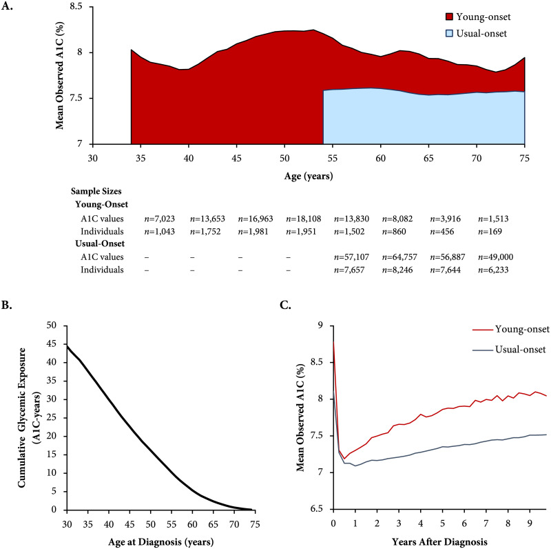
In the register, people with young-onset T2D had a higher mean observed A1C (mean of annual means 8.0% [64 mmol/mol], standard deviation 0.15%) versus those with usual-onset T2D (7.6% [59 mmol/mol], 0.03%) throughout the life span (p < 0.001; Fig 1). The cumulative glycemic exposure was >3 times higher for young- versus usual-onset T2D (41.0 [39.1–42.8] versus 12.1 [11.8–12.3] A1C-years [each A1C-year equivalent to one year with an 8% (64 mmol/mol) average A1C]). In the population cohort, mean observed A1C levels were more elevated among people with young-onset compared with usual-onset T2D, both at diagnosis and throughout the first decade after diagnosis.
Fig 1. Observed A1C and glycemic exposure among adults in the register and population cohorts.
These A1C values are not adjusted for medications. (A) Mean observed A1C across the age span (attained age) in the register cohort (2000–2016), stratified by age at diagnosis (smoothed using 3-year moving averages). The shaded areas indicate glycemic exposure in excess of 7%. Sample sizes are indicated for each 5-year age group. (B) Cumulative glycemic exposure in the register cohort (2000–2016), defined as area under the A1C curve in excess of 7%, by age at diagnosis. One A1C-year is equivalent to one year of exposure to an average A1C of 8%. For example, a person diagnosed with diabetes at age 30 years, with an average A1C of 8% from age 30 to 75 years, would have been exposed to a 1% excess in A1C over 45 years, which is equivalent to 45 A1C-years of glycemic exposure. (C) Mean observed A1C by years since type 2 diabetes diagnosis in the population cohort (2002–2016), stratified by age at diagnosis. A1C, hemoglobin A1c.
Glycemic deterioration in young-onset and usual-onset T2D
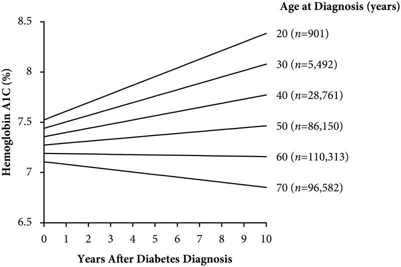
In the population cohort, glycemic deterioration differed significantly across age at diagnosis (Fig 2, Table C in S1 Appendix). Younger age at diagnosis was associated with faster glycemic deterioration (p-value for interaction <0.001). People aged ≤30 years at diagnosis had the most rapid deterioration (+0.08% [95% confidence interval 0.078 to 0.084%] per year for age 20 years at diagnosis) as compared with no deterioration in those aged 60 years at diagnosis (0.00% [−0.005 to −0.004%] per year), and glycemic improvement (−0.02% [−0.027 to −0.0244%] per year) in people aged 70 years at diagnosis. In a sensitivity analysis, we observed similar results allowing for a nonlinear relationship between age at diagnosis and glycemic deterioration (Fig E in S1 Appendix) and excluding A1C values from the first 6 to 12 months after diagnosis (Fig F in S1 Appendix).
Fig 2. Glycemic deterioration during the first decade after type 2 diabetes diagnosis.
Results are stratified by age at diagnosis (population cohort, Hong Kong Diabetes Surveillance Database, 2002–2016). Glycemic deterioration is the modeled slope of the A1C over time after adjusting for oral glucose-lowering drug prescriptions. The sample size (n) is indicated for each age group (age at diagnosis <25, 25–34, 35–44, 45–54, 55–64, ≥65 years). See Table C in S1 Appendix for numeric values. A1C, hemoglobin A1c.
Responses to OGLDs in young-onset and usual-onset T2D
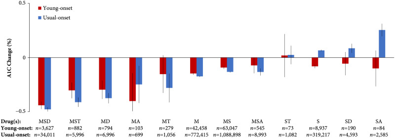
There was a statistically significant difference between people with young- and usual-onset T2D in their responses to OGLDs (p-value <0.001 for omnibus test across all combinations; Fig 3). Combinations containing metformin were associated with lower A1C values, but these decrements appeared slightly smaller for young-onset T2D. Metformin alone was associated with an A1C decrement of −0.15% (95% confidence interval −0.105% to −0.080%) in young-onset and −0.17% (−0.179% to −0.169%) in usual-onset T2D. The combination of metformin, a sulfonylurea and a dipeptidyl peptidase-4 inhibitor had the greatest A1C decrement (young-onset: −0.44% [−0.476% to −0.405%], usual-onset: −0.48% [−0.498% to −0.459%]). The combination of metformin and an α-glucosidase inhibitor (class exclusively consisting of acarbose) was associated with a greater A1C decrement in young-onset (−0.40% [−0.660% to −0.144%]) than usual-onset T2D (−0.25% [−0.420% to −0.077%]).
Fig 3. A1C responses to oral glucose-lowering drugs among people with young- and usual-onset type 2 diabetes.
These insulin-naive individuals were observed during the first decade after diabetes diagnosis (population cohort, Hong Kong Diabetes Surveillance Database, 2002–2016). Sample sizes (number of A1C measurements) are indicated for each combination. Error bars indicate 95% confidence intervals. Differences between young- and usual-onset type 2 diabetes were statistically significant across all combinations (omnibus test p < 0.001). A, acarbose; D, dipeptidyl peptidase-4 inhibitor; M, metformin; S, sulfonylurea; T, thiazolidinedione.
Most combinations containing a sulfonylurea (without metformin) were associated with reduced A1C values in young-onset T2D (−0.08% [−0.099% to −0.065%] for sulfonylurea alone) but increased A1C values in usual-onset T2D (+0.06% [+0.059% to +0.072%] for sulfonylurea alone). The combination of a sulfonylurea and an α-glucosidase inhibitor was associated with the largest A1C decrement in young-onset T2D (−0.10% [−0.266% to 0.064%]; usual-onset: +0.25% [+0.196% to +0.312%]). Sensitivity analyses excluding A1C values from the first 6 and 12 months after diagnosis yielded relatively similar findings for metformin-based combinations, although combinations containing a sulfonylurea were associated with increased A1C values in young-onset T2D (Fig G in S1 Appendix). Adjustment for the baseline A1C with each OGLD combination yielded similar results to the main analysis (Fig H in S1 Appendix).
Discussion
In this large population- and register-based study, we found that people with young-onset T2D had poorly controlled hyperglycemia throughout their life span, resulting in more than triple the cumulative glycemic exposure versus usual-onset T2D. This disparity was driven by rapid glycemic deterioration, which was particularly steep among people diagnosed with T2D before age 30 years. Conversely, we revealed that T2D diagnosis after age 60 years was associated with glycemic improvement—a novel finding that has not been previously reported to our knowledge. In this real-world study, people with young-onset T2D had slightly smaller A1C decrements compared with usual-onset T2D for most combinations of OGLDs including metformin, whereas young-onset T2D was unexpectedly associated with greater responsiveness to sulfonylureas and α-glucosidase inhibitors than usual-onset T2D. Although most OGLD combinations appear to lower A1C by similar decrements across age at diagnosis, the rapidity of glycemic deterioration in young-onset T2D suggests that early combination therapy [33] and aggressive treatment escalation are needed to reduce the massive excess in glycemic exposure that we observed.
Excess glycemic exposure in young-onset T2D
Our study is the first, to our knowledge, to describe the greater than 3-fold disparity in lifetime glycemic exposure between young- and usual-onset T2D. We estimated that people with young-onset T2D will accumulate nearly 40 A1C-years of glycemic exposure by age 75 years. Considering that every 10 A1C-years predicts a 25% increase in relative risk of MACE [2], this excess glycemic exposure is consistent with the high complication risks of young-onset T2D that we [6,7] and others [8,9] have reported. In the United Kingdom Prospective Diabetes Study [1], early reduction of glycemic exposure in T2D has been proven to prevent complications in future decades. Our real-world findings emphasize the urgent need for a paradigm shift toward early and intensive glycemic management to improve survival in the young-onset T2D population.
Rapid glycemic deterioration in young-onset T2D
Glycemic deterioration was particularly rapid in young-onset T2D, peaking at 0.09% per year for an age at diagnosis of 20 years. This rapid progression is thought to be caused by declining β-cell function in the setting of peripheral and hepatic insulin resistance [34,35]. Because of genetic factors, impaired β-cell secretion is an especially important driver of T2D in East Asians [36]. Our findings are consistent with the 20% to 35% per year decline in β-cell function reported among children aged 10 to 17 years in the Treatment Options for type 2 Diabetes in Adolescents and Youth study [34]. By contrast, we observed glycemic improvement among adults diagnosed at age 60 years or above. This finding supports the principle of therapy de-intensification among older adults [37], who may be predisposed to developing a milder phenotype of T2D [38,39]. However, previous studies of usual-onset T2D in European populations reported different results. In Scotland, glycemic deterioration rates were 0.09% and 0.20% per year for ages at diagnosis of 70 and 50 years, respectively, according to the Genetics of Diabetes Audit and Research in Tayside Study [13]. In A Diabetes Outcome Progression Trial, the rate of glycemic deterioration in a largely European cohort was 0.07%–0.14% per year [12]. It is unclear why these rates differed from our study. Rates of β-cell decline might be lower among older Chinese people [40] compared with Europeans [41]. Lifestyle might also play a role, as Hong Kong has the world’s most physically active population [42,43].
Differential responses to OGLDs between young- and usual-onset T2D
In this observational study, A1C decrements associated with OGLD combinations were relatively modest compared with clinical trials. Although we adjusted for adherence using time-varying covariates based on prescription dispensing records, this discrepancy may relate to people not consuming their dispensed medications [44,45]. Rather than focusing on the absolute magnitude of A1C decrements, we compared relative changes in A1C across different ages at diagnosis and drug classes to identify any potentially informative response differences in this real-world setting. In both young- and usual-onset T2D, the combination of metformin, a sulfonylurea and a DDP-4 inhibitor was associated with the greatest A1C reduction. These findings are consistent with evidence suggesting that dipeptidyl peptidase-4 inhibitors lower blood glucose especially well among of East Asians and other people with low body mass indices [46,47]. This combination might be particularly effective in the context of β-cell dysfunction in East Asians, especially in the presence of obesity [48]. Thiazolidinediones were associated with relatively large A1C decrements when used in combination with metformin and a sulfonylurea, which is consistent with the findings of the Treatment Options for type 2 Diabetes in Adolescents and Youth study [34].
Overall, young-onset T2D was associated with slightly smaller A1C decrements versus usual-onset T2D for most—but not all—regimens. As observed in previous studies [12], combinations containing a sulfonylurea were associated with increased A1C values in usual-onset T2D (0.06%–0.25%). However, in young-onset T2D, sulfonylureas were associated with reduced A1C values overall (−0.08% to −0.10%) and relatively smaller A1C increases compared with usual-onset T2D when excluding the initial 6-month period after diagnosis. The reason for this differential response is unclear. In our setting, we previously reported that 5% of people in a young-onset T2D cohort actually had maturity onset diabetes of the young type 3 [49]—which is known to respond well to sulfonylureas [50]—although genetic data were unavailable in the present study. Additionally, α-glucosidase inhibitors, which reduce glycemic excursion and demand on β-cells, were associated with greater A1C reductions in young- versus usual-onset T2D, albeit with wide confidence intervals. This class is efficacious and well tolerated in East Asians [51–54] and merits further investigation among people with young-onset T2D and poor β-cell reserve.
Strengths and limitations
Strengths of our study include its large sample size, complementary cohorts, and long follow-up duration. Unlike previous studies, our detailed register enabled us to capture A1C levels across the lifetime and quantify glycemic exposure at various ages at diagnosis. Using a territory-wide laboratory database containing over 3 million A1C measurements and 2.8 million dispensing records, we were able to demonstrate the important associations between age at diagnosis and glycemic deterioration and responses to OGLDs. Future studies are required to determine whether these findings extend to sodium-glucose cotransporter-2 inhibitors and glucagon-like peptide 1 receptor agonists, which were not widely available in our setting during the study period. As this was a real-world study, we could not confirm whether people consumed the medications dispensed. We cannot exclude the possibility of confounding by indication or by unmeasured variables. Further research is needed to confirm whether these findings are generalizable outside of Hong Kong’s healthcare system and to other ethnic groups. The differential responses to OGLDs between young- and usual-onset T2D are hypothesis-generating, and experimental studies are needed to confirm these findings.
Conclusion
Young-onset T2D progresses rapidly and remains poorly controlled throughout the life span, resulting in more than triple the lifetime glycemic exposure versus usual-onset T2D. The larger A1C reductions associated with sulfonylureas and α-glucosidase inhibitors in young-onset T2D emphasize the importance of β-cell dysfunction in this condition and calls for more precise phenotyping to personalize therapy [55–58]. Pending such evidence, our data suggest that most OGLD regimens are associated with lower glucose levels in young-onset T2D. However, better strategies are needed to apply and rapidly escalate these treatments to overcome therapeutic inertia and mitigate the aggressive trajectory of glycemic deterioration in this high-risk population—especially considering the lack of pharmacologic approaches to improve underlying β-cell function [17]. Given the complex biological and behavioral determinants of young-onset T2D, it is especially important to deploy these strategies in the context of integrated team-based care, disease classification, and patient empowerment to achieve better outcomes [59].
Supporting information
(PDF)
(PDF)
(DOCX)
Acknowledgments
We thank Dr. Allan Detsky for his useful comments on an earlier version of this manuscript and the Hong Kong Hospital Authority for providing the data for this study. Part of this work was presented at the American Diabetes Association’s 79th Scientific Sessions in San Francisco, California, June 7–11, 2019.
Abbreviations
- A1C
hemoglobin A1c
- MACE
major adverse cardiovascular events
- OGLD
oral glucose-lowering drug
- T2D
type 2 diabetes
Data Availability
Due to local laws and regulations regarding the use and distribution of personal data, the data used in the present study cannot be deposited in a public repository. Data access can be applied for through the Data Sharing Portal of the Hong Kong Hospital Authority (https://www3.ha.org.hk/data/DCL/Index/).
Funding Statement
CK is supported by the Canadian Institutes of Health Research (https://cihr-irsc.gc.ca/) Canada Graduate Scholarship and Michael Smith Foreign Study Supplements, the University of Toronto Clinician Investigator Program (https://cip.utoronto.ca/), the Canadian Society of Endocrinology and Metabolism (https://www.endo-metab.ca/) Dr. Fernand Labrie Research Fellowship Grant, and the Royal College of Physicians and Surgeons of Canada (http://www.royalcollege.ca/) Detweiler Traveling Fellowship. (Grant numbers not applicable.) The funders had no role in study design, data collection and analysis, decision to publish, or preparation of the manuscript.
References
- 1.Holman RR, Paul SK, Bethel MA, Matthews DR, Neil HAW. 10-Year Follow-up of Intensive Glucose Control in Type 2 Diabetes. New England Journal of Medicine. 2008;359(15):1577–89. 10.1056/NEJMoa0806470 [DOI] [PubMed] [Google Scholar]
- 2.Roussel R, Steg PG, Mohammedi K, Marre M, Potier L. Prevention of cardiovascular disease through reduction of glycaemic exposure in type 2 diabetes: A perspective on glucose-lowering interventions. Diabetes Obes Metab. 2018. February 1;20(2):238–44. 10.1111/dom.13033 [DOI] [PubMed] [Google Scholar]
- 3.Brown JB, Nichols GA, Perry A. The Burden of Treatment Failure in Type 2 Diabetes. Diabetes Care. 2004. July 1;27(7):1535–40. 10.2337/diacare.27.7.1535 [DOI] [PubMed] [Google Scholar]
- 4.Nichols GA, Rosales AG, Perrin NA, Fortmann SP. The Association Between Different A1C-Based Measures of Glycemia and Risk of Cardiovascular Disease Hospitalization. Diabetes Care. 2014. January;37(1):167–72. 10.2337/dc13-1300 [DOI] [PMC free article] [PubMed] [Google Scholar]
- 5.van Wijngaarden RPT, Overbeek JA, Heintjes EM, Schubert A, Diels J, Straatman H, et al. Relation Between Different Measures of Glycemic Exposure and Microvascular and Macrovascular Complications in Patients with Type 2 Diabetes Mellitus: An Observational Cohort Study. Diabetes Ther. 2017. October 1;8(5):1097–109. 10.1007/s13300-017-0301-4 [DOI] [PMC free article] [PubMed] [Google Scholar]
- 6.Chan JCN, Lau ESH, Luk AOY, Cheung KKT, Kong APS, Yu LWL, et al. Premature Mortality and Comorbidities in Young-onset Diabetes: A 7-Year Prospective Analysis. The American Journal of Medicine. 2014. July 1;127(7):616–24. 10.1016/j.amjmed.2014.03.018 [DOI] [PubMed] [Google Scholar]
- 7.Ke C, Lau E, Shah BR, Stukel TA, Ma RC, So W-Y, et al. Excess Burden of Mental Illness and Hospitalization in Young-Onset Type 2 Diabetes: A Population-Based Cohort Study. Annals of Internal Medicine. 2019. February 5;170(3):145–54. 10.7326/M18-1900 [DOI] [PubMed] [Google Scholar]
- 8.Pavkov ME, Bennett PH, Knowler WC, Krakoff J, Sievers ML, Nelson RG. Effect of Youth-Onset Type 2 Diabetes Mellitus on Incidence of End-Stage Renal Disease and Mortality in Young and Middle-Aged Pima Indians. JAMA. 2006. July 26;296(4):421–6. 10.1001/jama.296.4.421 [DOI] [PubMed] [Google Scholar]
- 9.Steinarsson AO, Rawshani A, Gudbjörnsdottir S, Franzén S, Svensson A-M, Sattar N. Short-term progression of cardiometabolic risk factors in relation to age at type 2 diabetes diagnosis: a longitudinal observational study of 100,606 individuals from the Swedish National Diabetes Register. Diabetologia. 2018. March 1;61(3):599–606. 10.1007/s00125-017-4532-8 [DOI] [PMC free article] [PubMed] [Google Scholar]
- 10.Yeung RO, Zhang Y, Luk A, Yang W, Sobrepena L, Yoon K-H, et al. Metabolic profiles and treatment gaps in young-onset type 2 diabetes in Asia (the JADE programme): a cross-sectional study of a prospective cohort. The Lancet Diabetes & Endocrinology. 2014. January 12;2(12):935–43. [DOI] [PubMed] [Google Scholar]
- 11.van Raalte DH, Diamant M. Glucolipotoxicity and beta cells in type 2 diabetes mellitus: Target for durable therapy? Diabetes Research and Clinical Practice. 2011. August 1;93:S37–46. 10.1016/S0168-8227(11)70012-2 [DOI] [PubMed] [Google Scholar]
- 12.Kahn SE, Haffner SM, Heise MA, Herman WH, Holman RR, Jones NP, et al. Glycemic Durability of Rosiglitazone, Metformin, or Glyburide Monotherapy. New England Journal of Medicine. 2006. December 7;355(23):2427–43. 10.1056/NEJMoa066224 [DOI] [PubMed] [Google Scholar]
- 13.Donnelly LA, Zhou K, Doney ASF, Jennison C, Franks PW, Pearson ER. Rates of glycaemic deterioration in a real-world population with type 2 diabetes. Diabetologia. 2018. March;61(3):607–15. 10.1007/s00125-017-4519-5 [DOI] [PMC free article] [PubMed] [Google Scholar]
- 14.Wallace TM, Matthews DR. Coefficient of failure: a methodology for examining longitudinal β-cell function in Type 2 diabetes. Diabetic Medicine. 2002. June;19(6):465–9. 10.1046/j.1464-5491.2002.00718.x [DOI] [PubMed] [Google Scholar]
- 15.Ke C, Shah BR, Luk AO, Di Ruggiero E, Chan JCN. Cardiovascular Outcomes Trials in Type 2 Diabetes: Time to Include Young Adults. Diabetes, Obesity and Metabolism. 2020. January 20;22(1):3–5. 10.1111/dom.13874 [DOI] [PubMed] [Google Scholar]
- 16.TODAY Study Group, Zeitler P, Hirst K, Pyle L, Linder B, Copeland K, et al. A clinical trial to maintain glycemic control in youth with type 2 diabetes. N Engl J Med. 2012. June 14;366(24):2247–56. 10.1056/NEJMoa1109333 [DOI] [PMC free article] [PubMed] [Google Scholar]
- 17.Consortium RISE. Effects of Treatment of Impaired Glucose Tolerance or Recently Diagnosed Type 2 Diabetes With Metformin Alone or in Combination With Insulin Glargine on β-Cell Function: Comparison of Responses In Youth And Adults. Diabetes. 2019. August;68(8):1670–80. 10.2337/db19-0299 [DOI] [PMC free article] [PubMed] [Google Scholar]
- 18.2016 Population By-census Office. 2016 Population By-Census—Main Results [Internet]. Hong Kong: Census and Statistics Department, Hong Kong Special Administrative Region Government. 2017 Nov [cited 2019 Jan 7]. https://www.bycensus2016.gov.hk/data/16bc-main-results.pdf
- 19.Quan J, Li TK, Pang H, Choi CH, Siu SC, Tang SY, et al. Diabetes incidence and prevalence in Hong Kong, China during 2006–2014. Diabet Med. 2017. July 1;34(7):902–8. 10.1111/dme.13284 [DOI] [PubMed] [Google Scholar]
- 20.Lau IT. A Clinical Practice Guideline to Guide a System Approach to Diabetes Care in Hong Kong. Diabetes Metab J. 2017. April;41(2):81–8. 10.4093/dmj.2017.41.2.81 [DOI] [PMC free article] [PubMed] [Google Scholar]
- 21.Leung GM, Wong IO.L., Chan W-S, Choi S, Lo S-V. The ecology of health care in Hong Kong. Social Science & Medicine. 2005. August;61(3):577–90. [DOI] [PubMed] [Google Scholar]
- 22.Legislative Council Panel on Health Services. Enhancements of Medical Fee Waiver Mechanism of Hospital Authority [Internet]. Hong Kong: Legislative Council Panel on Health Services; 2017 Jul [cited 2020 Jun 8] p. 8. (LC Paper). Report No.: CB(2)1843/16-17(02). https://www.legco.gov.hk/yr16-17/english/panels/hs/papers/hs20170717cb2-1843-2-e.pdf
- 23.Chan JCN, So W, Ma RCW, Tong PCY, Wong R, Yang X. The Complexity of Vascular and Non-Vascular Complications of Diabetes: The Hong Kong Diabetes Registry. Curr Cardiovasc Risk Rep. 2011. June;5(3):230–9. 10.1007/s12170-011-0172-6 [DOI] [PMC free article] [PubMed] [Google Scholar]
- 24.Hospital Authority. Hospital Authority Drug Formulary [Internet]. 15.5. Hong Kong: Hospital Authority; 2020 [cited 2020 Jun 9]. https://www.ha.org.hk/hadf/Portals/0/Docs/HADF_List/External%20list%2020190413/6%20%20%20ENDOCRINE%20SYSTEM.pdf
- 25.Ke C, Stukel TA, Luk A, Shah BR, Jha P, Lau E, et al. Development and validation of algorithms to classify type 1 and 2 diabetes according to age at diagnosis using electronic health records. BMC Med Res Methodol. 2020. February 24;20(35):1–15. [DOI] [PMC free article] [PubMed] [Google Scholar]
- 26.Levy J, Atkinson AB, Bell PM, McCance DR, Hadden DR. Beta-cell deterioration determines the onset and rate of progression of secondary dietary failure in Type 2 diabetes mellitus: the 10-year follow-up of the Belfast Diet Study. Diabetic Medicine. 1998. April 1;15(4):290–6. [DOI] [PubMed] [Google Scholar]
- 27.Sherifali D, Nerenberg K, Pullenayegum E, Cheng JE, Gerstein HC. The Effect of Oral Antidiabetic Agents on A1C Levels: A systematic review and meta-analysis. Diabetes Care. 2010. August 1;33(8):1859–64. 10.2337/dc09-1727 [DOI] [PMC free article] [PubMed] [Google Scholar]
- 28.Mearns ES, Sobieraj DM, White CM, Saulsberry WJ, Kohn CG, Doleh Y, et al. Comparative Efficacy and Safety of Antidiabetic Drug Regimens Added to Metformin Monotherapy in Patients with Type 2 Diabetes: A Network Meta-Analysis. PLoS ONE. 2015. April 28;10(4):e0125879 10.1371/journal.pone.0125879 [DOI] [PMC free article] [PubMed] [Google Scholar]
- 29.Maruthur NM, Tseng E, Hutfless S, Wilson LM, Suarez-Cuervo C, Berger Z, et al. Diabetes Medications as Monotherapy or Metformin-Based Combination Therapy for Type 2 Diabetes: A Systematic Review and Meta-analysis. Annals of Internal Medicine. 2016. June 7;164(11):740 10.7326/M15-2650 [DOI] [PubMed] [Google Scholar]
- 30.Alberti KGMM Eckel RH, Grundy SM Zimmet PZ, Cleeman JI Donato KA, et al. Harmonizing the Metabolic Syndrome: A Joint Interim Statement of the International Diabetes Federation Task Force on Epidemiology and Prevention; National Heart, Lung, and Blood Institute; American Heart Association; World Heart Federation; International Atherosclerosis Society; and International Association for the Study of Obesity. Circulation. 2009. October 20;120(16):1640–5. 10.1161/CIRCULATIONAHA.109.192644 [DOI] [PubMed] [Google Scholar]
- 31.Harrell FE. SAS macros and data step programs useful in survival analysis and logistic regression [Internet]. Duke University Medical Center: Takima West Corporation; 1991 [cited 2018 Dec 28]. http://biostat.mc.vanderbilt.edu/wiki/pub/Main/SasMacros/survrisk.txt
- 32.Turner RC, McCarthy ST, Holman RR, Harris E. Beta-cell function improved by supplementing basal insulin secretion in mild diabetes. Br Med J. 1976. May 22;1(6020):1252–4. 10.1136/bmj.1.6020.1252 [DOI] [PMC free article] [PubMed] [Google Scholar]
- 33.Matthews DR, Paldánius PM, Proot P, Chiang Y, Stumvoll M, Del Prato S. Glycaemic durability of an early combination therapy with vildagliptin and metformin versus sequential metformin monotherapy in newly diagnosed type 2 diabetes (VERIFY): a 5-year, multicentre, randomised, double-blind trial. The Lancet. 2019. October 26;394(10208):1519–29. [DOI] [PubMed] [Google Scholar]
- 34.TODAY Study Group. Effects of metformin, metformin plus rosiglitazone, and metformin plus lifestyle on insulin sensitivity and β-cell function in TODAY. Diabetes Care. 2013. June;36(6):1749–57. 10.2337/dc12-2393 [DOI] [PMC free article] [PubMed] [Google Scholar]
- 35.Lascar N, Brown J, Pattison H, Barnett AH, Bailey CJ, Bellary S. Type 2 diabetes in adolescents and young adults. The Lancet Diabetes & Endocrinology. 2018. January;6(1):69–80. [DOI] [PubMed] [Google Scholar]
- 36.Ma RCW, Chan JCN. Type 2 diabetes in East Asians: similarities and differences with populations in Europe and the United States. Ann NY Acad Sci. 2013. April 1;1281(1):64–91. [DOI] [PMC free article] [PubMed] [Google Scholar]
- 37.Lipska KJ, Ross JS, Miao Y, Shah ND, Lee SJ, Steinman MA. Potential Overtreatment of Diabetes Mellitus in Older Adults With Tight Glycemic Control. JAMA Intern Med. 2015. March 1;175(3):356–62. 10.1001/jamainternmed.2014.7345 [DOI] [PMC free article] [PubMed] [Google Scholar]
- 38.Ahlqvist E, Storm P, Käräjämäki A, Martinell M, Dorkhan M, Carlsson A, et al. Novel subgroups of adult-onset diabetes and their association with outcomes: a data-driven cluster analysis of six variables. The Lancet Diabetes & Endocrinology. 2018;6(5):361–369. 10.1016/S2213-8587(18)30051-2 [DOI] [PubMed] [Google Scholar]
- 39.Wu H, Lau ESH, Yang A, Ma RCW, Kong APS, Chow E, et al. Trends in diabetes-related complications in Hong Kong, 2001–2016: a retrospective cohort study. Cardiovascular Diabetology. 2020. May 12;19(1):60 10.1186/s12933-020-01039-y [DOI] [PMC free article] [PubMed] [Google Scholar]
- 40.Gao Z, Yan W, Fang Z, Zhang Z, Yuan L, Wang X, et al. Annual decline in β-cell function in patients with type 2 diabetes in China. Diabetes Metab Res Rev. 2020. June 8;e3364 10.1002/dmrr.3364 [DOI] [PubMed] [Google Scholar]
- 41.U. K. Prospective Diabetes Study Group. U.K. Prospective Diabetes Study 16: Overview of 6 Years’ Therapy of Type II Diabetes: A Progressive Disease. Diabetes. 1995. November 1;44(11):1249–58. [PubMed] [Google Scholar]
- 42.Althoff T, Sosič R, Hicks JL, King AC, Delp SL, Leskovec J. Large-scale physical activity data reveal worldwide activity inequality. Nature. 2017. July 10;547(7663):336–9. 10.1038/nature23018 [DOI] [PMC free article] [PubMed] [Google Scholar]
- 43.Lu Y, Chen L, Yang Y, Gou Z. The Association of Built Environment and Physical Activity in Older Adults: Using a Citywide Public Housing Scheme to Reduce Residential Self-Selection Bias. Int J Environ Res Public Health. 2018. September 10;15(9):1973 10.3390/ijerph15091973 [DOI] [PMC free article] [PubMed] [Google Scholar]
- 44.Edelman SV, Polonsky WH. Type 2 Diabetes in the Real World: The Elusive Nature of Glycemic Control. Diabetes Care. 2017. November 1;40(11):1425–32. 10.2337/dc16-1974 [DOI] [PubMed] [Google Scholar]
- 45.Carls GS, Tuttle E, Tan R-D, Huynh J, Yee J, Edelman SV, et al. Understanding the Gap Between Efficacy in Randomized Controlled Trials and Effectiveness in Real-World Use of GLP-1 RA and DPP-4 Therapies in Patients With Type 2 Diabetes. Diabetes Care. 2017. November 1;40(11):1469–78. 10.2337/dc16-2725 [DOI] [PubMed] [Google Scholar]
- 46.Kim YG, Hahn S, Oh TJ, Kwak SH, Park KS, Cho YM. Differences in the glucose-lowering efficacy of dipeptidyl peptidase-4 inhibitors between Asians and non-Asians: a systematic review and meta-analysis. Diabetologia. 2013. April 1;56(4):696–708. 10.1007/s00125-012-2827-3 [DOI] [PubMed] [Google Scholar]
- 47.Davis TME, Mulder H, Lokhnygina Y, Aschner P, Chuang L-M, Grado CAR, et al. Effect of race on the glycaemic response to sitagliptin: Insights from the Trial Evaluating Cardiovascular Outcomes with Sitagliptin (TECOS). Diabetes, Obesity and Metabolism. 2018;20(6):1427–34. 10.1111/dom.13242 [DOI] [PubMed] [Google Scholar]
- 48.Ohn JH, Kwak SH, Cho YM, Lim S, Jang HC, Park KS, et al. 10-year trajectory of β-cell function and insulin sensitivity in the development of type 2 diabetes: a community-based prospective cohort study. The Lancet Diabetes & Endocrinology. 2016. January 1;4(1):27–34. [DOI] [PubMed] [Google Scholar]
- 49.Ng MCY, Lee S-C, Ko GTC, Li JKY, So W-Y, Hashim Y, et al. Familial Early-Onset Type 2 Diabetes in Chinese Patients: Obesity and genetics have more significant roles than autoimmunity. Diabetes Care. 2001. April 1;24(4):663–71. 10.2337/diacare.24.4.663 [DOI] [PubMed] [Google Scholar]
- 50.Hattersley AT, Patel KA. Precision diabetes: learning from monogenic diabetes. Diabetologia. 2017. May 1;60(5):769–77. 10.1007/s00125-017-4226-2 [DOI] [PMC free article] [PubMed] [Google Scholar]
- 51.Holman RR, Coleman RL, Chan JCN, Chiasson J-L, Feng H, Ge J, et al. Effects of acarbose on cardiovascular and diabetes outcomes in patients with coronary heart disease and impaired glucose tolerance (ACE): a randomised, double-blind, placebo-controlled trial. The Lancet Diabetes & Endocrinology. 2017. November;5(11):877–86. [DOI] [PubMed] [Google Scholar]
- 52.Chiasson J-L, Josse RG, Gomis R, Hanefeld M, Karasik A, Laakso M. Acarbose for prevention of type 2 diabetes mellitus: the STOP-NIDDM randomised trial. The Lancet. 2002. June 15;359(9323):2072–7. [DOI] [PubMed] [Google Scholar]
- 53.Lam KS, Tiu SC, Tsang MW, Ip TP, Tam SC. Acarbose in NIDDM patients with poor control on conventional oral agents. A 24-week placebo-controlled study. Diabetes Care. 1998. July;21(7):1154–8. 10.2337/diacare.21.7.1154 [DOI] [PubMed] [Google Scholar]
- 54.Chan JC, Chan KW, Ho LL, Fuh MM, Horn LC, Sheaves R, et al. An Asian multicenter clinical trial to assess the efficacy and tolerability of acarbose compared with placebo in type 2 diabetic patients previously treated with diet. Asian Acarbose Study Group. Diabetes Care. 1998. July;21(7):1058–61. 10.2337/diacare.21.7.1058 [DOI] [PubMed] [Google Scholar]
- 55.Davies MJ, D’Alessio DA, Fradkin J, Kernan WN, Mathieu C, Mingrone G, et al. Management of Hyperglycemia in Type 2 Diabetes, 2018. A Consensus Report by the American Diabetes Association (ADA) and the European Association for the Study of Diabetes (EASD). Diabetes Care. 2018. December;41(12):2669–701. 10.2337/dci18-0033 [DOI] [PMC free article] [PubMed] [Google Scholar]
- 56.American Diabetes Association. 6. Glycemic Targets: Standards of Medical Care in Diabetes—2019. Diabetes Care. 2019. January 1;42(Supplement 1):S61–70. [DOI] [PubMed] [Google Scholar]
- 57.Imran SA, Agarwal G, Bajaj HS, Ross S. Targets for Glycemic Control. Canadian Journal of Diabetes. 2018. April 1;42:S42–6. 10.1016/j.jcjd.2017.10.030 [DOI] [PubMed] [Google Scholar]
- 58.National Institute for Health and Care Excellence. Type 2 diabetes in adults: management [Internet]. National Institute for Health and Care Excellence; 2015 Dec [cited 2019 Feb 26] p. 1–44. https://www.nice.org.uk/guidance/ng28/chapter/1-Recommendations#hba1c-measurement-and-targets
- 59.Lim LL, Lau ESH, Kong APS, Davies MJ, Levitt NS, Eliasson B, et al. Aspects of Multicomponent Integrated Care Promote Sustained Improvement in Surrogate Clinical Outcomes: A Systematic Review and Meta-analysis. Diabetes Care. 2018. June 1;41(6):1312–20. 10.2337/dc17-2010 [DOI] [PMC free article] [PubMed] [Google Scholar]


