. 2020 Sep 3;9:e56571. doi: 10.7554/eLife.56571

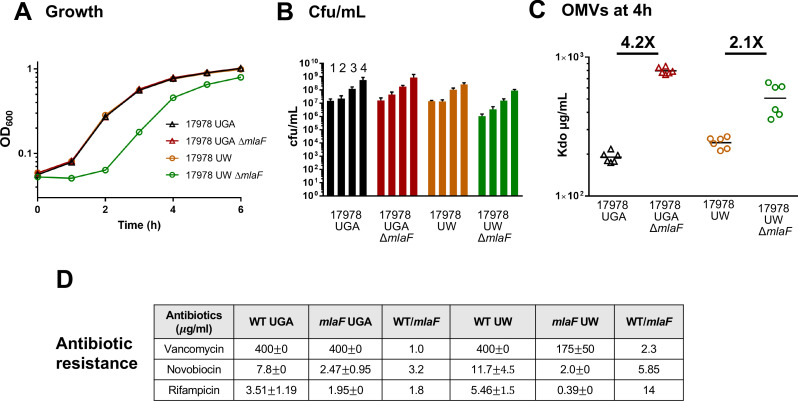
Figure 1. ΔmlaF mutant from the UW group exhibits significant growth defects.

(A) Growth curves of WT and ΔmlaF deletion mutants from the UGA lab (triangles) and UW lab (circles). Standard deviation error bars, if smaller than symbols, are not shown. (B) Colony-forming units (cfu) of cultures at hours 1–4. (C) Outer membrane vesicle (OMV) quantification of WT and ΔmlaF deletion mutants harvested at 4 hr and quantified by measuring 3-deoxy-D-manno-oct-2-ulosonic acid (Kdo) levels using the Purpald assay. Growth curves were performed in biological triplicate. OMV assays were performed in biological duplicate and quantified in technical triplicates. (D) MICs for vancomycin, novobiocin, and rifampicin for both UGA and UW strains. MICs were calculated as described in the Materials and methods.

Figure 1.

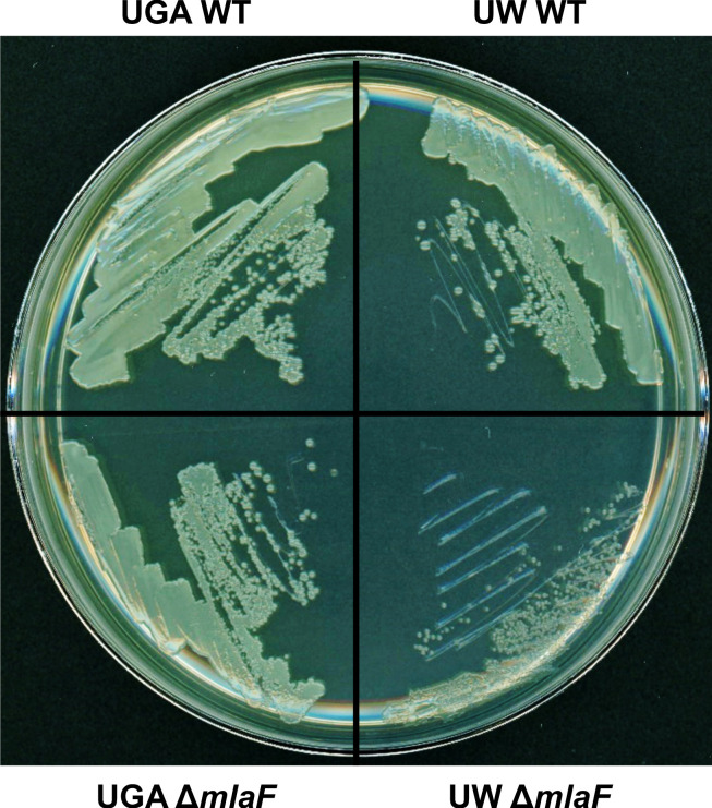
Figure 1—figure supplement 1. UW ΔmlaF has a distinct colony morphology.

Figure 1—figure supplement 1.

Top left – UGA WT grown on LB + 1.5% agar. Bottom left – UGA ΔmlaF. Top right – UW WT. Bottom right – UW ΔmlaF.
Figure 1—figure supplement 2. OMVs collected from stationary phase cultures at 24 hr.
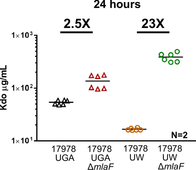
Figure 1—figure supplement 2.

The UW ΔmlaF strain exhibits lysis in stationary phase, artificially inflating OMVs with cellular debris when collected after 24 hr.
Figure 1—figure supplement 3. obgE* alleles are selected against in the absence of Mla.

Figure 1—figure supplement 3.

(A) Growth curves of ΔmlaC (blue) or ΔmlaF (green) mutants generated in the UW WT. Any strains generated by our hands in this background are annotated with MJPX, with X indicating an independently generated clone. For comparison, the UW WT and UW ΔmlaF provided to us are displayed as black and orange circles respectively. Growth curves were performed in biological triplicate. (B) Table displaying frequency of obgE* to obgE reversion. Both UGA strains contain the WT N258 allele. Both UW strains contain the obgE* allele of N258I. When ΔmlaC strains were generated in the UW WT, obgE* reverted to obgE 12/12 times tested. When ΔmlaF strains were generated in the UW WT, obgE* reverted to obgE 3/3 times tested. SNP presence was determined by sanger sequencing.