A 66-year-old female patient received a diagnosis of breast cancer. She underwent bilateral mastectomies and was indicated for postoperative chemotherapy. Pretreatment transthoracic echocardiography (TTE) revealed a normal systolic left ventricular (LV) function [LV ejection fraction (LVEF), 65%]. Adjuvant combination chemotherapy with epirubicin (total 400 mg/m2) and cyclophosphamide (total 2000 mg/m2) was administered. Then, she received docetaxel (total 300 mg/m2), followed by total 104 mg/kg trastuzumab.
Fifteen months after the initiation of her chemotherapy, she was admitted to a hospital and diagnosed with acute decompensated heart failure. LVEF determined by TTE fell to 40%, and she was diagnosed with cancer therapy-related cardiac dysfunction (CTRCD).
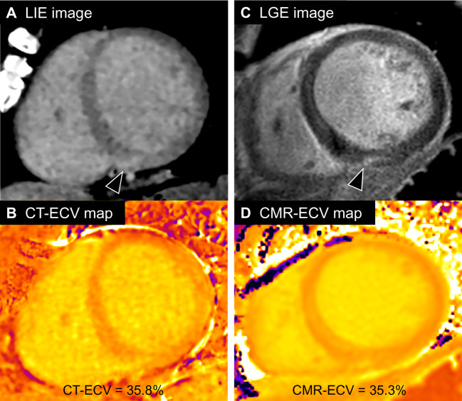
Obstructive coronary artery disease was excluded by coronary computed tomography (CT) angiography. Delayed phase cardiac CT revealed right ventricular insertion point late iodine enhancement (LIE) (Panel A: LIE image, arrowhead) which represents focal myocardial fibrosis, and myocardial global extracellular volume fraction (ECV) by CT was 35.8% (Panel B: CT-ECV map), suggesting the presence of diffuse fibrosis. These findings were consistent with the cardiac magnetic resonance imaging (CMR) results [Panel C: late gadolinium enhancement (LGE) image and Panel D: CMR-ECV map, global ECV = 35.3%]. Cardiac CT was helpful in detection and quantification of myocardial fibrosis that can be a feature of CTRCD.
CT imaging allows evaluation of CTRCD utilizing LIE and ECV analyses. This novel CT imaging can be performed in conjunction with standard coronary artery evaluation and cancer follow-up. We believe this technology could be useful for the ‘one-stop shop’ evaluation of CTRCD.
Consent: The author/s confirm that written consent for submission and publication of this case report including image(s) and associated text has been obtained from the patient in line with COPE guidance.


