Abstract
Background
The Child–Turcotte–Pugh score (CTP) is the most commonly used tool to assess hepatic reserve and predict survival in hepatocellular cancer (HCC). The CTP stratification accuracy has been questioned and attempts have been made to improve the objectivity of the system. Serum insulin-like growth factor-1 (IGF-1)-CTP has been proposed to improve CTP prognostic accuracy. We aimed to validate the IGF-CTP score in our patient population.
Patients and Methods
A total of 84 diagnosed HCC patients were enrolled prospectively. IGF-CTP scores in addition to CTP scores were calculated. C-index was used to compare the prognostic significance of the two scoring systems and overall survival (OS).
Results
Cirrhosis was present in 48 (57.1%) patients, 35 (41.7%) patients were non-cirrhotic, 36 (42.8%) patients were hepatitis B (HBV) positive, and 8 (9.5%) patients were hepatitis C (HCV) positive. Serum IGF-1 levels were significantly lower in cirrhotic compared with non-cirrhotic patients (p=0.04). There was a significant difference in OS rates of patients with serum IGF-1 level <50 ng/mL and patients with serum IGF-1 levels ≥50 ng/mL (p=0.02); the OS rates were 6.5 and 14.8 months, respectively (p=0.02). The median OS of all patients was 7.38 months (95% CI: 5.89–13.79). The estimated C-index for CTP and IGF-CTP scores was 0.605 (95% CI: 0.538, 0.672) and 0.599 (95% CI: 0.543, 0.655), respectively. The U statistics indicated that the C-indices between two scoring systems are not statistically different (P= 0.91).
Conclusion
This study evaluated IGF-1 levels and the IGF-CTP classification in a predominantly HBV (+) cohort of HCC patients with 41.7% non-cirrhotic. Although the prognostic value was similar, among patients with CTP-A, class those reclassified as IGF-CTP B had shorter OS than those with IGF-CTP score A. Thus, further validations of IGF-CTP score in similar populations may add additional value as a stratification tool to predict HCC outcome.
Keywords: Child–Turcotte–Pugh, cirrhosis, hepatocellular carcinoma, insulin-like growth factor-1
Introduction
Hepatocellular carcinoma (HCC) is the sixth most common cancer and the fourth leading cause of cancer-related mortality worldwide.1 It usually develops in patients with chronic liver disease or cirrhosis. Despite advances in prevention, screening and treatment strategies, overall survival remains poor, and a 5-year survival rate is still under 20%.2 Treatment decisions for HCC are challenging and are commonly based on an assessment of hepatic reserve based on clinical and laboratory findings. There are several staging and prognostic scoring systems considered in the decision making for the treatment of HCC such as TNM, the Barcelona Clinic Liver Cancer (BCLC), Okuda, cancer of the Liver Italian Program (CLIP), Child–Turcotte–Pugh (CTP), Model for End-Stage Liver Disease (MELD), and ALBI score, etc.3–7 The BCLC staging system is the most commonly used staging system, and it classifies patients into five categories; very early (0), early (A), intermediate (B), advanced (C), and terminal (D). The factors that are considered in the BCLC staging system are patient performance status, tumor size, number of nodules, major vascular invasion status, presence or absence of extrahepatic spread (lymph node involvement or metastasis), and the CTP score.6 The MELD is a chronic liver disease severity scoring system that uses a patient’s laboratory values which include serum bilirubin, serum creatinine, and the international normalized ratio (INR).8 In this way, 3-month survival rates are predicted based on these laboratory results. The CTP classification system has been a standard classification system over decades for assessing the hepatic reserve to determine the prognosis of cirrhotic patients and to help patient selection for routine therapy and clinical trial enrollment and stratification.9 Despite the presence of objective parameters such as serum bilirubin, serum albumin, and the international normalized ratio of prothrombin time (INR) in the CTP system, there are two subjective parameters which include ascites and encephalopathy. These scores can be subjective and may change on a daily basis under the influence of medications and nutritional status. Our team integrated insulin-like growth factor-1 (IGF-1) into CTP and developed and validated IGF-CTP classification system in 2014.10 In this newly proposed classification system, two subjective parameters in the CTP scoring system, ascites and encephalopathy, were replaced by serum IGF-1 level.10 Most of the circulating IGF-1 is produced by the liver, and therefore, circulating IGF-1 level reflects hepatic synthetic function. The relationship between IGF-1 level and hepatic function has been reported by several studies that demonstrated the correlation between the severity of cirrhosis and the development of HCC and low serum IGF-1 concentration.11–13 Furthermore, the levels of IGF-1 are significantly lower in patients with cirrhosis CTP C than CTP A and B. Additionally, correlation with other advanced cirrhotic and portal hypertension parameters such as albumin level, INR value, and spleen size also has been reported.14–16 Similarly, a significant decrease in IGF-1 concentration was observed in patients with advanced HCC.17 Notably, the CTP classification system has been developed for cirrhotic patients, and eventually became the standard tool to assess hepatic reserve in HCC, despite its limitations. Our team developed and validated the IGF-CTP score and reported improved prediction of OS by the new IGF-CTP classification system. However, there is a need to validate the new scoring system in a different cohort of patients with HCC. This study is a validation study of the IGF-CTP system to determine its predictive value for OS in Turkish patients with predominantly HBV related HCC, and also to investigate the independent predictive and/or the prognostic role of serum level of IGF-1 when used alone in these patients.
Patients and Methods
Patients
The patients who were diagnosed with HCC in the clinic or admitted to our hospital with HCC diagnosis between November 2014 and May 2017 were included. Patients’ data were prospectively collected in the database at the Cancer Institute of the Hacettepe University, and serum samples were obtained at baseline, at the time of study enrollment. The patients with HCC diagnosis with either histopathologically or based on radiological findings that determined in AASLD (American Association for the Study of Liver Diseases) and EASL (European Association for the Study of Liver) guidelines were included.18,19 For cirrhotic patients with the absence of histological confirmation, the diagnosis must be based on the typical hallmark of HCC in imaging techniques that obtained by 4-phase multidetector CT scan or dynamic contrast-enhanced MRI (hypervascular in the arterial phase with washout in the portal venous or delayed phases). Other eligibility criteria were patients aged 18 years or older, and collected variables included level of serum Alfa-fetoprotein (AFP) concentrations, hematological and biochemical parameters, HCC stage based on BCLC staging system (stages 0, A, B, C or D), and the score of CTP classification. Liver cirrhosis in the patients with HCC was determined based on histological or clinical information or imaging and laboratory results, which reflected the hepatic reserve and classic clinical signs of cirrhosis such as non-malignant ascites, hepatic encephalopathy, thrombocytopenia, splenomegaly, and the presence of esophageal varices. Patients were excluded if they had additional cancer diagnosis other than HCC. The CTP score of the patients was assessed for all patients based on serum bilirubin, albumin, and INR as laboratory parameters, and ascites and hepatic encephalopathy as clinical findings. Treatment decisions of patients have been discussed in a multidisciplinary manner at the institution liver tumor board to discuss patients’ candidacy for local and systemic treatment modalities which were then recommended based on the guidelines. Concurrent treatment with both systemic and local approaches was included as study variables. Laboratory parameters, including serum IGF-1, were obtained, and survival rates were calculated. The Body Mass Index (BMI) was calculated as weight in kilograms divided by the square of height in meters, and classified according to the International Classification of WHO as underweight, healthy weight, overweight and obesity. The patient defined as underweight if BMI was in the range of 15 to 19.9, healthy weight if the BMI was 20 to 24.9, overweight if BMI was 25 to 29.9, and obese if it was 30 to 35 or greater.
Laboratory Parameters and Serum IGF-1 Measurement
Laboratory results were obtained at the time of initial HCC diagnosis or the time of patients’ inclusion in the study. Blood samples for IGF-1 measurements were collected and stored at −80ºC until the end of the study. To quantify circulating IGF-1 levels, serum samples were analyzed in duplicate using the human IGF-1 ELISA Kit (Elabscience, catalog no: E-EL-H0086) according to the manufacturer’s instructions. IGF-CTP scores were calculated and assigned class A, B or C based on serum albumin, bilirubin, and prothrombin time and plasma IGF-1 level replaced ascites and encephalopathy grading. Firstly, the optimal serum IGF-1 ranges of patients were determined and formed three distinct groups related to survival time as used in the first study: more than 50 ng/mL = 1 point; 26 to 50 ng/mL = 2 points and less than 26 ng/mL = 3 points. Thereafter, we used the new IGF score (IGF-CTP) which replacing the acid and encephalopathy grading with the plasma IGF-1 value.
Ethical Aspects
The study was designed and conducted following the Helsinki declaration. Approval of the study was granted from the Ethics and institutional research committees of the Hacettepe University Faculty of Medicine.
Assessment of Survival Outcomes
The primary outcome of interest was overall survival (OS) defined as the time from the blood draw date to death or censorship, in which individuals lost to follow-up were censored at the date they were last known to be alive. Additionally, the analyses were also performed for the OS from the date of diagnosis to the date of death or the last follow-up date. OS was calculated for all patients.
Statistical Analysis
The Log rank test was used to compare the OS. Univariate Cox model, C-index and U-statistics were used to compare the prognostic performance of the new (IGF-CTP) and original Child–Turcotte–Pugh systems. Differences in patients’ characteristics were compared, and categorical variables, number of patients and percentage of patients in each category were provided. Chi-Square (X2) or Fisher’s exact test was used to test for statistical differences between the treatment groups. Survival rates were estimated by the Kaplan–Meier method and the Log rank test was used to compare OS rates between groups. Univariable and multivariable associations between survival and the covariates were investigated using the Cox proportional hazards model. Hazard ratios (HRs) with 95% confidence intervals (CIs) were calculated. All tests were 2-sided with a significance level of 0.05. Analyses were performed using SPSS version 22 statistical software (IBM Corporation, Somers, New York, USA).
Results
Baseline Patient Characteristics
From November 2014 to May 2017, a total of 84 patients with HCC were enrolled. All patients were informed about the purpose of the study and only patients who consented and agreed to provide blood samples for further evaluation were enrolled in the study. Thirteen (15.5%) of the patients were female and 71 (84.5%) of patients were male, the median age was 64 years (range; 19–90 years). Fifty-eight (69%) patients were CTP A, 22 (26.2%) were CTP B and 3 (3.6%) were CTP C. Among HCC patients, HBV positive patients were much more common than HCV positive patients. In terms of the type of hepatitis, 36 (42.9%) patients were HBV positive, 48 (57.1%) were HBV negative, 8 (9.5%) were HCV positive and 76 (90.5%) remained patients were HCV negative. Among those whose cirrhosis status was known, 48 of the patients were cirrhotic and 35 were non-cirrhotic. Overall survival time was computed as the period from the blood collection date to the date of death or the last follow-up date, whichever occurred first.
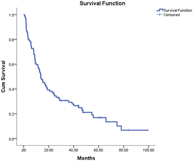
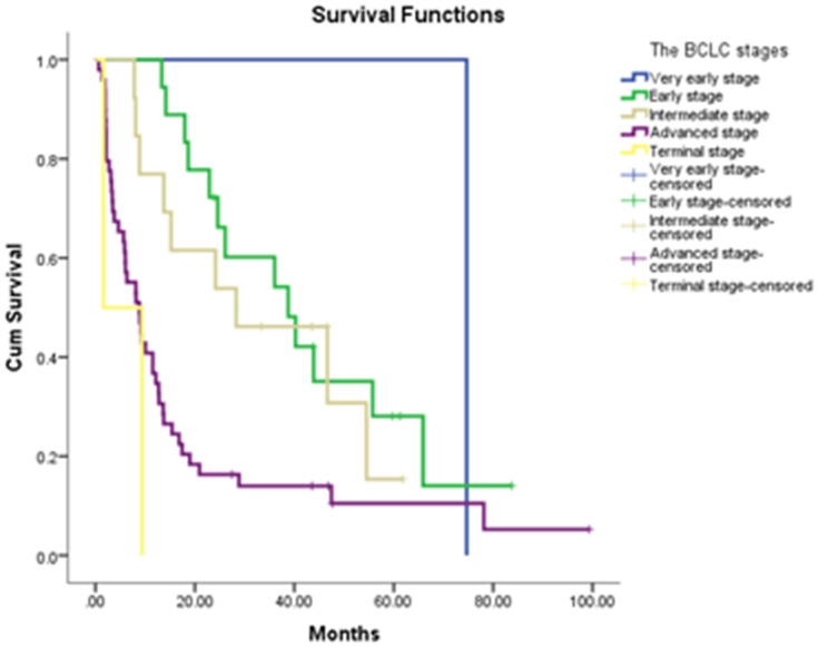
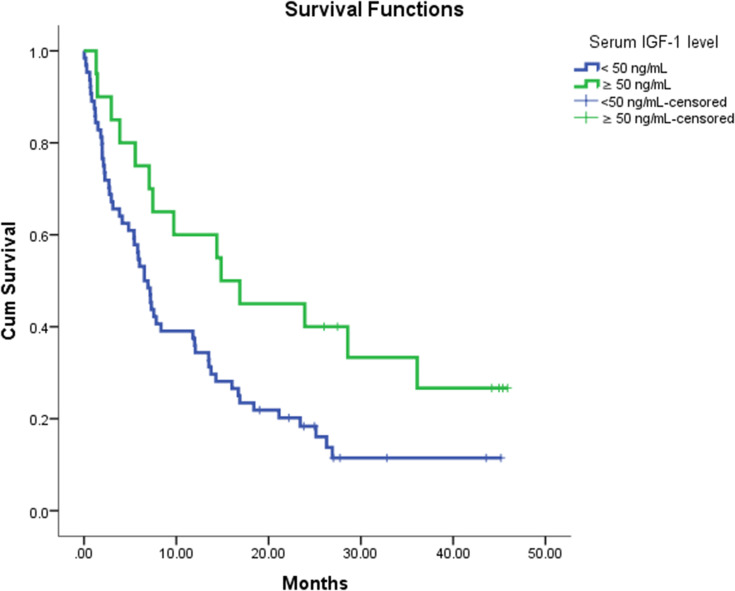
Sixty-nine of the 84 patients died. The estimated median survival was 7.38 months (95% CI: 5.89–13.79 months). The estimated C-index for Child–Turcotte–Pugh score system was 0.605 (95% CI: 0.538, 0.672), and the estimated C-index was 0.599 (95% CI: 0.543, 0.655) for the IGF-integrated score system. The U statistics indicated that the C-indexes between two score systems were not statistically different (P-value = 0.91). In the general population, the estimated median overall survival rate was 13.7 months (95% CI: 9.54–17.92 months) (Figure 1). In terms of OS rates according to the BCLC staging system, there was a statistically significant difference between stages. The median OS of very early stage patients was 74.6 months, 38.7 months for early-stage, 28.3 months for intermediate stage, 8.8 months for advanced stage, and 1.6 months for terminal stage patients (p<0.001) (Figure 2). According to serum IGF-1 levels, using a cutoff of 50 (which is the lowest normal IGF-1 level per IGF-CTP score), there was a significant difference between median OS rates of patients with serum IGF-1 level <50 ng/mL and patients with serum IGF-1 levels ≥50 ng/mL; 6.5 and 14.8 months, respectively (p=0.02) (Figure 3).
Figure 1.
Kaplan-Meier curve shows the median overall survival rate in general patient population.
Figure 2.
Kaplan-Meier curve demonstrates the survival rates of HCC patients according to the BCLC staging system.
Figure 3.
Kaplan-Meier curve shows the the survival rates of HCC patients according to serum IGF-1 level.
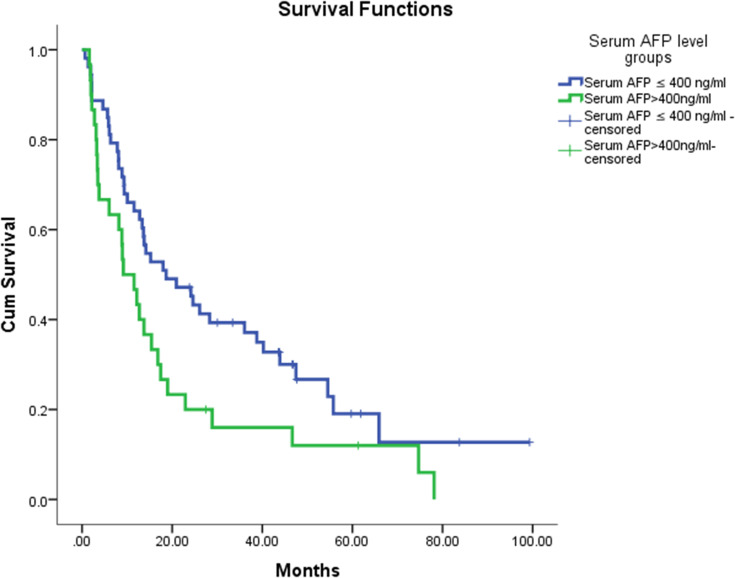
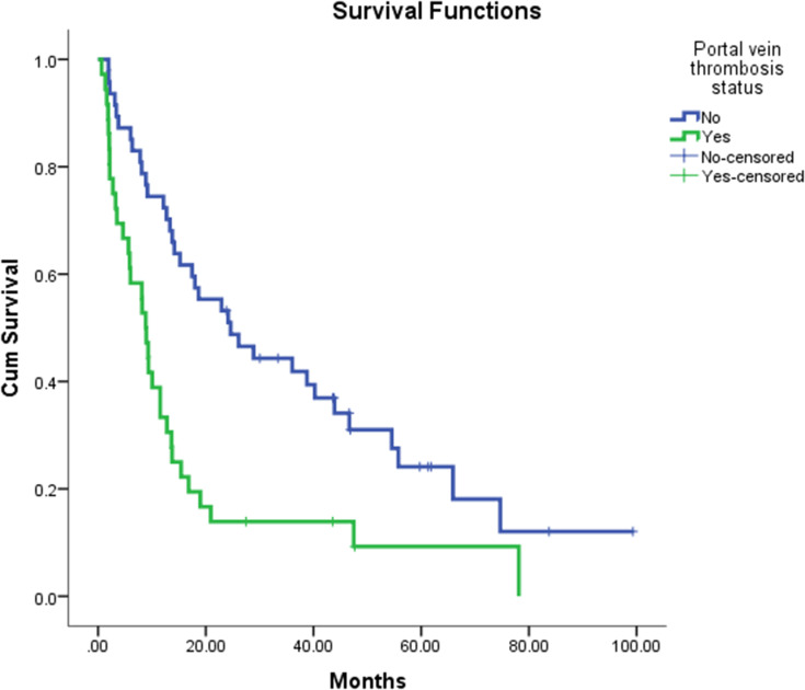
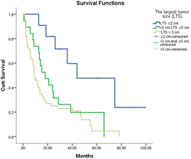
Table 1 displays the number of patients classified by the original CTP score system and by the IGF-modified score system. Table 2 displays the results of the Log rank test to compare OS between groups with IGF ≤ 26 and IGF > 26 as well as IGF-CPS A vs B (ie old A new A vs old A new B) for Child-Pugh “A” and “B” patients separately. IGF-CTP further stratified CTP A patients in the prognostic outcome. Twenty-four patients with CTP-A were reclassified as IGF-CTP A and has a median OS of 16.48 (95% CI=7.47, NA), 34 patients were reclassified as IGF-CTP B had a shorter median OS of 7.38 (95% CI=6.55, 16.91). Among CTP B patients, two were reclassified as IGF-CTP A with a median OS of 19.4 (95% CI=14.87, NA) while 13 were reclassified as IGF-CTP B with a shorter median OS of 5.43 (95% CI=1.81, NA). Two patient groups were created according to their serum AFP levels, group A represented patients with serum AFP levels ≤400 ng/mL and group B represented the patients with AFP level >400 ng/mL. There was a statistically significant difference between OS rates of group A and group B, the median OS rate was 18.6 and 9.1 months, respectively (p=0.032) (Figure 4). As a prognostic factor for HCC patients, tumor-related vascular invasion status was evaluated, and 36 patients (42.8%) had vascular invasion, 47 (56%) did not have, and 1 (1.2%) patient was with unknown status. There was a statistically significant difference between OS rates of patients without vs with tumor vascular invasion, the median OS rate was 24.6 and 8.8 months, respectively (p<0.001) (Figure 5). Several laboratory parameters related to the liver and HCC were analyzed, the median LDH was 250.5 (76–1073) U/L, ALT was 35 (8–268) U/L, AST was 50 (16–620) U/L, ALP was 153.5 (56–1222) U/L, GGT was 144.5 (25–1719) U/L, and AFP was 128.5 (1.2–286.748) U/L. There was a negative correlation between serum IGF-1 levels and MELD score, patients who had higher MELD scores tended to have lower IGF-1 (p=0.03). The first treatments that HCC patients had after diagnosis were divided into six groups that included radiofrequency ablation (RFA)/microwave ablation (MWA), transarterial chemoembolization (TACE)/transarterial radioembolization (TARE), systemic cytotoxic therapy, tyrosine kinase inhibitor treatment (TKI), surgical treatment, and best supportive care (BSC) groups. Among 75 patients who had the first-line treatment data, 7 (8.3%) of the patients had RFA/MWA, 22 (26.2%) of the patients had TACE/TARE, 24 (28.6%) of the patients had systemic cytotoxic treatment, 9 (10.7%) of the patients had TKI treatment, 9 (10.7%) of the patients had tumor resection at the initial and 4 (4.8%) of the patients followed with BSC (Table 3). We used univariate cox regression analysis to determine the first-line treatment modalities that may be related to patients’ survival. Similarly, this analysis was conducted to determine the effect of serum IGF-1 levels, serum AFP levels, portal vein thrombosis status, AST, ALT, bilirubin, metastatic status, first-line treatment modality, and gender on survival as well (Table 4). Patients were divided into three groups according to tumor size, ≤2 cm, >2 to ≤5 cm, and >5 cm, respectively. There were 11 patients in ≤2 cm group, 19 patients in >2 to ≤5 cm group, and 52 patients in the >5 cm groups. The median OS was 43.8 months in the ≤2 cm group, 17.9 months in the >2 cm to ≤5 cm group, and 9.2 months in the >5 cm group. There was a significant difference between the groups in terms of overall survival (p=0.005) (Figure 6). According to patient BMI scores, 30 patients were classified as healthy overweight, 30 patients were overweight, and 15 patients were classified as obese. The diabetes rate was 26%. There was no relationship between patients’ BMI classes and IGF-1. The median follow-up time as the time from diagnosis of HCC date to death or censorship was 59.7 months (range 37.9–81.4 months). Additionally, the median follow-up time as the time from blood collection date to death or censorship was 32.8 months (range 7.9–57.6 months).
Table 1.
Child–Pugh–Turcotte (CTP) Score Vs IGF-Child–Pugh–Turcotte Score Class
| IGF-CPT (CPG New) | Child–Pugh–Turcotte Class | |||
|---|---|---|---|---|
| A | B | C | Total | |
| A | 24 | 2 | 0 | 26 |
| B | 34 | 13 | 0 | 47 |
| C | 0 | 7 | 3 | 10 |
| Total | 58 | 22 | 3 | 83 |
| Frequency Missing = 1 | ||||
Table 2.
Compare OS Between Groups of IGF-1 ≤26 Ng/mL Vs IGF-1 >26 Ng/mL, IGF-CTP a Vs B in Each Categorical of Child–Pugh Score System
| Scoring System | Level | N | Event | Median OS (95% CI) | OS Rate at 1 Year (95% CI) | P-value | |
|---|---|---|---|---|---|---|---|
| Child Pugh “A” | Child Pugh “A” | 58 | 44 | 11.94 (7.07, 18.42) | 0.5 (0.39, 0.65) | ||
| IGF-1 | 0>26 | 31 | 23 | 11.81 (5.56, 28.62) | 0.48 (0.34, 0.7) | 0.7768 | |
| 1≤26 | 27 | 21 | 12.07 (6.55, 26.32) | 0.52 (0.36, 0.75) | |||
| IGF-CTP | A | 24 | 16 | 16.48 (7.47, NA) | 0.58 (0.42, 0.82) | 0.1292 | |
| B | 34 | 28 | 7.38 (6.55, 16.91) | 0.44 (0.3, 0.64) | |||
| Group | Old A new A | 24 | 16 | 16.48 (7.47, NA) | 0.58 (0.42, 0.82) | 0.1292 | |
| Old A new B | 34 | 28 | 7.38 (6.55, 16.91) | 0.44 (0.3, 0.64) | |||
| Child Pugh “B” | Child Pugh “B” | 22 | 21 | 6.48 (2.11, 13.52) | 0.32 (0.17, 0.59) | ||
| IGF-1 | 0>26 | 7 | 7 | 9.74 (2.11, NA) | 0.43 (0.18,1) | 0.4913 | |
| 1≤26 | 15 | 14 | 6.02 (1.94, 13.52) | 0.27 (0.12, 0.62) | |||
| IGF-CTP | A | 2 | 2 | 19.41 (14.87, NA) | 1 (1, 1) | 0.0747 | |
| B | 13 | 13 | 5.43 (1.81, NA) | 0.15 (0.04, 0.55) | |||
| C | 7 | 6 | 6.94 (1.97, NA) | 0.43 (0.18, 1) | |||
| Group | Old B new A | 2 | 2 | 19.41 (14.87, NA) | 1 (1, 1) | 0.0747 | |
| Old B new B | 13 | 13 | 5.43 (1.81, NA) | 0.15 (0.04, 0.55) | |||
| Old B new C | 7 | 6 | 6.94 (1.97, NA) | 0.43 (0.18, 1) |
Figure 4.
Kaplan-Meier curve illustrates the survival rates of HCC patients according to serum AFP level.
Figure 5.
Kaplan-Meier curve shows the survival rates of HCC patients according to portal vein thrombosis status.
Table 3.
Demographic and Clinical Characteristics of the Patients at Baseline
| Number | Percentage | |||
|---|---|---|---|---|
| Total patients (n) | 84 | 100% | ||
| Median age of all patients | 64 (19–90) | 100% | ||
| Median age | Female | 65 (29–85) | 19% | |
| Male | 64 (19–90) | 81% | ||
| Gender | Female | 13 | 15.5% | |
| Male | 71 | 84.5% | ||
| Serum AFP level | ≤400 | 53 | 63.1% | |
| >400 | 30 | 35.7% | ||
| Not reported | 1 | 1.2% | ||
| Child–Turcotte–Pugh classes | A | 58 | 69% | |
| B | 22 | 26.2% | ||
| C | 3 | 3.6% | ||
| Not reported | 1 | 1.2% | ||
| Portal vein invasion | No | 47 | 56% | |
| Yes | 36 | 42.8% | ||
| Not reported | 1 | 1.2% | ||
| Treatment groups as the first-line | Surgery | 9 | 10.7% | |
| RFA or MWA | 7 | 8.3% | ||
| TACE or TARE | 22 | 26.2% | ||
| Systemic cytotoxic treatment | 24 | 28.6% | ||
| Tyrosine Kinase inhibitor | 9 | 10.7% | ||
| BSC | 4 | 4.8% | ||
| Not reported | 9 | 10.7% | ||
| Hepatitis Infection | HBV | Positive | 36 | 42.9% |
| Negative | 48 | 57.1% | ||
| HCV | Positive | 8 | 9.6% | |
| Negative | 76 | 90.4% | ||
| The BCLC stage | Very early | 1 | 1.2% | |
| Early | 18 | 21.4% | ||
| Intermediate | 13 | 15.5% | ||
| Advanced | 50 | 59.5% | ||
| Terminal | 2 | 2.4% | ||
| Cirrhosis status | No | 35 | 41.7% | |
| Yes | 48 | 57.1% | ||
| Not reported | 1 | 1.2% | ||
| Diabetes | No | 46 | 54.8% | |
| Yes | 22 | 26.2% | ||
| Not reported | 16 | 19% | ||
| Body mass index (BMI) | Healthy weight | 30 | 35.7% | |
| Overweight | 30 | 35.7% | ||
| Obese | 15 | 17.9% | ||
| Not reported | 9 | 10.7% | ||
Table 4.
Cox Regression Analysis and Prognostic Factors for Survival
| Hazard Ratio (95% CI)¥ | P value | |
|---|---|---|
| IGF-1≥50 vs IGF-1<50 | 0.50 (0.27–0.91) | 0.024* |
| AFP >400 vs AFP ≤400 | 1.95 (1.19–3.18) | 0.008* |
| Male vs female | 0.78 (0.40–1.48) | 0.44 |
| Portal vein invasion positive vs negative | 2.36 (1.45–3.85) | 0.001* |
| AST >45 vs AST ≤45 | 2.64(1.57–4.44) | <0.001* |
| ALT >40 vs ALT ≤40 | 1.70(1.05–2.76) | 0.031* |
| Bilirubin >2 vs bilirubin ≤2 | 2.17(1.18–4.02) | 0.013* |
| Metastatic status, positive vs negative | 2.18 (1.24–3.84) | 0.007* |
| Surgery vs BSC | 0.05 (0.01–0.21) | <0.001* |
| Systemic cytotoxic treatment vs BSC | 0.23 (0.07–0.71) | 0.011* |
| Tyrosine Kinase vs BSC | 0.29 (0.08–1.03) | 0.056 |
| RFA or MWA vs BSC | 0.14 (0.04–0.51) | 0.003* |
| TACE or TARE vs BSC | 0.14 (0.04–0.45) | 0.001* |
Notes: *Statistically significant. p<0.05 was considered as significant.
Abbreviations: IGF-1, insulin-like growth factor 1; BCLC, Barcelona Clinic Liver Cancer; AFP, α-fetoprotein; ALT, alanine transaminase; AST, aspartate transaminase; INR, international normalized ratio; TACE, transarterial chemoembolization; TARE, transarterial radioembolization; RFA, radiofrequency ablation; MWA, micro wave ablation; TKIs, tyrosine kinase inhibitors; BSC, best supportive care.
Figure 6.
Kaplan-Meier curve demonstrates the survival rates of HCC patients according to the largest tumor size.
Discussion
In this study, we validated serum IGF-1 levels in an HCC patient population as a serum marker of the liver reserve for the prediction of patient survival and risk stratification. Overall, the IGF-CTP classification system that replaces encephalopathy and ascites with serum IGF-1 levels provided similar survival prediction ability to CTP and did not lead to more precise predictions compared to the original CTP classification in our HCC patients as reported previously.10,20 However, our 4-blood parameter score remained easier to calculate and more objective.
Notably, IGF-1 has a crucial regulating role upon the proliferation and differentiation of the cell. IGF-1 is a peptide hormone with strong mitogenic effects on both normal and cancerous cells,21,22 and also activated IGF-1 inhibits cell apoptosis.23 There are several studies that have reported the association between high serum levels of IGF-I and increased risks of different types of cancer such as those of the prostate, breast, esophagus, colon, and lung.24–28 The relationship between IGF-1 and HCC is different, unlike other cancers types; patients with HCC have lower serum IGF-1 levels than healthy controls.29 One of the possible reasons is the hepatic synthesis that is the major source of circulating IGF-1, therefore, advanced cirrhosis and/or HCC tumors suppress normal hepatic function by replacing normal hepatocytes. This was also evident in our study in patients with versus those without cirrhosis, Table 5. Previous studies reported the association between low serum IGF-1 levels in HCC patients and extensive liver involvement, vascular invasion, and shorter OS.30 In our study, the mean serum IGF-1 level was 36.3 ng/mL (standard deviation (SD) ± 38.97 ng/mL), which was consistent with prior reports (two studies and one meta-analysis) of significantly decreased serum levels of IGF-1 in patients with HCC.30–32 In these studies, patients with lower serum IGF-1 levels were associated with worse prognosis (survival outcomes) than patients with higher IGF-1 levels. Similarly, our study demonstrated that HCC patients with lower serum IGF-1 had a worse OS than patients with higher IGF-1. Additionally, serum IGF-1 levels were found to be an independent predictor of survival in univariate analysis of our patient cohort.
Table 5.
Serum Level of IGF-1 According to Different Characteristics of HCC Patients
| Patients Characteristics | Groups of Variable | The Mean Serum Level of IGF-1(range) | P value |
|---|---|---|---|
| Age | ≤60 | 42.8 (1.3–143.5) | 0.3 |
| >60 | 32.7 (2.9–141.3) | ||
| Gender | Female | 38.5 (3.9–132.1) | 0.8 |
| Male | 35.9 (1.3–143.5) | ||
| Cirrhosis status | No | 46.2 (1.3–141.3) | 0.04* |
| Yes | 29.1 (2.1–143.5) | ||
| The largest tumour size | ≤5cm | 40 (2.9–143.5) | 0.6 |
| >5 cm | 35.2 (1.3–141.3) | ||
| Number of Tumour lesions | Uninodularity | 36.01(3.1–138.7) | 0.9 |
| Multinodularity | 37.11 (1.3–143.5) | ||
| Lymph node involvement | No | 38.4 (2.9–143.5) | 0.4 |
| Yes | 30.7 (1.3–138.7) | ||
| Distant metastasis | No | 39.6 (2.1–143.5) | 0.12 |
| Yes | 23.4 (1.3–118) | ||
| Vascular invasion | No | 38.8 (2.9–143.5) | 0.6 |
| Yes | 33.9 (1.3–138.7) | ||
| Serum AFP level | ≤400 | 39.7 (2.9–143.5) | 0.3 |
| >400 | 30.5 (1.3–118) | ||
| Serum ALT value | ≤40 | 35.3 (2.9–141.3) | 0.8 |
| >40 | 37.6 (1.3–143.5) | ||
| Serum AST level | ≤45 | 46.1 (2.9–143.5) | 0.054 |
| >45 | 29.2 (1.3–131.3) | ||
| Serum Bilirubin | ≤2 | 38.9 (2.9–143.5) | 0.3 |
| >2 | 27.2(1.3–131.3) | ||
| Hepatitis C | Negative | 36.7(1.3–143.5) | 0.8 |
| Positive | 33.2 (2.10–132.1) | ||
| Hepatitis B | Negative | 34.3(2.10–132.1) | 0.6 |
| Positive | 39.1(1.3–143.5) | ||
| Albumin | ≤3.5 | 21.1 (1.3–88.1) | 0.009* |
| >3.5 | 44.6 (2.9–143.5) | ||
| INR | ≤1.2 | 41.5 (2.1–143.5) | 0.018* |
| >1.2 | 17.2 (1.3–75.3) | ||
| The BCLC stage | Very early | 41.8 (41.8 −41.8) | 0.5 |
| Early | 47.1 (2.9–143.5) | ||
| Intermediate | 30.4 (6.3–132.1) | ||
| Advanced | 35.2 (2.1–138.7) | ||
| Terminal | 2.8 (1.3–4.4) | ||
| Metastatic status | No | 39.6 (2.1–143.5) | 0.12 |
| Yes | 23.4 (1.3–118.0) | ||
| Child–Turcotte–Pugh class | A | 43.1 (2.9–143.5) | 0.054 |
| B | 24.4 (2.1–131.3) | ||
| C | 4.2 (1.3–6.7) | ||
| First-line treatment modality | Surgery | 77.4 (10.8–143.5) | 0.019* |
| RFA or MWA | 19.6 (2.9–41.8) | ||
| TACE or TARE | 35.2 (3.1–138.7) | ||
| Cytotoxic treatment | 39.4 (4.2–131.3) | ||
| TKIs | 38.3 (3.9–108.7) | ||
| BSC | 4.2 (1.3–9.7) |
Notes: *Statistically significant, p<0.05 was considered as significant.
Abbreviations: IGF-1, insulin-like growth factor 1; BCLC, Barcelona Clinic Liver Cancer; AFP, α-fetoprotein; ALT, alanine transaminase; AST, aspartate transaminase; INR, international normalized ratio; TACE, transarterial chemoembolization; TARE, transarterial radioembolization; RFA, radiofrequency ablation; MWA, micro wave ablation; TKIs, tyrosine kinase inhibitors; BSC, best supportive care.
Moreover, serum IGF-1 levels varied in patients based on their CTP classes. Cirrhotic patients with preserved liver functions defined as CTP class A tended to have higher IGF-1 levels than patients with advanced-stage CTP class B and C.11,33 Therefore, serum IGF-1 has been considered as a surrogate marker for hepatic reserve in cirrhotic patients. Consistent with these findings, we found that IGF-1 levels changed significantly according to the presence or absence of cirrhosis of the patients in our study, Table 5.
In our study, serum IGF-1 levels were associated with albumin, INR, and MELD score, and additionally, for the association between AST and IGF-1, there was a trend towards being significant. Although not statistically significant in all categories, possibly due to the small number of patients in our study which was not powered to assess specific parameter correlation, these results were consistent with two previous studies.30,31 Therefore, in contrast to Kaseb et al (2014) study, the current study shows that among tumor patient’s characteristics, tumor nodularity (uni- or multi-nodularity), largest tumor size, tumor metastatic status, and the BCLC stage had no significant correlation with serum IGF-1 levels; however, first-line treatment modality, which was significantly different in terms of survival rates, had a significant correlation with serum IGF-1 levels in the present study which is consistent with IGF-1 predictive value in HCC. Furthermore, low serum IGF-1 levels have been reported to be associated with extensive liver involvement and vascular invasion in patients with HCC.30 In the present study, the vascular invasion positive patients had lower IGF-1 levels than vascular invasion negative patients; however, it was not statistically significant for the same reason of being underpowered.
IGF-CTP classification has been developed by Kaseb et al (2014) based on replacing the two subjective parameters (ascites and encephalopathy) of the original CTP classification system with blood IGF-1 which made it a totally objective score. The IGF-CTP classification has been studied in three independent cohorts to predict survival in patients with HCC compared to the original CTP classification. The first validation study was done with 100 Egyptian patients with HCC, the difference between IGF-CTP and original CTP systems was statistically significant, and the IGF-CTP score was validated as a survival predictor in this cohort.20 In this study, the rate of reclassified CTP class A as IGF-CTP class B was 32.5%, and rate of IGF-CTP class B was significantly worse than OS rate of IGF-CTP class A. The second study was done with 393 Korean patients with HCC, and the vast majority (78.9%) of patients in the study were hepatitis B virus-positive.34 Although there was a trend towards a better prediction of survival by the IGF-CTP classification system compared to the original CTP system, the difference (14%) between the two classification systems was not statistically significant. However, the Korean study included 71.5% of early-intermediate stage HCC patients, defined as BCLC stages 0-B, who are classic candidates for surgery, transplant, ablation, and local therapy procedures, which may have affected their survival independently, Table 6. The third validation study was performed on 216 German patients with HCC. In contrast to the Egyptian and Korean cohorts, the most common factor that caused liver disease was alcohol. In the German cohort, the patient reclassification rate was 35.6% when the IGF-CTP system was used. The IGF-CTP score allocated the majority of patients into high-risk group C. This reassignment did not improve the prediction of OS, and the C-index analysis showed no relevant improvement in prediction.31 However, the study population was very heterogeneous since it included 28.3% early-intermediate stage patients, defined as BCLC stages 0-B, in addition to 20% of terminal stage patients, defined as BCLC stage D, which may have independently affected patients survival as well, Table 6. Similarly, in our current cohort, 38.1% were classified as BCLC stages 0-B, while the majority of the US validation cohort (Kaseb et al, 2014) were classified as advanced HCC, BCLC stage C; 76.8%. In our current cohort, 51.8% of patients were reclassified, and the majority of reclassified patients were CTP class A, who classified to IGF-CTP class B. The IGF-CTP classification system in our study was not superior to original CTP score in terms of prediction of OS, and the estimated C-index for CTP score system was 0.605 (95% CI: 0.538, 0.672), and the estimated C-index was 0.599 (95% CI: 0.543, 0.655) for the IGF-CTP system, the difference was not statistically significant.
Table 6.
Comparative Characteristics of Patient Populations of Validation Studies and Original Study
| Cohort | Number of Patient | Viral Hepatitis | Cirrhosis | CTP Classes | The BCLC Stages | OS Month | ||||||||
|---|---|---|---|---|---|---|---|---|---|---|---|---|---|---|
| HBV Positive (%) | HCV Positive (%) | Yes (%) |
No (%) |
A (%) |
B (%) |
C (%) |
0 (%) |
A (%) |
B (%) |
C (%) |
D (%) |
|||
| US training (9) | 310 | 44.8* | 44.8* | 62.6 | 37.4 | 71.8 | 25.6 | 2.6 | 6.5 | 8.7 | 9.7 | 63.2 | 7.4 | 13.2 |
| US validation (9) | 155 | 50.3* | 50.3* | 63.6 | 36.4 | 81.3 | 16.1 | 2.6 | 1.3 | 8.4 | 11 | 76.8 | 2.5 | 15.7 |
| Egyptian (19) | 100 | 0 | 100 | 87 | 13 | 40 | 32 | 28 | 0 | 1 | 8 | 60 | 31 | 8.6 |
| Korean (33) | 393 | 78.9 | 12.2 | 48.9 | 51.1 | 85 | 14.5 | 0.5 | 20.9 | 40.2 | 9.4 | 29 | 0.5 | NR |
| German (30) | 216 | 13.0 | 11.6 | 80.1 | 19.9 | 50 | 32.4 | 17.6 | 0 | 16.7 | 11.6 | 51.4 | 20.4 | 11.4 |
| Turkish (current study) | 84 | 42.8 | 9.5 | 57.8 | 42.2 | 69.9 | 26.5 | 3.6 | 1.2 | 21.4 | 15.5 | 59.5 | 2.4 | 7.3 |
Note: *The rate of HCV and HBV has not been reported separately.
Abbreviations: NR, not reached; HCV, hepatitis C; HBV, hepatitis B; CTP, Child–Turcotte–Pugh; The BCLC, the Barcelona Clinic Liver Cancer.
The potential explanation for the results of our study that appear to be different from previous studies that analyzed the IGF-CTP system is that our patients might have had different characteristics and disease stage. For example, 100% of Egyptian patients and 91.1% of Korean patients had viral hepatitis, while only 51.8% had hepatitis in our study. The main difference between the German study and others is that the majority of the causes of liver disease were non-viral, which may have affected the prediction power of the IGF-CTP system. Another reason for the different results obtained in the studies is the inconsistency between patient distributions in the BCLC and also CTP classes and HCC treatment modalities. Among the CTP classes of the studies, the most distinctly different patient distribution was found in CTP C; the rate of patients with CTP C was 2.6% in US validation study, 28% in the Egyptian cohort, 0.5% in the Korean cohort, 17.6% in the German cohort, and 3.6% in the Turkish cohort. Therefore, most of the studies were underpowered to test the superiority of IGF-CTP. Therefore, in future validation, focusing on specific disease stages, such as BCLC stage C, Child-Pugh A which is the most commonly treated population with systemic therapy per international guidelines will carry the best potential to assess the utility of the objective IGF-CTP score.
In conclusion, the IGF-CTP system, which has been recently developed and validated, is a new, promising and reliably objective classification system for hepatic reserve risk stratification of patients with HCC and predicting their OS rates. Our study validated the independent value of IGF-1 in predicting survival in HCC; however, it did not show the superiority of IGF-CTP over original CTP. Nonetheless, the IGF-CTP classification system still needs to be validated by future studies that should focus on homogeneous patient populations in terms of the HCC stage and therapeutic modality tested to assess the prognostic and predictive ability of IGF-CTP score. This is critical to improving risk stratification of HCC patients which is essential to the selection of patients for active therapy in routine practice and patients’ stratification in clinical trials, given the limitation of original CTP, the current standard in assessing hepatic reserve in HCC.
Acknowledgments
Thanks a lot to all employees of Hacettepe University Cancer Institute and special thanks to the chief of the institute, Professor Ayse Kars.
Abbreviations
HCC, Hepatocellular carcinoma; IGF-1, Serum insulin-like growth factor-1; The BCLC, Barcelona Clinic Liver Cancer; CLIP, cancer of the Liver Italian Program; CTP, Child–Turcotte–Pugh; MELD, Model for End-Stage Liver Disease; AFP, alfa-fetoprotein; TACE, transarterial chemoembolization; TARE, transarterial radioembolization; RFA, radiofrequency ablation; MWA, micro wave ablation; TKIs, tyrosine kinase inhibitors; BSC, best supportive care; HBV, hepatitis B virus; HCV, hepatitis C virus.
Author contributions
All authors made substantial contributions to conception and design, acquisition of data, or analysis and interpretation of data; took part in drafting the article or revising it critically for important intellectual content; agreed on the journal to which the article will be submitted; gave final approval of the version to be published; and agree to be accountable for all aspects of the work.
Disclosure
The authors have no commercial associations of support that might pose a conflict of interest. The authors report no conflicts of interest for this work.
References
- 1.Bray F, Ferlay J, Soerjomataram I, Siegel RL, Torre LA, Jemal A. Global cancer statistics 2018: GLOBOCAN estimates of incidence and mortality worldwide for 36 cancers in 185 countries. CA Cancer J Clin. 2018;68:394–424. doi: 10.3322/caac.21492 [DOI] [PubMed] [Google Scholar]
- 2.Jemal A, Bray F, Center MM, Ferlay J, Ward E, Forman D. Global cancer statistics. CA Cancer J Clin. 2011;61:69–90. doi: 10.3322/caac.20107 [DOI] [PubMed] [Google Scholar]
- 3.Amin MB, Edge SB. AJCC Cancer Staging Manual. Springer; 2017. [Google Scholar]
- 4.Okuda K, Ohtsuki T, Obata H, et al. Natural history of hepatocellular carcinoma and prognosis in relation to treatment. Study of 850 patients. Cancer. 1985;56:918–928. doi: [DOI] [PubMed] [Google Scholar]
- 5.Llovet JM, Bruix J. Prospective validation of the Cancer of the Liver Italian Program (CLIP) score: a new prognostic system for patients with cirrhosis and hepatocellular carcinoma. Hepatology. 2000;32:679–680. doi: 10.1053/jhep.2000.16475 [DOI] [PubMed] [Google Scholar]
- 6.Llovet JM, Bru C, Bruix J. Prognosis of hepatocellular carcinoma: the BCLC staging classification. Semin Liver Dis. 1999;19:329–338. doi: 10.1055/s-2007-1007122 [DOI] [PubMed] [Google Scholar]
- 7.Johnson PJ, Berhane S, Kagebayashi C, et al. Assessment of liver function in patients with hepatocellular carcinoma: a new evidence-based approach-the ALBI grade. J Clin Oncol. 2015;33:550–558. doi: 10.1200/JCO.2014.57.9151 [DOI] [PMC free article] [PubMed] [Google Scholar]
- 8.Freeman RB Jr, Wiesner RH, Harper A, et al. The new liver allocation system: moving toward evidence-based transplantation policy. Liver Transpl. 2002;8:851–858. doi: 10.1053/jlts.2002.35927 [DOI] [PubMed] [Google Scholar]
- 9.Vauthey JN, Dixon E, Abdalla EK, et al. Pretreatment assessment of hepatocellular carcinoma: expert consensus statement. HPB. 2010;12:289–299. doi: 10.1111/j.1477-2574.2010.00181.x [DOI] [PMC free article] [PubMed] [Google Scholar]
- 10.Kaseb AO, Xiao L, Hassan MM, et al. Development and validation of insulin-like growth factor-1 score to assess hepatic reserve in hepatocellular carcinoma. J Natl Cancer Inst. 2014;106. doi: 10.1093/jnci/dju088 [DOI] [PMC free article] [PubMed] [Google Scholar]
- 11.Abdel-Wahab R, Shehata S, Hassan MM, et al. Type I insulin-like growth factor as a liver reserve assessment tool in hepatocellular carcinoma. J Hepatocell Carcinoma. 2015;2:131–142. doi: 10.2147/JHC.S81309 [DOI] [PMC free article] [PubMed] [Google Scholar]
- 12.Takahashi Y. The role of growth hormone and insulin-like growth factor-i in the Liver. Int J Mol Sci. 2017;18:1447. doi: 10.3390/ijms18071447 [DOI] [PMC free article] [PubMed] [Google Scholar]
- 13.Ichikawa T, Nakao K, Hamasaki K, et al. Role of growth hormone, insulin-like growth factor 1 and insulin-like growth factor-binding protein 3 in development of non-alcoholic fatty liver disease. Hepatol Int. 2007;1:287–294. doi: 10.1007/s12072-007-9007-4 [DOI] [PMC free article] [PubMed] [Google Scholar]
- 14.Colakoglu O, Taskiran B, Colakoglu G, Kizildag S, Ari Ozcan F, Unsal B. Serum insulin like growth factor-1 (IGF-1) and insulin like growth factor binding protein-3 (IGFBP-3) levels in liver cirrhosis. Turk J Gastroenterol. 2007;18:245–249. [PubMed] [Google Scholar]
- 15.Ronsoni MF, Lazzarotto C, Fayad L, et al. IGF-I and IGFBP-3 serum levels in patients hospitalized for complications of liver cirrhosis. Ann Hepatol. 2013;12:456–463. doi: 10.1016/S1665-2681(19)31009-9 [DOI] [PubMed] [Google Scholar]
- 16.Khoshnood A, Nasiri Toosi M, Faravash MJ, et al. A survey of correlation between insulin-like growth factor-I (igf-I) levels and severity of liver cirrhosis. Hepat Mon. 2013;13:e6181. doi: 10.5812/hepatmon.6181 [DOI] [PMC free article] [PubMed] [Google Scholar]
- 17.Mazziotti G, Sorvillo F, Morisco F, et al. Serum insulin-like growth factor I evaluation as a useful tool for predicting the risk of developing hepatocellular carcinoma in patients with hepatitis C virus-related cirrhosis: a prospective study. Cancer. 2002;95:2539–2545. doi: 10.1002/cncr.11002 [DOI] [PubMed] [Google Scholar]
- 18.Liver EAFTSOT, Forner A, Llovet JM. EASL clinical practice guidelines: management of hepatocellular carcinoma. J Hepatol. 2018;69:182–236. doi: 10.1016/j.jhep.2018.03.019 [DOI] [PubMed] [Google Scholar]
- 19.Heimbach JK, Kulik LM, Finn RS, et al. AASLD guidelines for the treatment of hepatocellular carcinoma. Hepatology. 2018;67:358–380. doi: 10.1002/hep.29086 [DOI] [PubMed] [Google Scholar]
- 20.Abdel-Wahab R, Shehata S, Hassan MM, et al. Validation of an IGF-CTP scoring system for assessing hepatic reserve in Egyptian patients with hepatocellular carcinoma. Oncotarget. 2015;6:21193–21207. doi: 10.18632/oncotarget.4176 [DOI] [PMC free article] [PubMed] [Google Scholar]
- 21.Macaulay V. Insulin-like growth factors and cancer. Br J Cancer. 1992;65:311. doi: 10.1038/bjc.1992.65 [DOI] [PMC free article] [PubMed] [Google Scholar]
- 22.Jones JI, Clemmons DR. Insulin-like growth factors and their binding proteins: biological actions. Endocr Rev. 1995;16:3–34. [DOI] [PubMed] [Google Scholar]
- 23.Pollak M. Insulin and insulin-like growth factor signalling in neoplasia. Nat Rev Cancer. 2008;8:915–928. [DOI] [PubMed] [Google Scholar]
- 24.Chan JM, Stampfer MJ, Giovannucci E, et al. Plasma insulin-like growth factor-I and prostate cancer risk: a prospective study. Science. 1998;279:563–566. doi: 10.1126/science.279.5350.563 [DOI] [PubMed] [Google Scholar]
- 25.Hankinson SE, Willett WC, Colditz GA, et al. Circulating concentrations of insulin-like growth factor-I and risk of breast cancer. Lancet. 1998;351:1393–1396. doi: 10.1016/S0140-6736(97)10384-1 [DOI] [PubMed] [Google Scholar]
- 26.Yu H, Spitz MR, Mistry J, Gu J, Hong WK, Wu X. Plasma levels of insulin-like growth factor-I and lung cancer risk: a case-control analysis. J Natl Cancer Inst. 1999;91:151–156. doi: 10.1093/jnci/91.2.151 [DOI] [PubMed] [Google Scholar]
- 27.Sohda M, Kato H, Miyazaki T, et al. The role of insulin-like growth factor 1 and insulin-like growth factor binding protein 3 in human esophageal cancer. Anticancer Res. 2004;24:3029–3034. [PubMed] [Google Scholar]
- 28.Ma J, Pollak MN, Giovannucci E, et al. Prospective study of colorectal cancer risk in men and plasma levels of insulin-like growth factor (IGF)-I and IGF-binding protein-3. J Natl Cancer Inst. 1999;91:620–625. doi: 10.1093/jnci/91.7.620 [DOI] [PubMed] [Google Scholar]
- 29.Su WW, Lee KT, Yeh YT, et al. Association of circulating insulin-like growth factor 1 with hepatocellular carcinoma: one cross-sectional correlation study. J Clin Lab Anal. 2010;24:195–200. doi: 10.1002/jcla.20320 [DOI] [PMC free article] [PubMed] [Google Scholar]
- 30.Kaseb AO, Morris JS, Hassan MM, et al. Clinical and prognostic implications of plasma insulin-like growth factor-1 and vascular endothelial growth factor in patients with hepatocellular carcinoma. J Clin Oncol. 2011;29:3892–3899. doi: 10.1200/JCO.2011.36.0636 [DOI] [PMC free article] [PubMed] [Google Scholar]
- 31.Huber Y, Bierling F, Labenz C, et al. Validation of insulin-like growth factor-1 as a prognostic parameter in patients with hepatocellular carcinoma in a European cohort. BMC Cancer. 2018;18(1):774. doi: 10.1186/s12885-018-4677-y [DOI] [PMC free article] [PubMed] [Google Scholar]
- 32.Wang J, Li YC, Deng M, et al. Serum insulin-like growth factor-1 and its binding protein 3 as prognostic factors for the incidence, progression, and outcome of hepatocellular carcinoma: a systematic review and meta-analysis. Oncotarget. 2017;8:81098–81108. doi: 10.18632/oncotarget.19186 [DOI] [PMC free article] [PubMed] [Google Scholar]
- 33.Assy N, Pruzansky Y, Gaitini D, Shen Orr Z, Hochberg Z, Baruch Y. Growth hormone-stimulated IGF-1 generation in cirrhosis reflects hepatocellular dysfunction. J Hepatol. 2008;49:34–42. doi: 10.1016/j.jhep.2008.02.013 [DOI] [PubMed] [Google Scholar]
- 34.Lee DH, Lee JH, Jung YJ, et al. Validation of a modified child-turcotte-pugh classification system utilizing insulin-like growth factor-1 for patients with hepatocellular carcinoma in an HBV endemic area. PLoS One. 2017;12:e0170394. doi: 10.1371/journal.pone.0170394 [DOI] [PMC free article] [PubMed] [Google Scholar]


