Significance
Immune evasion is a hallmark of Yersinia pestis pathogenesis, including loss of pathogen-associated patterns recognized by Toll-like receptors. During its life cycle, Y. pestis alternates between mammalian hosts and arthropod transmission vectors and concurrently remodels its membrane, specifically modifying the structure of the lipid A portion of its lipopolysaccharide recognized by TLR4-MD2. Genomic analysis identified a single-nucleotide polymorphism that results in a premature stop in translation of the lipid A acyltransferase pagP, resulting in synthesis of a stealthy, hypoacylated lipid A structure absent in other Yersiniaceae. This provides evidence of lipid A as a crucial determinant in Y. pestis infectivity, pathogenesis, and host innate immune evasion and represents one of the earliest identified adaptations of Y. pestis from Yersinia pseudotuberculosis.
Keywords: lipid A, evolution, pathogenesis, immune evasion, Yersinia
Abstract
Immune evasion through membrane remodeling is a hallmark of Yersinia pestis pathogenesis. Yersinia remodels its membrane during its life cycle as it alternates between mammalian hosts (37 °C) and ambient (21 °C to 26 °C) temperatures of the arthropod transmission vector or external environment. This shift in growth temperature induces changes in number and length of acyl groups on the lipid A portion of lipopolysaccharide (LPS) for the enteric pathogens Yersinia pseudotuberculosis (Ypt) and Yersinia enterocolitica (Ye), as well as the causative agent of plague, Yersinia pestis (Yp). Addition of a C16 fatty acid (palmitate) to lipid A by the outer membrane acyltransferase enzyme PagP occurs in immunostimulatory Ypt and Ye strains, but not in immune-evasive Yp. Analysis of Yp pagP gene sequences identified a single-nucleotide polymorphism that results in a premature stop in translation, yielding a truncated, nonfunctional enzyme. Upon repair of this polymorphism to the sequence present in Ypt and Ye, lipid A isolated from a Yp pagP+ strain synthesized two structures with the C16 fatty acids located in acyloxyacyl linkage at the 2′ and 3′ positions of the diglucosamine backbone. Structural modifications were confirmed by mass spectrometry and gas chromatography. With the genotypic restoration of PagP enzymatic activity in Yp, a significant increase in lipid A endotoxicity mediated through the MyD88 and TRIF/TRAM arms of the TLR4-signaling pathway was observed. Discovery and repair of an evolutionarily lost lipid A modifying enzyme provides evidence of lipid A as a crucial determinant in Yp infectivity, pathogenesis, and host innate immune evasion.
Yersinia pestis (Yp) is the causative agent of plague and responsible for the Black Death, one of the deadliest pandemics in human history (1). The Yp life cycle includes an arthropod vector, such as fleas, which feed on wild or domestic rodents that subsequently come into contact with humans (2, 3). In humans, Yp can cause three primary disease manifestations: bubonic, septicemic, and pneumonic plague. Bubonic plague is characterized by the appearance of bubos that result from inflammation of the lymph nodes draining the area associated with flea bites. These bubos form infection foci that allow the bacteria to spread out systemically, resulting in septicemic plague, which is 100% fatal if left untreated. Pneumonic plague occurs when the bacteria reach the lungs via the blood stream and can be spread person-to-person through aerosol droplets (4, 5).
Lipopolysaccharide (LPS) or endotoxin is the major component in the outer leaflet of the outer membrane of Gram-negative bacteria. LPS is composed of three structural regions: O-antigen, core polysaccharide, and lipid A (6). Due to mutations in the O-antigen gene pseudocluster, Yp isolates lack an O-antigen and produce lipid A capped by a core oligosaccharide (lipooligosaccharide [LOS]) (7). The immunostimulatory property of LPS lies in the lipid A component, which can be released into the environment after cell lysis for detection by the serum LPS-binding protein, thereby triggering an innate immune response via the TRL4/MD2/CD14 complex (8). The potent endotoxicity of Escherichia coli lipid A is a key etiologic step of sepsis and septic shock (9). The molecular structure of E. coli lipid A consists of a β-(1′,6)–linked diglucosamine backbone, phosphate groups at the 1′ and 4′ positions, and various fatty acid chains connected to the diglucosamine backbone depending on the bacterial species and growth conditions (10–13). Immune stimulation depends on the lipid A structure, which can be modified in a number of ways including, but not limited to, the addition of carbohydrate moieties, addition or removal of phosphate groups, as well as variation in length, number, and order of fatty acid chains (11–13). Each bacterial pathogen can modify their lipid A to exploit environmental niches to enhance the ability to colonize, spread to different tissue, and/or avoid the host’s immune defense.
Yp is noted for its ability to evade the mammalian innate immune response, which allows it to colonize and infect a host before an effective immune response is mounted, leading to increased mortality (14, 15). A number of virulence factors have been shown to suppress the innate immune response and prevent activation of the adaptive immune response including the following: the capsular-like F1 antigen (16); LcrV, a protein involved in the formation of the type 3 secretion system (T3SS) needle tip (17, 18); the outer membrane protein Ail (19, 20); the T3SS effector Yersinia outer proteins (Yops) (21–24); and many others (14, 25, 26). Structural modifications to lipid A, the membrane anchor of LPS, also play a role in immune evasion (14, 27, 28).
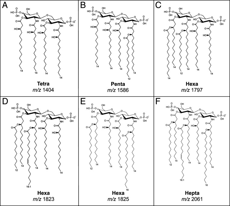
When grown at 21 °C to 28 °C, which mimics the temperature in an arthropod vector, Yp predominantly produces a hexa-acylated lipid A molecule (Fig. 1); however, when the temperature shifts to 37 °C (the mammalian host temperature), lipid A production shifts to generate a hypo-acylated lipid A (Fig. 1, tetra-acylated) (28, 29). Like E. coli and other enterobacteria, hexa-acylated lipid A from Yp grown at a reduced temperature is a potent immune stimulator of the human TLR4-signaling pathway, whereas tetra-acylated lipid A is a weak agonist (6). This shift in lipid A composition results in decreased recognition mediated via TLR4, thereby dampening TLR4-driven innate immune responses and resulting in the ability of Yp to evade host defenses in the early stages of infection (28–32).
Fig. 1.
Proposed structures of Yersinia lipid A molecules. Yersinia species are capable of producing a variety of lipid A structures that are influenced by their surrounding environments. (A) Y. pestis grown at 37 °C. (B) Y. pseudotuberculosis grown at 37 °C. (C) Y. enterocolitica grown at 37 °C. (D) Y. pestis, Y. pseudotuberculosis, and Y. enterocolitica grown <26 °C, (E, F) PagP+ palmitoylated lipid A species.
Lipid A extracted from Yp at mammalian temperature displays minimal endotoxic properties, providing a potential mechanism for the bacteria to avoid the host immune system (33). PagP is an eight-stranded antiparallel β-barrel acyltransferase present in the outer membrane in a limited number of Gram-negative organisms (34). Enzymatically, PagP transfers palmitate (C16) from a phospholipid donor, delivered into the external leaflet of the outer membrane to the hydroxyl group of the R-3-hydroxymyristate (C14-OH) chain on lipid A. This reaction is unique because it occurs in the outer membrane without access to thiol-activated precursors located in the bacterial cytoplasm (34). PagP activity and palmitoylation of lipid A has been implicated in many processes including resistance to cationic antimicrobial peptides in Salmonella, increased biofilm formation in Pseudomonas aeruginosa, and infectivity in Bordetella bronchiseptica (35–38). Interestingly, lipid A isolated from Y. pestis is devoid of palmitoylated lipid A species, whereas it is observed in closely related Yersinia pseudotuberculosis (Ypt) or Yersinia enterocolitica (Ye) species, raising the possibility of potential functional differences in PagP between these species (32). Analysis of the pagP gene sequence from all sequenced Yp genomes identified a single-nucleotide polymorphism (SNP) that resulted in a premature stop in translation of the Yp pagP gene, yielding a nonfunctional enzyme.
In this study, we examine the impact of this SNP causing premature loss of translation by restoring it in the Yp-KIM6 pagP gene (a BSL-2 version of Yp) to the sequence present in all Ypt and Ye isolates. We demonstrate that Yp encoding a functional PagP results in a hexa-acylated lipid A structure with increased immunostimulatory properties, as compared to the wild type (WT) KIM6 tetra-acylated lipid A when grown at 37 °C. This increase in innate immune recognition via the TLR4-MD-2–signaling pathway could have implications in determining how downstream adaptive immune responses are altered and represents one of the earliest identified adaptations of Yp from Ypt.
Results
Bioinformatic Analyses of the pagP Gene.
To identify differences in the pagP gene sequence among Yersinia species, a bioinformatics approach was utilized. Comparison of the pagP gene sequence from 19 individual Yersinia species identified two SNPs specific to the Yp isolates examined, when compared to the pagP of Y. pseudotuberculosis (SI Appendix, Fig. S1). The first Yp-specific SNP is synonymous and occurs at base 15, whereas the second (A→G) is located at base 599 and results in a premature stop codon (W→*) that truncates the PagP peptide by three amino acids at the C-terminal end of the protein in all Yp isolates. These amino acids are not part of any known functional motif; however, the truncated version of the polypeptide is likely to be structurally destabilized or not inserted in the outer membrane. This SNP was present in all Yp isolates examined, including those that are considered ancestral, suggesting that the truncating pagP SNP may be a very early evolutionary feature of Yp as a species. These mutations were examined in the genomic data available in GenBank (39) as of March 2, 2020, and found to present with the same distribution along species lines. Finally, one additional SNP was specific to Yp Angola (base 424 [T→G]) that results in an apparently functionally neutral amino acid (aa) change (aa 142, W→G).
Modeling Suggests that Yp SNP Results in Structurally Altered PagP Protein.
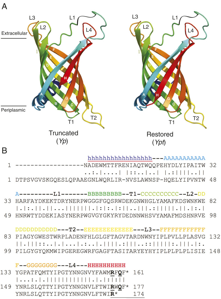
The amino acid sequence of the Yp PagP protein differs from other Yersinia species by only three C-terminal amino acid residues: tryptophan (W), glutamine (Q), and phenylalanine (F). The truncation is apparently sufficient to eliminate PagP function in Yp isolates yet lies in the terminal region of the protein, suggesting that the remainder of the protein could be synthesized but nonfunctional. To understand potential structural differences between Yp and other Yersinia species PagP proteins, the amino acid sequences were three-dimensionally modeled using the fold recognition bioinformatic software I-TASSER (15). The predicted secondary and tertiary structures (Fig. 2) reveal a β-barrel structure consistent with previous reports of PagP structure (34). Importantly, the final strand of the β-barrel (strand H, red; Fig. 2A) is incomplete in the Yp model (“Truncated” PagP), yielding an incomplete and structurally altered protein. In the full-length, non-Yp PagP model (“Restored” PagP or Pag+), the final strand is complete and is able to form a compete β-barrel structure, allowing for folding of a functional enzyme. Additionally, the secondary structural elements of PagP were mapped onto the primary structure of E. coli PagP (Fig. 2B). The conserved penultimate glutamine residue (shown in boldface) is buried in the PagP interior while the flanking aromatic amino acid residues contribute to the continuous hydrophobic membrane surface potential. Without the three C-terminal residues, the new C terminus becomes the conserved interior arginine residue (shown in boldface). This arginine normally contacts several interior water molecules and the amide side chain of the penultimate glutamine residue. Therefore, the C-terminal three-amino-acid deletion exposes the arginine to the hydrophobic membrane surface, moves the α-carboxylate moiety from the membrane periphery to the membrane interior, and removes hydrogen-bonding interactions with three neighboring peptide bonds: two in β-strand A and one in β-strand G. These predictions suggest that a full-length protein is necessary for PagP function and that loss of function in Yp is due to structural alteration of the PagP protein that results from the SNP-induced early truncation.
Fig. 2.
Predicted tertiary and secondary structure of PagP homologs from Y. pestis and Y. pseudotuberculosis. (A) The molecular model is shown in A without the C-terminal three amino acid residues in β-strand H (Truncated–Y. pestis) and with these residues in place (Restored–Y. pseudotuberculosis). (B) The primary structure of E. coli PagP is presented with its known secondary structural elements indicated above and with the sequence of Y. pseudotuberculosis PagP aligned below.
Growth Temperature Impacts Lipid A Structures Isolated from WT Yp.
It has been previously demonstrated that growth temperature affects the overall structure of Yp lipid A (27, 29, 32). Lipid A isolated after growth at environmental temperatures results in the synthesis of a hexa-acylated structure consisting of a bisphosphorylated diglucosamine backbone with ester- and amide-linked 3-OH C14 fatty acids (29, 32). This base structure is further modified by the addition of acyloxyacyl-linked palmitoleic acid (C16:1) at the 2′ position and lauric acid at the 3′ position (Fig. 1). Yp lipid A isolated after growth at mammalian temperature is tetra-acylated, lacking these additional fatty acids (Fig. 1).
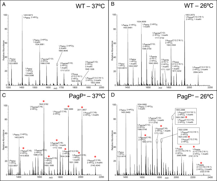
To determine the role of PagP in the biosynthesis of lipid A, we repaired the pagP SNP at base 599 to correct the Yp defect. The Yp BSL-2, nonselect agent KIM6 strain with a functional PagP enzyme was designated as Yp-KIM6-PagP+. To investigate temperature-specific lipid A remodeling in the presence and absence of PagP, WT Yp-KIM6 and Yp-KIM6-PagP+ were grown at mammalian (37 °C) and vector (26 °C) temperatures, and lipid A was isolated and analyzed by mass spectrometry (MS). After growth at 37 °C (Fig. 3C), we observed three major bisphosphoryl lipid A species in the Yp-KIM6-PagP+ strain: penta-acylated with one C16 fatty acid (m/z 1642) and hexa-acylated with two C16 fatty acids either without (m/z 1880) or with one aminoarabinose moiety (m/z 2011). Comparatively, WT Yp-KIM6 grown at 37 °C (Fig. 3A) contained lipid A species that lacked the C16 addition (higher m/z species m/z 1822 and m/z 1953 contain the C16:1 addition, which is mediated by LpxP, a lipid A palmitoleoyl transferase) (32). Unexpectedly, these data indicate that the functional Yp pagP gene encodes an enzyme capable of transferring two acyloxyacyl-linked palmitate chains at the 2′ and 3′ positions of lipid A.
Fig. 3.
MS analysis of PagP+ strains reveals lipid A palmitoylation. Negative ion mode ESI LTQ-FT mass spectrum of lipid A from (A) Yp-WT at 37 °C, (B) Yp-WT-KIM6 at 26 °C, (C) Yp-KIM6-PagP+ at 37 °C, and (D) Yp-KIM6-PagP+ at 26 °C. All ions are singly deprotonated. Shorthand notation is as follows: Lipid A (LA); subscript indicates number of acyl chains; C12 indicates lauric acid; C16:1 indicates palmitoleic acid; HPO3 indicates phosphate group; and Ara4N indicates aminoarabinose. Tetra-acylated Yp lipid A contains four primary 3-hydroxymyristic acid C14(3-OH) acyl chains and is identified as LAtetra. Yp lipid A can also be penta-acylated ([LApenta(C12)] or [LApenta(C16:1)]) or hexa-acylated (LAhexa[C12, C16:1]) with four primary C14(3-OH) acyl chains and secondary acyl chains of C12 and C16:1, respectively. Yp lipid A is predominantly diphosphorylated (2 HPO3) and can be modified with the addition of one or two aminoarabinose (Ara4N) groups. In addition to the lipid A structures present in Yp-KIM6 at 37 °C, Yp-KIM6-PagP+ at 37 °C contain the addition of one or two palmitic acid (C16) acyl chains as secondary fatty acids, (LApenta[C16], and LAhexa[C12, C16], and LAhexa[2 C16]), respectively, whereas Yp-KIM6-PagP+ at 26 °C contains the addition of a palmitic acid (C16) acyl chain as a secondary fatty acid, (LApenta[C16], LAhexa[C12, C16], LAhepta[C12, C16, C16:1]), respectively. Red asterisks indicate palmitoylation. See SI Appendix for further details.
PagP enzymatic activity was also observed in lipid A isolated from Yp-KIM6-PagP+ grown at 26 °C (Fig. 3D; m/z 1642, 1773, 1824, 1955, 2060, 2191, and 2322). The increased complexity in the MS spectra is consistent with the temperature-regulated activity of LpxL (HtrB) and LpxP that modifies the base tetra-acylated structure (m/z 1404, Fig. 3A) with a C12 or C16:1 fatty acid, respectively (40). Complete characterization of the 37 °C and 26 °C lipid A structures are presented as SI Appendix, Methods, and Fig. S2). Fig. 1 describes lipid A structures corresponding with the MS results in Fig. 3.
Gas Chromatography Demonstrates that Yp-KIM6-PagP+ Strain Lipid A Is Palmitoylated.
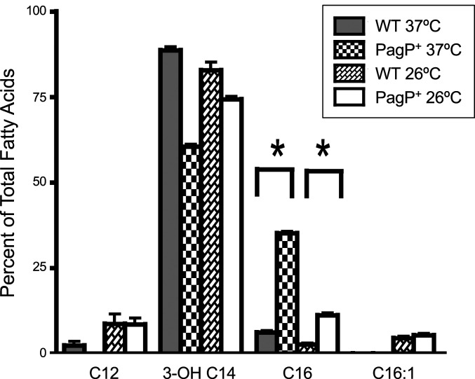
To determine the effect of repairing the pagP gene in Yp-KIM6, lipid A from Yp-KIM6 and Yp-KIM6-PagP+ strains grown at 37 °C and 26 °C was extracted and subjected to gas chromatographic-flame ionizing detector (GC-FID) analysis. Individual fatty acids were assigned, and amounts of each fatty acid present on lipid A were calculated in reference to a pentadecanoic acid internal standard (Fig. 4). Yp-KIM6-PagP+ grown at 37 °C had a 5.7-fold increase in the percentage of lipid A C16 acylation, as compared to Yp-KIM6 grown at the same temperature (35.2% ± 0.5 vs. 6.3% ± 0.4 total fatty acids) (Fig. 4). Y. pseudotuberculosis and Y. enterocolitica displayed similar levels of palmitoylation as the Yp-KIM6-PagP+ (29). Basal levels of C16 in the 37 °C grown samples are possibly due to phospholipid contamination. A similar relative fold difference was observed when the bacteria were grown at 26 °C (4.1-fold increase in Yp-KIM6-PagP; 11.1% ± 0.6 vs. 2.8% ± 0.3 total fatty acids) (Fig. 4). These data show a significant (P ≤ 0.005) increase in palmitoylation of lipid A in Yp-KIM6-PagP+ when grown at either temperature and provide evidence for a functional PagP enzyme in our Yp mutant strain.
Fig. 4.
Relative amounts of acyl groups (expressed as percentage of total fatty acid content) in lipid A from Yersinia strains grown at 37 °C or 26 °C. Data are from one representative experiment of three total experiments of n = 3. Error bars are SEM and represent the mean of triplicates; *P ≤ 0.005.
Yp PagP Is a Palmitoyltransferase.
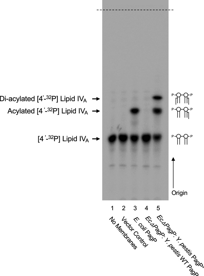
To confirm that PagP in the Yp-KIM6-PagP+ strain was the source of palmitoylation activity, membranes from E. coli expressing the Yp WT and PagP+ enzyme were isolated and assayed for palmitoyl transferase activity using the tetra-acylated lipid A precursor [4′-32P] lipid IVA as an acceptor molecule. Briefly, Yp PagP proteins (WT and PagP+) and as a positive control, E. coli PagP, were expressed from the low copy vector pWSK29 in E. coli strain W3110 ∆pagP. Membranes isolated from E. coli ∆pagP expressing the WT Yp pagP gene did not result in any reaction products (Fig. 5, lane 4). As expected, complementation with E. coli PagP resulted in the production of a single, faster-migrating reaction product due to the addition of a single palmitate (C16:0) molecule (Fig. 5, lane 3). However, addition of membranes from E. coli expressing Yp-PagP+ resulted in two reaction products corresponding to the addition of two palmitate groups (Fig. 5, lane 5). The slower-migrating species is similar in size to the product from the E. coli PagP control and is interpreted as a single palmitoylation event. The second, faster-migrating reaction product is interpreted as a structure that has undergone dipalmitoylation, again confirming that the functional pagP gene encodes an enzyme capable of transferring two acyloxyacyl-linked palmitate chains.
Fig. 5.
In vitro assay of Yp PagP heterologous expression in E. coli. Both Yp WT (lane 4) and Yp-KIM6-PagP+ (lane 5) enzymes were expressed in E. coli strain W3110 ∆PagP, and E. coli membranes were isolated for the in vitro assay. The 32P-labeled reaction products were separated by thin layer chromatography (TLC) and visualized by phosphorimaging analysis. Individual lipid A structures are shown next to their PagP enzymatic product with the “acylated” product having one and the “di-acylated” having two palmitates, respectively. WT E. coli W3110 with native E. coli PagP was also analyzed (lane 3). The solvent front is identified with the dashed black line.
Palmitoylated LPS Exhibits Greater Endotoxic Potential than Nonpalmitoylated LPS.
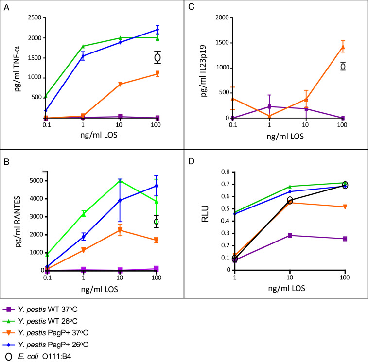
As the structure of the lipid A component of LPS is known to induce proinflammatory responses via the MD-2/TLR4 receptor complex, we hypothesized that the altered lipid A structure of the Yp-KIM6-PagP+ strain would display increased endotoxic potential. To demonstrate the endotoxic activity of the lipooligosaccharide (LOS) obtained from the Yp-KIM6 and Yp-KIM6-PagP+ strains, we measured cytokine production using promonocytic-vitamin D3 differentiated human THP-1 and histiocytic-phorbol myristate-acetate differentiated U937 cells. The cytokines chosen were TNF-α and IL23p19, both driven through NF-κB and RANTES, which are driven by a TRIF-TRAM response. As expected, LOS isolated from both Yp-KIM6 and Yp-KIM6-PagP+ cultures grown at 26 °C demonstrated high levels of endotoxic-like activity in THP-1 cells, which is attributed to the hexa-acylated lipid A structures present at this growth temperature. In contrast, LPS from Yp-KIM6-PagP+ grown at 37 °C was significantly more immunostimulatory than Yp-KIM6 grown at the same temperature for all cytokines measured (Fig. 6 A–C). Similar results were shown using U937 cells (SI Appendix, Fig. S3).
Fig. 6.
Lipid A from Yp-KIM6-PagP+ shows significantly increased endotoxicity. Enzyme-linked immunosorbent assays were run for (A) TNF-α, (B) RANTES, and (C) IL-23p19 from supernatants of THP-1 cells stimulated with Yp-KIM6 and Yp-KIM6-PagP+ LPS both grown at 37 °C or 26 °C. E. coli O111:B4 LPS was used as a control. All stimulations were for 20 h. Data are from one representative experiment of three total experiments of n = 3. Error bars are SEM and represent the mean of triplicates. (D) Luminescence measurements read as relative light units (RLUs) from HEK293 cells transiently expressing human or murine TLR4/MD-2 stimulated with Yp-KIM6 and Yp-KIM6-PagP+ LPS grown at 37 °C or 26 °C. E. coli O111:B4 LPS was used as a control. All stimulations were for 4 h. Data are from one representative experiment of three total experiments of n = 3. Error bars are SEM and represent the mean of triplicates.
To further demonstrate that human cells differentially respond to Yp-KIM6-PagP+ LOS, as compared to Yp-KIM6 LOS, we transiently transfected HEK-293 cells with human TLR4/MD2 followed by LPS stimulation. Results shown in Fig. 6D show a significant increase in NF-κB gene-driven response to Yp-KIM6-PagP+ LPS grown at 37 °C when compared to Yp-KIM6 LPS grown at the same temperature. LPS grown at 26 °C showed similarly high levels of a NF-κB gene-driven response for both strains. The innate immune response of the LOS purified from Y. pestis wild-type (tetra-acylated) and PagP+ strains was also measured and compared to E. coli LPS in both mouse and human primary cells. Mouse ex vivo splenocytes from C57BL6 and BALB/c backgrounds and human peripheral blood mononuclear cells from multiple healthy donors (n = 3) were evaluated for seven immune cytokine readouts (SI Appendix, Fig. S4). Results from these experiments support the conclusion that repairing the PagP enzyme in Y. pestis augments the immune response elicited by LOS. In summary, we demonstrate that mammalian temperature Yp-KIM6-PagP+ LPS can activate both the MyD88 and TRIF/TRAM arms of the TLR4-signaling pathway, which supports the finding that the repaired strain results in production of LPS with higher immunostimulatory potential.
Discussion
Since its recent divergence from Y. pseudotuberculosis (Ypt) ∼20,000 y ago, the Y. pestis (Yp) genome has undergone a significant number of gene inactivations, which likely played a prominent role in its pathogenic potential (27). In fact, as many as 13% of Ypt genes are no longer functional in Yp, whereas only 32 chromosomal genes and two virulence plasmids, important for pathogenesis, have been acquired since speciation (41, 42). Genes can become lost over time through genetic drift (43); however, genes may also remain inactivated if the loss allows for increased survival and propagation. We propose that pagP was under such selective pressures and that the lack of a functional PagP resulted in increased survival by evasion of the innate immune response, a critical step in pathogenesis.
PagP has been extensively studied because it is one of the few lipid A modification enzymes known to exist in the Gram-negative bacterial outer membrane (34, 44, 45). While the biological role of PagP has been studied in several bacterial species, the comparison of PagP homologs and the palmitoylation activities shown in this study provide interesting insights regarding the evolution in a particular organism. The Yp pagP gene sequence aligns closely with the pagP sequences from other Gram-negative bacteria, including the region coding the conserved active site His33 and Ser77 residues of E. coli PagP (46, 47). Ypt and Yp pagP homologs share 99% identity at the DNA level. They differ in the presence of a G-to-A transition mutation in Yp that converts the codon for Trp200 (TGG) into an amber stop codon (TAG), leading to a truncation of three carboxyl terminal amino acid residues. Interestingly, all Yp pagP sequences in the public domain contained the same SNP at position 599, suggesting that this mutation is advantageous and consistent across the Yp population from any of the past global pandemics.
Inspection of the E. coli PagP structure implicates the last three amino acids in the formation of hydrogen bonds that close the first and final β-strands into a β-barrel in the outer membrane (OM). These last three residues are expected to be particularly important in PagP because the β-bulge in the first β-strand changes the registration of hydrogen bonding with the final β-strand and causes sensitivity of the outer leaflet hydrogen bonds to the detergent environment (47). Consequently, the last three amino acids in the PagP sequence contribute two key hydrogen bonds in the inner-leaflet–exposed region that close the β-barrel structure. The absence of these three residues likely exposes the carboxyl-terminus and polar β-barrel interior region unfavorably to the phospholipid hydrocarbon chains in the OM inner leaflet. The thermodynamic cost of exposing charged amino acid residues to the membrane interior using the outer membrane β-barrel phospholipase OMPLA as a model has been established (48). Although the penalty for exposing the α-carboxylate moiety to the membrane interior was not determined explicitly, it can be approximated from the penalty for exposing the side chain of aspartic acid to the membrane interior, which was measured to be 1.38 kcal/mol. The C-terminal arginine side chain was reported to contribute 2.14 kcal/mol (48). It is well known that the per-residue free energy cost for disrupting peptide H-bonds in a membrane is about 4 kcal/mol (49). Without accounting for any stabilization contributed by the aromatic side chains of the penultimate tryptophan and C-terminal phenylalanine residues, the thermodynamic cost of deleting the C-terminal three amino acid residues from Ypt PagP (as is the case in Yp PagP), and of exposing the polar α-carboxylate and guanidinium moieties to the nonpolar membrane environment, is conservatively estimated to be about 16 kcal/mol. Additionally, PagP and many outer membrane proteins in general possess a carboxyl-terminal phenylalanine residue, which has been shown to be critical for correct OM assembly in studies of the PhoE porin (50, 51).
To demonstrate that the premature stop was responsible for gene inactivation, we repaired the SNP in the nonvirulent BSL-2 Yp-KIM6+ strain to that present in Ypt sequences. Lipid A isolated from the Yp-KIM6-PagP+ strain grown at 26 °C and 37 °C revealed the presence of palmitoylated lipid A at both temperatures (Fig. 2). Additionally, GC-FID results show a quantitative increase in palmitoylation of Yp-KIM6-PagP+ lipid A, as compared to Yp-KIM6, further demonstrating a functionally repaired PagP protein. Interestingly, the lipid A profiles at 37 °C contained spectra representing hexa-acylated lipid A with two palmitate chains. This indicates the addition of two palmitate moieties on the same lipid A molecule. Both Ypt and Ye have a single palmitate as their base structure is modified by the addition of acyloxyacyl-linked lauric acid (C12) at the 3′ position. To confirm these results, we examined the detailed structure of doubly palmitoylated hexa-acylated lipid A using MS. The two palmitate residues were found attached to the hydroxyl group of R-3-hydroxymyristate chains at positions 2′ and 3′ (Fig. 1). The position of palmitoylation resembles the combination of each single palmitoylation in E. coli, B. bronchiseptica, and P. aeruginosa, which occur at positions 2′ and 3′, respectively (29, 34). PagP enzymatic activity was confirmed by the membrane reconstitution experiment that demonstrated that two C16 fatty acids were added to tetra-acylated lipid A and suggests that the repaired enzyme is able to add two palmitate residues, as there are no other fatty acids to hinder their respective additions to the primary acyl chains at positions 2′ and 3′.
Temperature-dependent lipid A modification has been proposed to be a key modulator in attenuation of Yp virulence and can alter its recognition by the host immune system (29). When the PagP function is restored, the resulting lipid A becomes more proinflammatory due to enhanced TRL4-mediated signaling (Fig. 6). The LPS from Yp-KIM6-PagP+ grown at 37 °C and utilized in a whole-cell stimulation assay shows a marked increase in both extracellular, MyD88-dependent, endosomal, and TRIF/TRAM-dependent signaling, providing evidence that hexa-acylated LPS produced at the mammalian temperature from Yp-KIM-PagP+ was more stimulatory than the predominantly tetra-acylated form from Yp-KIM6.
This study provides evidence that lipid A modifying enzymes can be used to engineer lipid A molecules that may have significant therapeutic use as potential vaccine adjuvants or antisepsis agonists. TLR4 ligands have now been described in the literature as having the ability to be used as adjuvants in multicomponent vaccines, suggesting decreased virulence via immune recognition (52–56). As LOS isolated from Yp-KIM6-PagP+ can stimulate human cells through both the MyD88 and the TRIM/TRAF arms of the TLR4-signaling pathway, it may represent a candidate for vaccine adjuvant development.
Materials and Methods
Detailed materials and methods describing bacterial strains, culture conditions, Y. pestis PagP mutagenesis, LPS purification and lipid A isolation, mass spectrometry, membrane experiments, cell stimulations, and bioinformatics can be found in SI Appendix.
Supplementary Material
Acknowledgments
We thank Francesca Gardner for review and critique of the manuscript. This work was supported in part by NIH Grants AI123820 and GM111066 (to R.K.E. and D.R.G.), U19AI110820 (to D.A.R.), AI129940 (to M.S.T.), AI150098 (to M.S.T.), and AI138576 (to M.S.T.); the International Centre for Cancer Vaccine Science project of the International Research Agendas program of the Foundation for Polish Science cofinanced by the European Union under the European Regional Development Fund (MAB/2017/03) at the University of Gdansk (to D.R.G.); and by Canadian Institutes of Health Research Grant MOP-125979 (to R.E.B.).
Footnotes
The authors declare no competing interest.
This article is a PNAS Direct Submission.
This article contains supporting information online at https://www.pnas.org/lookup/suppl/doi:10.1073/pnas.1917504117/-/DCSupplemental.
Data Availability.
All study data are included in the article and SI Appendix. All GenBank and protein sequence data are publicly available. All experimental data will be made available upon request.
References
- 1.Perry R. D., Fetherston J. D., Yersinia pestis: Etiologic agent of plague. Clin. Microbiol. Rev. 10, 35–66 (1997). [DOI] [PMC free article] [PubMed] [Google Scholar]
- 2.Cavanaugh D. C., Randall R., The role of multiplication of Pasteurella pestis in mononuclear phagocytes in the pathogenesis of flea-borne plague. J. Immunol. 83, 348–363 (1959). [PubMed] [Google Scholar]
- 3.Hinnebusch B. J.et al., Role of Yersinia murine toxin in survival of Yersinia pestis in the midgut of the flea vector. Science 296, 733–735 (2002). [DOI] [PubMed] [Google Scholar]
- 4.Erickson D. L.et al., Acute oral toxicity of Yersinia pseudotuberculosis to fleas: Implications for the evolution of vector-borne transmission of plague. Cell. Microbiol. 9, 2658–2666 (2007). [DOI] [PubMed] [Google Scholar]
- 5.Meyer K. F., Pneumonic plague. Bacteriol. Rev. 25, 249–261 (1961). [DOI] [PMC free article] [PubMed] [Google Scholar]
- 6.Miller S. I., Ernst R. K., Bader M. W., LPS, TLR4 and infectious disease diversity. Nat. Rev. Microbiol. 3, 36–46 (2005). [DOI] [PubMed] [Google Scholar]
- 7.Prior J. L.et al., The failure of different strains of Yersinia pestis to produce lipopolysaccharide O-antigen under different growth conditions is due to mutations in the O-antigen gene cluster. FEMS Microbiol. Lett. 197, 229–233 (2001). [DOI] [PubMed] [Google Scholar]
- 8.Hajjar A. M., Ernst R. K., Tsai J. H., Wilson C. B., Miller S. I., Human Toll-like receptor 4 recognizes host-specific LPS modifications. Nat. Immunol. 3, 354–359 (2002). [DOI] [PubMed] [Google Scholar]
- 9.Coats S. R., Do C. T., Karimi-Naser L. M., Braham P. H., Darveau R. P., Antagonistic lipopolysaccharides block E. coli lipopolysaccharide function at human TLR4 via interaction with the human MD-2 lipopolysaccharide binding site. Cell. Microbiol. 9, 1191–1202 (2007). [DOI] [PubMed] [Google Scholar]
- 10.Han Y.et al., Construction of monophosphoryl lipid A producing Escherichia coli mutants and comparison of immuno-stimulatory activities of their lipopolysaccharides. Mar. Drugs 11, 363–376 (2013). [DOI] [PMC free article] [PubMed] [Google Scholar]
- 11.Raetz C. R., Reynolds C. M., Trent M. S., Bishop R. E., Lipid A modification systems in gram-negative bacteria. Annu. Rev. Biochem. 76, 295–329 (2007). [DOI] [PMC free article] [PubMed] [Google Scholar]
- 12.Raetz C. R., Whitfield C., Lipopolysaccharide endotoxins. Annu. Rev. Biochem. 71, 635–700 (2002). [DOI] [PMC free article] [PubMed] [Google Scholar]
- 13.Simpson B. W., Trent M. S., Pushing the envelope: LPS modifications and their consequences. Nat. Rev. Microbiol. 17, 403–416 (2019). [DOI] [PMC free article] [PubMed] [Google Scholar]
- 14.Demeure C.et al., Yersinia pestis and plague: An updated view on evolution, virulence determinants, immune subversion, vaccination and diagnostics. Microbes Infect. 21, 202–212 (2019). [DOI] [PubMed] [Google Scholar]
- 15.Yang J.et al., The I-TASSER Suite: Protein structure and function prediction. Nat. Methods 12, 7–8 (2015). [DOI] [PMC free article] [PubMed] [Google Scholar]
- 16.Du Y., Rosqvist R., Forsberg A., Role of fraction 1 antigen of Yersinia pestis in inhibition of phagocytosis. Infect. Immun. 70, 1453–1460 (2002). [DOI] [PMC free article] [PubMed] [Google Scholar]
- 17.Sing A.et al., Yersinia V-antigen exploits toll-like receptor 2 and CD14 for interleukin 10-mediated immunosuppression. J. Exp. Med. 196, 1017–1024 (2002). [DOI] [PMC free article] [PubMed] [Google Scholar]
- 18.Sodhi A., Sharma R. K., Batra H. V., Yersinia rLcrV and rYopB inhibits the activation of murine peritoneal macrophages in vitro. Immunol. Lett. 99, 146–152 (2005). [DOI] [PubMed] [Google Scholar]
- 19.Anisimov A. P.et al., Intraspecies and temperature-dependent variations in susceptibility of Yersinia pestis to the bactericidal action of serum and to polymyxin B. Infect. Immun. 73, 7324–7331 (2005). [DOI] [PMC free article] [PubMed] [Google Scholar]
- 20.Kolodziejek A. M., Hovde C. J., Minnich S. A., Yersinia pestis Ail: Multiple roles of a single protein. Front. Cell. Infect. Microbiol. 2, 103 (2012). [DOI] [PMC free article] [PubMed] [Google Scholar]
- 21.Cornelis G. R., Yersinia type III secretion: Send in the effectors. J. Cell Biol. 158, 401–408 (2002). [DOI] [PMC free article] [PubMed] [Google Scholar]
- 22.Grosdent N., Maridonneau-Parini I., Sory M. P., Cornelis G. R., Role of Yops and adhesins in resistance of Yersinia enterocolitica to phagocytosis. Infect. Immun. 70, 4165–4176 (2002). [DOI] [PMC free article] [PubMed] [Google Scholar]
- 23.Juris S. J., Shao F., Dixon J. E., Yersinia effectors target mammalian signalling pathways. Cell. Microbiol. 4, 201–211 (2002). [DOI] [PubMed] [Google Scholar]
- 24.Navarro L., Alto N. M., Dixon J. E., Functions of the Yersinia effector proteins in inhibiting host immune responses. Curr. Opin. Microbiol. 8, 21–27 (2005). [DOI] [PubMed] [Google Scholar]
- 25.Atkinson S., Williams P., Yersinia virulence factors: A sophisticated arsenal for combating host defences. F1000 Res. 5, F1000 Faculty Rev-1370 (2016). [DOI] [PMC free article] [PubMed] [Google Scholar]
- 26.Li B., Yang R., Interaction between Yersinia pestis and the host immune system. Infect. Immun. 76, 1804–1811 (2008). [DOI] [PMC free article] [PubMed] [Google Scholar]
- 27.Achtman M.et al., Yersinia pestis, the cause of plague, is a recently emerged clone of Yersinia pseudotuberculosis. Proc. Natl. Acad. Sci. U.S.A. 96, 14043–14048 (1999). [DOI] [PMC free article] [PubMed] [Google Scholar]
- 28.Matsuura M., Structural modifications of bacterial lipopolysaccharide that facilitate Gram-negative bacteria evasion of host innate immunity. Front. Immunol. 4, 109 (2013). [DOI] [PMC free article] [PubMed] [Google Scholar]
- 29.Rebeil R., Ernst R. K., Gowen B. B., Miller S. I., Hinnebusch B. J., Variation in lipid A structure in the pathogenic yersiniae. Mol. Microbiol. 52, 1363–1373 (2004). [DOI] [PubMed] [Google Scholar]
- 30.Hajjar A. M.et al., Humanized TLR4/MD-2 mice reveal LPS recognition differentially impacts susceptibility to Yersinia pestis and Salmonella enterica. PLoS Pathog. 8, e1002963 (2012). [DOI] [PMC free article] [PubMed] [Google Scholar]
- 31.Montminy S. W.et al., Virulence factors of Yersinia pestis are overcome by a strong lipopolysaccharide response. Nat. Immunol. 7, 1066–1073 (2006). [DOI] [PubMed] [Google Scholar]
- 32.Rebeil R.et al., Characterization of late acyltransferase genes of Yersinia pestis and their role in temperature-dependent lipid A variation. J. Bacteriol. 188, 1381–1388 (2006). [DOI] [PMC free article] [PubMed] [Google Scholar]
- 33.Goodin J. L.et al., Purification and protective efficacy of monomeric and modified Yersinia pestis capsular F1-V antigen fusion proteins for vaccination against plague. Protein Expr. Purif. 53, 63–79 (2007). [DOI] [PMC free article] [PubMed] [Google Scholar]
- 34.Bishop R. E., The lipid A palmitoyltransferase PagP: Molecular mechanisms and role in bacterial pathogenesis. Mol. Microbiol. 57, 900–912 (2005). [DOI] [PubMed] [Google Scholar]
- 35.Chalabaev S.et al., Biofilms formed by gram-negative bacteria undergo increased lipid a palmitoylation, enhancing in vivo survival. MBio 5, e01116-14 (2014). [DOI] [PMC free article] [PubMed] [Google Scholar]
- 36.Guo L.et al., Lipid A acylation and bacterial resistance against vertebrate antimicrobial peptides. Cell 95, 189–198 (1998). [DOI] [PubMed] [Google Scholar]
- 37.Pilione M. R., Pishko E. J., Preston A., Maskell D. J., Harvill E. T., pagP is required for resistance to antibody-mediated complement lysis during Bordetella bronchiseptica respiratory infection. Infect. Immun. 72, 2837–2842 (2004). [DOI] [PMC free article] [PubMed] [Google Scholar]
- 38.Preston A.et al., Bordetella bronchiseptica PagP is a Bvg-regulated lipid A palmitoyl transferase that is required for persistent colonization of the mouse respiratory tract. Mol. Microbiol. 48, 725–736 (2003). [DOI] [PubMed] [Google Scholar]
- 39.Benson D. A.et al., GenBank. Nucleic Acids Res. 43, D30–D35 (2015). [DOI] [PMC free article] [PubMed] [Google Scholar]
- 40.Hittle L. E.et al., Site-specific activity of the acyltransferases HtrB1 and HtrB2 in Pseudomonas aeruginosa lipid A biosynthesis. Pathog. Dis. 73, ftv053 (2015). [DOI] [PMC free article] [PubMed] [Google Scholar]
- 41.Eppinger M.et al., The complete genome sequence of Yersinia pseudotuberculosis IP31758, the causative agent of Far East scarlet-like fever. PLoS Genet. 3, e142 (2007). [DOI] [PMC free article] [PubMed] [Google Scholar]
- 42.Parkhill J.et al., Genome sequence of Yersinia pestis, the causative agent of plague. Nature 413, 523–527 (2001). [DOI] [PubMed] [Google Scholar]
- 43.Holt K. E.et al., High-throughput sequencing provides insights into genome variation and evolution in Salmonella Typhi. Nat. Genet. 40, 987–993 (2008). [DOI] [PMC free article] [PubMed] [Google Scholar]
- 44.Bishop R. E., Structural biology of membrane-intrinsic beta-barrel enzymes: Sentinels of the bacterial outer membrane. Biochim. Biophys. Acta 1778, 1881–1896 (2008). [DOI] [PMC free article] [PubMed] [Google Scholar]
- 45.Bishop R. E.et al., Transfer of palmitate from phospholipids to lipid A in outer membranes of Gram-negative bacteria. EMBO J. 19, 5071–5080 (2000). [DOI] [PMC free article] [PubMed] [Google Scholar]
- 46.Ahn V. E.et al., A hydrocarbon ruler measures palmitate in the enzymatic acylation of endotoxin. EMBO J. 23, 2931–2941 (2004). [DOI] [PMC free article] [PubMed] [Google Scholar]
- 47.Hwang P. M.et al., Solution structure and dynamics of the outer membrane enzyme PagP by NMR. Proc. Natl. Acad. Sci. U.S.A. 99, 13560–13565 (2002). [DOI] [PMC free article] [PubMed] [Google Scholar]
- 48.Moon C. P., Fleming K. G., Side-chain hydrophobicity scale derived from transmembrane protein folding into lipid bilayers. Proc. Natl. Acad. Sci. U.S.A. 108, 10174–10177 (2011). [DOI] [PMC free article] [PubMed] [Google Scholar]
- 49.White S. H., How hydrogen bonds shape membrane protein structure. Adv. Protein Chem. 72, 157–172 (2005). [DOI] [PubMed] [Google Scholar]
- 50.de Cock H., van Blokland S., Tommassen J., In vitro insertion and assembly of outer membrane protein PhoE of Escherichia coli K-12 into the outer membrane. Role of Triton X-100. J. Biol. Chem. 271, 12885–12890 (1996). [DOI] [PubMed] [Google Scholar]
- 51.Struyvé M., Moons M., Tommassen J., Carboxy-terminal phenylalanine is essential for the correct assembly of a bacterial outer membrane protein. J. Mol. Biol. 218, 141–148 (1991). [DOI] [PubMed] [Google Scholar]
- 52.Airhart C. L.et al., Induction of innate immunity by lipid A mimetics increases survival from pneumonic plague. Microbiology 154, 2131–2138 (2008). [DOI] [PubMed] [Google Scholar]
- 53.Airhart C. L.et al., Lipid A mimetics are potent adjuvants for an intranasal pneumonic plague vaccine. Vaccine 26, 5554–5561 (2008). [DOI] [PMC free article] [PubMed] [Google Scholar]
- 54.Fischer N. O.et al., Colocalized delivery of adjuvant and antigen using nanolipoprotein particles enhances the immune response to recombinant antigens. J. Am. Chem. Soc. 135, 2044–2047 (2013). [DOI] [PubMed] [Google Scholar]
- 55.Gregg K. A.et al., Rationally eesigned TLR4 ligands for vaccine adjuvant discovery. MBio 8, e00492-17 (2017). [DOI] [PMC free article] [PubMed] [Google Scholar]
- 56.Needham B. D.et al., Modulating the innate immune response by combinatorial engineering of endotoxin. Proc. Natl. Acad. Sci. U.S.A. 110, 1464–1469 (2013). [DOI] [PMC free article] [PubMed] [Google Scholar]
Associated Data
This section collects any data citations, data availability statements, or supplementary materials included in this article.
Supplementary Materials
Data Availability Statement
All study data are included in the article and SI Appendix. All GenBank and protein sequence data are publicly available. All experimental data will be made available upon request.


