Abstract
Objectives:
To systematically review the literature and to summarize all evidence related to the diagnosis and management of patulous eustachian tube.
Methods:
The present study was conducted in accordance with the Preferred Reporting Items for Systematic Reviews and Meta-Analyses (PRISMA) statement.
Results:
Overall, 59 articles were retrieved and included in the analysis. Studies investigating treatments enrolled 1279 patients collectively, with follow-up duration varying from few days and up to 2 years. Eight studies reported medical treatments with intranasal saline instillation as the most frequently studied option. Other studies reported various surgical treatments varying from simple tympanostomy to invasive procedures targeting the orifice of the ET or the anatomical features surrounding it. In addition, 10 studies including 367 subjects investigated different diagnostic methods.
Conclusion:
Currently, there is a wide spectrum of diagnostic and therapeutic interventions with minimal clinical efficacy, a persistent lack of systematic guidelines, and several gaps in previous research endeavours.
PROSPERO REG. NO. CRD: 1644000
Keywords: patulous eustachian tube, management, diagnosis
Patulous eustachian tube (PET), a benign, largely idiopathic, condition, was first described in 1867. It has adverse effects that can markedly affect quality of life such as abnormal autophony, breathing, or hearing.1 Several factors have been reported to be causative, including pregnancy, neurological disorders, weight loss and radiotherapy, and some medications including oral contraceptives and diuretics.2 In terms of pathophysiology, all stressors that distort normal closure of the eustachian tube (ET) during the resting state can result in PET. This includes conditions affecting the elasticity, surface tension, and opening pressure of the ET.2
According to experts, there is currently a lack of universal consensus regarding symptom scores, tests, and guidelines to diagnose PET. Meanwhile, the diagnostic approach relies primarily on clinical assessment of the presenting symptoms.3 This non-systematic approach makes the identification of these cases challenging. Symptoms that should raise suspicion include aural fullness, popping, and discomfort or pain, in addition to autophony of breathing or voice. Signs include tympanic membrane retraction and signs of negative middle ear pressure.3,4 Further studies have been recommended to enhance the diagnostic approach to this condition.
Despite the modest rarity of PET and the hurdles to diagnose, it also represents a challenging condition for patients and clinicians due to limited therapeutic options.5 Current approaches vary depending on the severity of symptoms and range from informative reassurance to invasive interventions. Some cases may need combined or surgical treatment such as those with no clinical improvement and persistent movement of the tympanic membrane.6 To date, there have been limited research papers with variable outcome measures and no definite recommendations that outline a precise evidence-based, patient-centered therapeutic guideline.3,5
Given the insufficient diagnostic and therapeutic guidelines, we aim to systematically review the literature and to summarize all evidence related to the diagnosis and management of PET.
Methods
This study has followed the Preferred Reporting Items for Systematic Reviews and Meta-Analyses (PRISMA) statement.7 The study protocol was applied for recording in PROSPERO. Provided the nature of the present study (example, systematic review), requirements for ethics review were waived.
Any primary study that reported or investigated diagnostic approaches or management options for PET were included. There were no restrictions regarding different populations, race, origin, age, ethnicity, language, or publication date. However, letters, editorial comments, theses, book chapters, non-human and experimental studies, and articles with no available full text were excluded. Studies with no extractable data were also excluded.
A computerized systematic search was performed for potentially eligible articles published up to December 2019 using the PubMed, Scopus, ISI Web of Science, Embase, Virtual Health Library (VHL), and Cochrane Central Register of Controlled Trials databases. The search term “patulous eustachian tube” was used to retrieve all relevant articles within the scope of the present study. A manual inspection of the reference section of all competent reports was also considered to recognize any further articles. Search results were retrieved and duplicates were removed using EndNote X7.4 (EndNote, USA) for Windows (Microsoft Corporation, Redmond, WA, USA). Two independent reviewers screened potentially relevant articles and included all reports that met the research criteria. Initial screening of titles and abstracts was followed by full-text review. Disagreements were resolved by discussion and consensus between reviewers and among senior researchers.
Three authors independently collected the target outcomes from the eligible articles and filled it into blind Excel sheets. Any discrepancy was resolved by discussion to reach consensus. The extraction form was established using a pilot extraction of a few blindly selected studies. Extracted outcomes comprised demographic informations of the enrolled subjects. In addition, different types of therapeutic and diagnostic approaches, and follow-up period were analyzed. Complete, partial, and non-resolution rates were also compared. Descriptive statistics was carried out and the data were presented as numbers and proportions.
Results
The search of the medical literature retrieved 1062 titles (PubMed [n = 204]; VHL [n = 199]; Scopus [n=246], Embase [n=202], ISI Web of Science [n=204], Cochrane Central Register of Controlled Trials [n=7]). After removing 215 duplicates, 847 titles and abstracts were pooled for initial screening (Figure 1). Three reviewers working independently selected 71 articles for full-text screening. After discussion, 25 articles were excluded for irrelevance or improper design, among other reasons (Figure 1). Ultimately, 46 articles were eligible by screening and 13 were identified through the manual search, resulting in 59 articles included for data extraction and analysis.
Figure 1.
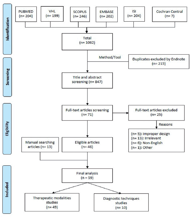
PRISMA flowchart of studies selection and screening. ISI: Web of Science, VHL: virtual health library
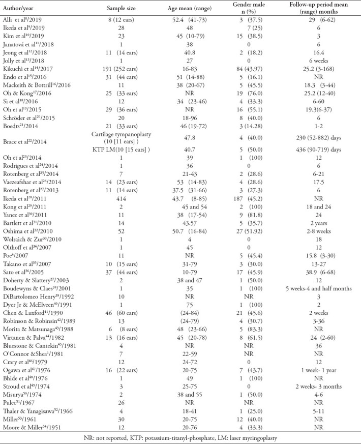
Of the 59 included studies, 49 investigated treatment options, while 10 described diagnostic approaches.8-54 The studies investigating treatments collectively enrolled 1279 patients (1442 ears), with follow-up intervals varying from 2 weeks to 2 years. Baseline demographics of the enrolled articles are summarized in Table 1.
Table 1.
Baseline characters of 49 studies reporting treatment of patulous eustachian tube.
Medical treatment approach
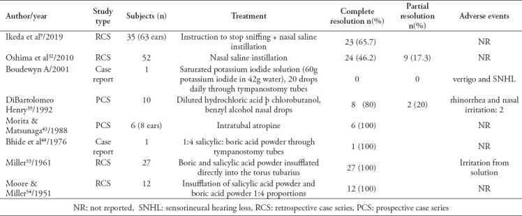
Eight articles described several non-surgical, intranasal or intratubal treatments (Table 2). Treatment options included guidance to stop sniffing and nasal saline instillation, irrigation using normal saline, potassium iodide, diluted hydrochloric acid, beta chlorobutanol, benzyl alcohol, atropine, 1:4 salicylic acid powder, boric acid powder. The latter was reported in 3 studies enrolling 40 patients, which led to a 100% resolution of PET symptoms.48,53,54 Irritation was the only side effect reported. More recent studies favored the use of topical normal saline with high efficacy (65.7% experienced complete resolution in the study by Ikeda9) and safety (example, no adverse events were reported with the use of saline drops).
Table 2.
Outcomes from 8 studies reporting treatment of patulous eustachian tube through medical approach.
Tympanic membrane mass loading approach
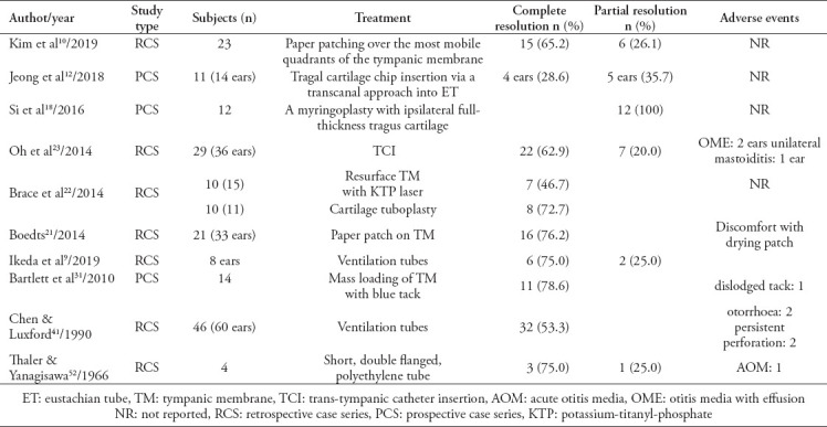
Several techniques were used to increase the mass of the tympanic membrane, which is aimed at decreasing its movement and, therefore, potentially improving PET symptoms. In the review, 10 studies reported different means to load the tympanic membrane, which are summarized in Table 3. These techniques included paper patching, loading with ventilation tubes, cartilage chip insertion, myringoplasty, potassium-titanyl-phosphate (KTP) laser, and loading with blue tack. Complete resolution was more frequently reported with the use of blue tack (78.6%) in a sample size of 11 subjects, and paper patching (65.2-76.2%) in 44 patients (56 ears). The side effects of paper patching were limited to mild to moderate discomfort.
Table 3.
Outcomes from 10 studies reporting treatment of patulous eustachian tube through tympanic membrane mass loading approach.
Eustachian tube occlusion approach
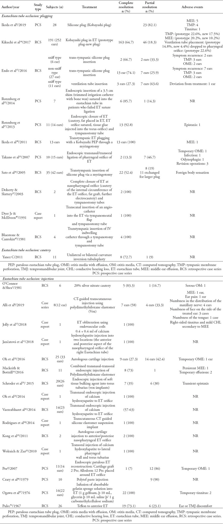
Ten studies investigated ET occlusion using several modalities including plugging (example, silicon plug, catheter with bone wax, and orifice suturing), cautery, and injections. The outcomes of these reports are elaborated in Table 4. The largest sample size was reported by Kikuchi et al,14 in which 252 ears were occluded using Kobayashi plug, with a complete resolution rate (CRR) of 64.7% and several reported side effects including tympanic membrane perforation (17.5%) and plug dropping toward the pharyngeal tube (22.6%). Cauterization was reported in 3 case series including 22 patients. Several techniques and resolution rates are summarized, with otitis media as the only reported side effect. Various types of injectable elements used for inducing ET occlusion are also summarized in Table 4. Those with the largest sample sizes included autologous cartilage injection (33 ears [CRR 27.3%]), soft-tissue bulking agent (26 ears [CRR, 35%]), calcium hydroxyapatite (23 ears [CRR 57-63%]), absorbable gelatin sponge solution (22 ears [CRR 100%]), and Teflon (26 ears [CRR 73.1%]). Otitis media with effusion, epistaxis, tinnitus, and temporomandibular joint discomfort were reported as adverse events post-injection.
Table 4.
Outcomes from 10 studies reporting treatment of patulous eustachian tube through eustachian tube occlusion.
Muscular techniques
Modulating the tone of the tensor veli palatine muscle can change the patency of the ET and was investigated as a potential therapeutic approach to PET. Appendix 1 summarizes relevant evidence. The most commonly investigated technique was tensor veli palatine transection or transposition with pterygoid hamulotomy, which was reported by Virtanen (44) on 16 ears, with a CRR of 56.2%.
Diagnostic approaches
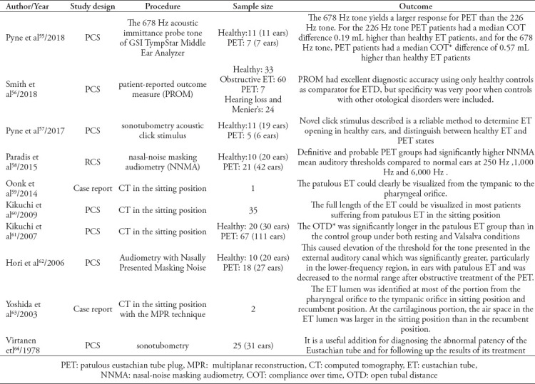
Ten studies including 367 subjects investigated various diagnostic methods.55-64 The various diagnostic options summarized in this review (Table 5) include the 678 Hz acoustic immittance probe tone of GSI TympStar Middle Ear Analyzer (Grason-Stadler, Eden Prairie, MN, USA), patient-reported outcome measure (PROM), sonotubometry acoustic click stimulus, nasal-noise masking audiometry (NNMA), computed tomography in the sitting position, and sonotubometry.
Table 5.
Summary of 10 studies reporting different applied diagnostic methods of patulous eustachian tube.
Discussion
Patulous eustachian tube is a modestly prevalent condition that impacts the quality of life of affected individuals. The recognition of and approach to this condition is usually challenging due to the highly subjective nature of the disease and various management options that fail to be consistent in terms of clinical efficacy.3 The present systematic review summarized therapeutic and diagnostic options published in the literature, updated previous work in this field, and included more studies and approaches compared with previous reviews. Although the diagnosis of PET is mainly clinical, several approaches have been proposed to confirm the diagnosis.3 This is significant because symptoms can be nonspecific or misleading in some instances, especially autophony, which is often incorrectly regarded as a pathognomonic sign of PET, while several other conditions may precipitate it, such as external ear canal occlusion, superior canal dehiscence, and foreign bodies such as hair or wax.1,65 These approaches are also important for persistent treatment-resistant cases and for research purposes.66 Thus, having a consistent diagnostic approach is important for clinical studies that investigate various management options, and for synthesizing comparative evidence. Meanwhile, there is still no gold standard for diagnosing PET. The most commonly reported approach is computed tomography in the sitting position with or without the Valsalva maneuver, which is used to detect any anatomical patency and measure open tubal distance.60,61 It remains; however, costly, and not readily available in all healthcare centers. Sonotubometry, tympanometry, and audiometry, along with other approaches, that may or may not need special equipment, were summarized in this review to emphasize the need for a tailored, case-specific approach that combines clinical assessment with diagnostic tests to detect PET. Given the results and nature of the included studies, this appears to be better than standard tests. More research is, nevertheless, needed to inform and develop future guidelines.
In terms of therapeutic approaches, several medical and surgical interventions were suggested, none of which, however, proved to be consistently effective.67 Our review highlights the lack of randomized controlled trials and the need for more interventional studies. Non-surgical interventions mainly include the intratympanic or intranasal administration of different agents. Most recent studies have focused on the role of saline instillation for symptom relief in those with PET.28,32 Intranasal saline was suggested as the initial step in the management plan due to its moderate to high efficacy and clean safety profile.32 However, confounders such as instructing patients to stop sniffing were reported, especially in those with PET and habitual sniffing, which need to be accounted for in future studies. Similar to previous systematic reviews, we failed to find any evidence supporting the use of nasal estrogen cream to treat PET.67
Patients with no clinical improvement and persistent movement of the tympanic membrane may be candidates for combination therapies or surgical treatment.6 The wide spectrum of surgical interventions mainly targets the tympanic membrane, ET orifice or the surrounding anatomical features, and range from simple to invasive procedures. Again, surgical approaches are limited by the lack of consistency of clinical efficacy, scant descriptions of the details of some procedures, and the primitive evidence that we still have in this area. For example, the outcome of “partial resolution” was not consistently defined among the studies.
Our results highlight several additional challenges that impede a more informed approach to PET. Some of these challenges are specific to the nature of the condition, such as the scarcity of objective outcomes and that symptoms can be self-limited, which is an important confounder in research studies. Other challenges leave room for improvement through further research and interventions. They include small sample sizes, high risk of bias, unmatched confounders, poorly defined follow-up periods, and poorly described intervention(s), in addition to the lack of clinical trials and guidelines for diagnosing and managing PET. The present systematic review was limited by the lack of quantitative meta-analysis and the exclusion of non-English articles due to a lack of native speakers.
In conclusion, the present systematic review is the most recent comprehensive investigation of diagnostic and therapeutic approaches to PET to date. It revealed a highly variable spectrum of choices, with a lack of systematic guidelines and several gaps in current research endeavours. As such, a case-specific, step-wise approach is recommended.
Acknowledgment
The authors gratefully acknowledge Editage (www.editage.com) for English language editing.
Footnotes
References
- 1.O'Connor AF, Shea JJ. Autophony and the patulous eustachian tube. Laryngoscope. 1981;91:1427–1435. doi: 10.1288/00005537-198109000-00003. [DOI] [PubMed] [Google Scholar]
- 2.Grimmer JF, Poe DS. Update on eustachian tube dysfunction and the patulous eustachian tube. Curr Opin Otolaryngol Head Neck Surg. 2005;13:277–282. doi: 10.1097/01.moo.0000176465.68128.45. [DOI] [PubMed] [Google Scholar]
- 3.Schilder A, Bhutta M, Butler C, Holy C, Levine L, Kvaerner K, et al. Eustachian tube dysfunction:consensus statement on definition, types, clinical presentation and diagnosis. Clin Otolaryngol. 2015;40:407–411. doi: 10.1111/coa.12475. [DOI] [PMC free article] [PubMed] [Google Scholar]
- 4.Poe DS. Diagnosis and management of the patulous eustachian tube. Otol Neurotol. 2007;28:668–677. doi: 10.1097/mao.0b013e31804d4998. [DOI] [PubMed] [Google Scholar]
- 5.Llewellyn A, Norman G, Harden M, Coatesworth A, Kimberling D, Schilder A, et al. Interventions for adult Eustachian tube dysfunction:a systematic review. Health Technol Assess. 2014;18:1–180, v-vi. doi: 10.3310/hta18460. [DOI] [PMC free article] [PubMed] [Google Scholar]
- 6.Hussein AA, Adams AS, Turner JH. Surgical management of Patulous eustachian tube:A systematic review. Laryngoscope. 2015;125:2193–2198. doi: 10.1002/lary.25168. [DOI] [PMC free article] [PubMed] [Google Scholar]
- 7.Moher D, Liberati A, Tetzlaff J, Altman DG. Preferred reporting items for systematic reviews and meta-analyses:the PRISMA statement. Ann Intern Med. 2009;151:264–269, W64. doi: 10.7326/0003-4819-151-4-200908180-00135. [DOI] [PubMed] [Google Scholar]
- 8.Alli A, Shukla R, Cook JL, Waddell A. A novel computed tomography guided, transcutaneous approach to treat refractory autophony in patients with a patulous Eustachian tube - A case series. J Laryngol Otol. 2019;133:201–204. doi: 10.1017/S0022215119000264. [DOI] [PubMed] [Google Scholar]
- 9.Ikeda R, Kawase T, Takata I, Suzuki Y, Sato T, Katori Y, et al. Width of Patulous Eustachian tube:comparison of assessment by Sonotubometry and Tubo-tympano-aerography. Otol Neurotol. 2019;40:e386–e392. doi: 10.1097/MAO.0000000000002141. [DOI] [PubMed] [Google Scholar]
- 10.Kim SJ, Shin SA, Lee HY, Park Y-H. Paper patching for patulous eustachian tube. Acta Otolaryngol. 2019;139:122–128. doi: 10.1080/00016489.2018.1541505. [DOI] [PubMed] [Google Scholar]
- 11.Jancatova D, Zelenik K, Kominek P, Formanek M. Saline injection to determine the volume required for personalised patulous Eustachian tube augmentation with long-standing material. J Laryngol Otol. 2018;132:1029–1031. doi: 10.1017/S0022215118001810. [DOI] [PubMed] [Google Scholar]
- 12.Jeong J, Nam J, Han SJ, Shin SH, Hwang K, Moon IS. Trans-tympanic cartilage chip insertion for intractable patulous Eustachian tube. J Audiol Otol. 2018;22:154–159. doi: 10.7874/jao.2018.00017. [DOI] [PMC free article] [PubMed] [Google Scholar]
- 13.Jolly K, Darr A, Chavda SV, Ahmed SK. Patulous Eustachian tube obliteration using endovascular coils:A novel technique. J Laryngol Otol. 2018;132:564–566. doi: 10.1017/S0022215118000725. [DOI] [PubMed] [Google Scholar]
- 14.Kikuchi T, Ikeda R, Oshima H, Takata I, Kawase T, Oshima T, et al. Effectiveness of Kobayashi plug for 252 ears with chronic patulous Eustachian tube. Acta Oto-Laryngologica. 2017;137:253–258. doi: 10.1080/00016489.2016.1231420. [DOI] [PubMed] [Google Scholar]
- 15.Endo S, Mizuta K, Takahashi G, Nakanishi H, Yamatodani T, Misawa K, et al. The effect of ventilation tube insertion or trans-tympanic silicone plug insertion on a patulous Eustachian tube. Acta Oto-Laryngologica. 2016;136:551–555. doi: 10.3109/00016489.2016.1143118. [DOI] [PubMed] [Google Scholar]
- 16.Mackeith SA, Bottrill ID. Polydimethylsiloxane elastomer injection in the management of the patulous eustachian tube. J Laryngol Otol. 2016;130:805–810. doi: 10.1017/S0022215116008215. [DOI] [PubMed] [Google Scholar]
- 17.Oh S, Lee IW, Goh EK, Kong SK. Endoscopic autologous cartilage injection for the patulous eustachian tube. Am J Otolaryngol. 2016;37:78–82. doi: 10.1016/j.amjoto.2015.12.002. [DOI] [PubMed] [Google Scholar]
- 18.Si Y, Chen Y, Li P, Jiang H, Xu G, Li Z, et al. Eardrum thickening approach for the treatment of patulous Eustachian tube. Eur Arch Otorhinolaryngol. 2016;273:3673–3678. doi: 10.1007/s00405-016-4022-5. [DOI] [PubMed] [Google Scholar]
- 19.Oh SJ, Lee IW, Goh EK, Kong SK. Trans-tympanic catheter insertion for treatment of patulous eustachian tube. Am J Otolaryngol. 2015;36:748–752. doi: 10.1016/j.amjoto.2015.07.003. [DOI] [PubMed] [Google Scholar]
- 20.Schroder S, Lehmann M, Sudhoff HH, Ebmeyer J. Treatment of the patulous Eustachian tube with soft-tissue bulking agent injections. Otol Neurotol. 2015;36:448–452. doi: 10.1097/MAO.0000000000000646. [DOI] [PubMed] [Google Scholar]
- 21.Boedts M. Paper patching of the tympanic membrane as a symptomatic treatment for patulous eustachian tube syndrome. J Laryngol Otol. 2014;128:228–235. doi: 10.1017/S0022215114000036. [DOI] [PubMed] [Google Scholar]
- 22.Brace MD, Horwich P, Kirkpatrick D, Bance M. Tympanic membrane manipulation to treat symptoms of patulous eustachian tube. Otol Neurotol. 2014;35:1201–1206. doi: 10.1097/MAO.0000000000000320. [DOI] [PubMed] [Google Scholar]
- 23.Oh SJ, Kang DW, Goh EK, Kong SK. Calcium hydroxylapatite injection for the patulous Eustachian tube. Am J Otolaryngol. 2014;35:443–444. doi: 10.1016/j.amjoto.2013.12.015. [DOI] [PubMed] [Google Scholar]
- 24.Rodrigues JC, Waddell A, Cook JL. A novel, computed tomography guided, trans-cutaneous approach to treat refractory autophony in a patient with a patulous eustachian tube. J Laryngol Otol. 2014;128:182–184. doi: 10.1017/S0022215113003599. [DOI] [PubMed] [Google Scholar]
- 25.Rotenberg B, Davidson B. Endoscopic transnasal shim technique for treatment of patulous eustachian tube. Laryngoscope. 2014;124:2466–2469. doi: 10.1002/lary.24751. [DOI] [PubMed] [Google Scholar]
- 26.Vaezeafshar R, Turner JH, Li G, Hwang PH. Endoscopic hydroxyapatite augmentation for patulous eustachian tube. Laryngoscope. 2014;124:62–66. doi: 10.1002/lary.24250. [DOI] [PubMed] [Google Scholar]
- 27.Rotenberg BW, Busato GM, Agrawal SK. Endoscopic ligation of the patulous eustachian tube as treatment for autophony. Laryngoscope. 2013;123:239–243. doi: 10.1002/lary.23635. [DOI] [PubMed] [Google Scholar]
- 28.Ikeda R, Oshima T, Oshima H, Miyazaki M, Kikuchi T, Kawase T, et al. Management of patulous eustachian tube with habitual sniffing. Otol Neurotol. 2011;32:790–793. doi: 10.1097/MAO.0b013e3182184e23. [DOI] [PubMed] [Google Scholar]
- 29.Kong SK, Lee IW, Goh EK, Park SH. Autologous cartilage injection for the patulous eustachian tube. Am J Otolaryngol. 2011;32:346–348. doi: 10.1016/j.amjoto.2010.03.008. [DOI] [PubMed] [Google Scholar]
- 30.Yanez C, Pirron JA, Mora N. Curvature inversion technique:A novel tuboplastic technique for patulous Eustachian tube - A preliminary report. Otolaryngol Head Neck Surg. 2011;145:446–451. doi: 10.1177/0194599811406347. [DOI] [PubMed] [Google Scholar]
- 31.Bartlett C, Pennings R, Ho A, Kirkpatrick D, Van Wijhe R, Bance M. Simple mass loading of the tympanic membrane to alleviate symptoms of patulous Eustachian tube. J Otolaryngol Head Neck Surg. 2010;39:259–268. [PubMed] [Google Scholar]
- 32.Oshima T, Kikuchi T, Kawase T, Kobayashi T. Nasal instillation of physiological saline for patulous eustachian tube. Acta Otolaryngol. 2010;130:550–553. doi: 10.3109/00016480903314009. [DOI] [PubMed] [Google Scholar]
- 33.Wolraich D, Zur KB. Use of calcium hydroxylapatite for management of recalcitrant otorrhea due to a patulous eustachian tube. Int J Pediatr Otorhinolaryngol. 2010;74:1455–1457. doi: 10.1016/j.ijporl.2010.09.023. [DOI] [PubMed] [Google Scholar]
- 34.Olthoff A, Laskawi R, Kruse E. Successful treatment of autophonia with botulinum toxin:Case report. Ann Otol Rhinol Laryngol. 2007;116:594–598. doi: 10.1177/000348940711600807. [DOI] [PubMed] [Google Scholar]
- 35.Takano A, Takahashi H, Hatachi K, Yoshida H, Kaieda S, Adachi T, et al. Ligation of eustachian tube for intractable patulous eustachian tube:A preliminary report. Eur Arch Otorhinolaryngol. 2007;264:353–357. doi: 10.1007/s00405-006-0185-9. [DOI] [PubMed] [Google Scholar]
- 36.Sato T, Kawase T, Yano H, Suetake M, Kobayashi T. Trans-tympanic silicone plug insertion for chronic patulous Eustachian tube. Acta Otolaryngol. 2005;125:1158–1163. doi: 10.1080/00016480510038167. [DOI] [PubMed] [Google Scholar]
- 37.Doherty JK, Slattery Iii WH. Autologous fat grafting for the refractory patulous Eustachian tube. Otolaryngol Head Neck Surg. 2003;128:88–91. doi: 10.1067/mhn.2003.34. [DOI] [PubMed] [Google Scholar]
- 38.Boudewyns A, Claes J. Acute cochleovestibular toxicity due to topical application of potassium iodide. Eur Arch Otorhinolaryngoly. 2001;258:109–111. doi: 10.1007/s004050100319. [DOI] [PubMed] [Google Scholar]
- 39.DiBartolomeo JR, Henry DF. A new medication to control patulous eustachian tube disorders. Am J Otol. 1992;13:323–327. [PubMed] [Google Scholar]
- 40.Dyer Jr RK, McElveen Jr JT. The patulous eustachian tube:Management options. Otolaryngol Head Neck Surg. 1991;105:832–835. doi: 10.1177/019459989110500610. [DOI] [PubMed] [Google Scholar]
- 41.Chen DA, Luxford WM. Myringotomy and tube for relief of patulous eustachian tube symptoms. Am J Otol. 1990;11:272–273. [PubMed] [Google Scholar]
- 42.Hazell JWP, Robinson PJ. Patulous Eustachian tube syndrome:The relationship with sensorineural hearing loss treatment by Eustachian tube diathermy. J Laryngol Otol. 1989;103:739–742. doi: 10.1017/s0022215100109946. [DOI] [PubMed] [Google Scholar]
- 43.Morita M, Matsunaga T. Effects of an anti-cholinergic on the function of patulous eustachian tube. Acta Otolaryngol Suppl. 1988;458:63–66. doi: 10.3109/00016488809125104. [DOI] [PubMed] [Google Scholar]
- 44.Virtanen H, Palva T. Surgical treatment of patulous eustachian tube. Arch Otolaryngol. 1982;108:735–739. doi: 10.1001/archotol.1982.00790590057016. [DOI] [PubMed] [Google Scholar]
- 45.Bluestone CD, Cantekin EI. “How I do it”--otology and neurotology. A specific issue and its solution. Management of the patulous Eustachian tube. Laryngoscope. 1981;91:149–152. doi: 10.1288/00005537-198101000-00023. [DOI] [PubMed] [Google Scholar]
- 46.Crary WG, Wexler M, Berliner K. The abnormally patent Eustachian tube:alterations in psychological status resulting from polytef paste injections. Arch Otolaryngol. 1979;105:21–23. doi: 10.1001/archotol.1979.00790130025006. [DOI] [PubMed] [Google Scholar]
- 47.Ogawa S, Satoh I, Tanaka H. Patulous Eustachian tube. A new treatment with infusion of absorbable gelatin sponge solution. Arch Otolaryngol. 1976;102:276–280. doi: 10.1001/archotol.1976.00780100062006. [DOI] [PubMed] [Google Scholar]
- 48.Bhide AR. Dizziness due to patulous eustachian tube. Indian J Otolaryngol. 1976;28:148. [Google Scholar]
- 49.Stroud MH, Spector GJ, Maisel RH. Patulous Eustachian tube syndrome. Preliminary report of the use of the tensor veli palatini transposition procedure. Arch Otolaryngol. 1974;99:419–421. doi: 10.1001/archotol.1974.00780030433006. [DOI] [PubMed] [Google Scholar]
- 50.Misurya VK. Surgical treatment of the abnormally patulous eustachian tube. J Laryngol Otol. 1974;88:877–883. doi: 10.1017/s0022215100079494. [DOI] [PubMed] [Google Scholar]
- 51.Pulec JL. Abnormally patent eustachian tubes:Treatment with injection of poly-tetrafluoroethylene (teflon) paste. Laryngoscope. 1967;77:1543–1554. doi: 10.1288/00005537-196708000-00019. [DOI] [PubMed] [Google Scholar]
- 52.Thaler S, Yanagisawa E. The abnormally patent eustachian tube. Arch Otolaryngol. 1966;84:418–421. doi: 10.1001/archotol.1966.00760030420009. [DOI] [PubMed] [Google Scholar]
- 53.Miller JB. Patulous Eustachian Tube:report of 30 cases. Arch Otolaryngol. 1961;73:310–321. doi: 10.1001/archotol.1961.00740020318011. [DOI] [PubMed] [Google Scholar]
- 54.Moore PM, Miller JB. Patulous Eustachian tube. AMA Arch Otolaryngol. 1951;54:643–650. doi: 10.1001/archotol.1951.03750120036006. [DOI] [PubMed] [Google Scholar]
- 55.Bance M, Tysome JR, Smith ME. Patulous Eustachian tube (PET), a practical overview. World J Otorhinolaryngol Head Neck Surg. 2019;5:137–142. doi: 10.1016/j.wjorl.2019.08.003. [DOI] [PMC free article] [PubMed] [Google Scholar]
- 56.Marcano Acuna M, Dalmau Galofre J, Balaguer Garcia R, Agostini Porras G. Uncommon aetiology for autophony:Patulous Eustachian tube. Acta Otorrinolaringol Esp. 2013;64:237–239. doi: 10.1016/j.otorri.2012.01.006. [DOI] [PubMed] [Google Scholar]
- 57.Pyne JM, Lawen TI, Floyd DD, Bance M. The 678 Hz acoustic immittance probe tone:a more definitive indicator of PET than the traditional 226 Hz method. J Otolaryngol Head Neck Surg. 2018;47:43. doi: 10.1186/s40463-018-0290-y. [DOI] [PMC free article] [PubMed] [Google Scholar]
- 58.Smith ME, Cochrane IL, Donnelly N, Axon PR, Tysome JR. The performance of patient-reported outcome measures as diagnostic tools for eustachian tube dysfunction. Otol Neurotol. 2018;39:1129–1138. doi: 10.1097/MAO.0000000000001931. [DOI] [PubMed] [Google Scholar]
- 59.Pyne JM, Amoako-Tuffour Y, Earle G, McIntyre G, Butler MB, Bance M. Transmission of a novel sonotubometry acoustic click stimulus in healthy and patulous eustachian tube subjects:A retrospective case -control study. J Otolaryngol Head Neck Surg. 2017;46:47. doi: 10.1186/s40463-017-0227-x. [DOI] [PMC free article] [PubMed] [Google Scholar]
- 60.Paradis J, Bance M. Assessment of nasal-noise masking audiometry as a diagnostic test for patulous Eustachian tube. Otol Neurotol. (e36) 2015;36 doi: 10.1097/MAO.0000000000000696. [DOI] [PubMed] [Google Scholar]
- 61.Oonk AM, Steens SC, Pennings RJ. Radiologic confirmation of patulous eustachian tube by recumbent computed tomography. Otol Neurotol. (e117) 2014;35 doi: 10.1097/MAO.0000000000000171. [DOI] [PubMed] [Google Scholar]
- 62.Kikuchi T, Oshima T, Hori Y, Kawase T, Kobayashi T. Three-dimensional computed tomography imaging of the eustachian tube lumen in patients with patulous eustachian tube. ORL J Otorhinolaryngol Relat Spec. 2009;71:312–316. doi: 10.1159/000265939. [DOI] [PubMed] [Google Scholar]
- 63.Kikuchi T, Oshima T, Ogura M, Hori Y, Kawase T, Kobayashi T. Three-dimensional computed tomography imaging in the sitting position for the diagnosis of patulous Eustachian tube. Otol Neurotol. 2007;28:199–203. doi: 10.1097/01.mao.0000253280.10501.72. [DOI] [PubMed] [Google Scholar]
- 64.Hori Y, Kawase T, Hasegawa J, et al. Audiometry with nasally presented masking noise:Novel diagnostic method for patulous eustachian tube. Otol Neurotol. 2006;27:596–599. doi: 10.1097/01.mao.0000226301.21080.1c. [DOI] [PubMed] [Google Scholar]
- 65.Yoshida H, Kobayashi T, Morikawa M, Hayashi K, Tsujii H, Sasaki Y. CT imaging of the patulous eustachian tube - Comparison between sitting and recumbent positions. Auris Nasus Larynx. 2003;30:135–140. doi: 10.1016/s0385-8146(03)00006-3. [DOI] [PubMed] [Google Scholar]
- 66.Virtanen H. Patulous Eustachian tube. Diagnostic evaluation by sonotubometry. Acta Otolaryngol. 1978;86:401–407. doi: 10.3109/00016487809107519. [DOI] [PubMed] [Google Scholar]
- 67.Bance M, Tysome JR, Smith ME. Patulous Eustachian tube (PET), a practical overview. World J Otorhinolaryngol Head Neck Surg. 2019;5:137–142. doi: 10.1016/j.wjorl.2019.08.003. [DOI] [PMC free article] [PubMed] [Google Scholar]
- 68.Marcano Acuna M, Dalmau Galofre J, Balaguer Garcia R, Agostini Porras G. Uncommon aetiology for autophony:Patulous eustachian tube. Acta Otorrinolaringol Esp. 2013;64:237–239. doi: 10.1016/j.otorri.2012.01.006. [DOI] [PubMed] [Google Scholar]
- 69.Luu K, Remillard A, Fandino M, Saxby A, Westerberg BD. Treatment effectiveness for symptoms of patulous Eustachian tube:A systematic review. Otol Neurotol. 2015;36:1593–1600. doi: 10.1097/MAO.0000000000000900. [DOI] [PubMed] [Google Scholar]


