Abstract
Characterizations and in vitro toxicity screening were performed on metal oxide engineered nanomaterials (ENMs) independently comprising ZnO, CuO, CeO2, Fe2O3, WO3, V2O5, TiO2, Al2O3 and MgO. Nanomaterials that exhibited the highest toxicity responses in the in vitro screening assays (ZnO, CuO, and V2O5) and the lesser explored material WO3 were tested for acute pulmonary toxicity in vivo. Female and male mice (C57Bl/6J) were exposed to aerosolized metal oxide ENMs in a nose-only exposure system and toxicity outcomes (biomarkers of cytotoxicity, immunotoxicity, inflammation, and lung histopathology) at 4 and 24 h after the start of exposure were assessed. The studies were performed as part of the NIEHS Nanomaterials Health Implications Research consortium with the purpose of investigating the effects of ENMs on various biological systems. ENMs were supplied by the Engineered Nanomaterials Resource and Coordination Core. Among the ENMs studied, the highest toxicity was observed for CuO and ZnO NPs in both in vitro and in vivo acute models. Compared to sham-exposed controls, there was a significant increase in bronchoalveolar lavage neutrophils and proinflammatory cytokines and a loss of macrophage viability at both 4 h and 24 h for ZnO and CuO but not seen for V2O5 or WO3. These effects were observed in both female and male mice. The cell viability performed after in vitro exposure to ENMs and assessment of lung inflammation after acute inhalation exposure in vivo were shown to be sensitive endpoints to predict ENM acute toxicity.
Keywords: Nanomaterials, Nanoparticles, In vitro toxicity, Inhalation toxicity, Metal oxides
1. Introduction
Nanomaterials have gained a significant amount of attention due to their unique properties distinguishing them from their bulk counterparts. In particular, nanomaterials have high surface area to volume ratios as a result of their small size; this increases the activity of many particles and elicits novel properties including varied optical, electrical, catalytic, and magnetic functionalities (Navrotsky, 2001; Lai et al., 2018; Louro et al., 2019). Nanomaterials occur in the environment as products of combustion (Holder et al., 2012) and are manufactured intentionally to achieve specific properties such as with engineered nanomaterials (ENMs). In the studies we report here, the focus of toxicity assessment was on metal oxide ENMs.
Metal oxide ENMs have common uses in a variety of applications including biomedical therapeutics, medical implants, bio-imaging, biosensing, and in the manufacture of cosmetics, pigments and electronic devices (Andreescu et al., 2012; Chavali and Nikolova, 2019). With their increased use in both consumer products and industrial processes, concern surrounding the toxicity of these materials is of increasing importance. This is especially the case for inhalable ENMs since pulmonary exposure is one of the most frequent unintentional routes of exposure (Ahamed et al., 2015). The toxicity of some ENMs (TiO2, ZnO, CuO, CeO2, and Fe2O3) has been extensively studied both in vitro and in vivo after inhalation exposure (Grassian et al., 2007; Adamcakova-Dodd et al., 2014; Pettibone et al., 2008; Schwotzer et al., 2017). However, there are limited toxicological data on other ENMs that have become commercially important more recently such as V2O5 and WO3. The toxicity of vanadium oxide NMs with different oxidation forms (V2O5, V2O3, or VO2) on the pulmonary system either in vitro or in vivo has received some attention (Worle-Knirsch et al., 2007; Kulkarni et al., 2014) but studies of WO3 nanoparticles (NPs) are limited (Prajapati et al., 2017).
The studies reported herein were performed as part of the NIEHS Nanomaterials Health Implications Research (NHIR) consortium with the purpose of investigating the effects of ENMs on various biological systems. The metal oxide ENMs used for our studies were chosen based on their abundance in a variety of applications (Golbamaki et al., 2015) as well as on an agreement of all NHIR Consortium members. All materials provided and characterized by the Engineered Nanomaterials Resource and Coordination Core (ERCC) were produced by either flame spray pyrolysis (FSP), a technique for cost-effective and scalable synthesis of nanomaterials that is used in the industry (Gröhn et al., 2014; Teoh et al., 2010), or were purchased from commercial sources (Supplementary Table 1). In vivo studies examining the toxicity of ENMs generally provide more meaningful data than in vitro studies, however, they are costly and time consuming especially when a large number of ENMs need to be evaluated (Bakand et al., 2005). Cost-effective in vitro approaches have significant potential for primary toxicity screening of metal oxide ENMs. Pal et al. assessed the toxicity of eight ENMs in vitro in human monocyte/macrophage (THP-1) cells (Pal et al., 2015). Liu et al. investigated the toxicity of seven metal oxide ENMs in RAW 264.7 murine macrophages and Beas-2B human bronchial epithelial cells (Liu et al., 2015). Rat alveolar macrophages were used in in vitro assay predicting the short-term inhalation toxicity of nanomaterials (Wiemann et al., 2016). Previous attempts at assessing the potential harmful effects of a wide range of ENMs have been made, however, it is difficult to collect and compare the data acquired by independent laboratories because of differences in experimental factors and techniques. Differences in nanomaterial dispersion techniques, cytotoxicity assays, cell lines, and the ENM properties themselves could affect the results of in vitro cytotoxicity assessments. A standard protocol for preparation and characterization on ENMs was developed by Deloid et al. to ensure consistent and reliable results between in vitro toxicity experiments of the same ENMs at different times and/or laboratories (DeLoid et al., 2017a). This protocol for ENM dispersion was applied in this study to minimize the variation between replicated experiments and to enable comparisons with other laboratories using the same protocol.
A cytotoxicity assay was conducted investigating several ENMs provided by the ERCC to screen the materials for their potential toxicities. Those ENMs exhibiting toxicity were further evaluated for cytotoxicity, immunotoxicity, and pulmonary inflammation in an acute exposure murine model established in our laboratory (Grassian et al., 2007; Adamcakova-Dodd et al., 2014; Pettibone et al., 2008), in both females and males. ENM concentrations used in the in vitro studies were determined based on common concentrations used in the literature and also the calculated doses per surface area of exposed cells that were equivalent to estimated doses deposited in the pulmonary region (alveolar surface area) in acute inhalation exposures in mice.
The aim of our studies was to establish a screening method for toxicity of inhaled ENMs using in vitro and in vivo acute protocols. In particular, we investigated if simple endpoints such as cell viability (after exposure to ENMs at in vitro submersed conditions) and lung inflammation (after acute inhalation exposure of mice) can serve as sensitive endpoints for toxicity prediction. The cytotoxicity of nine metal oxide ENMs (ZnO, CuO, CeO2, Fe2O3, WO3, V2O5, TiO2, Al2O3 and MgO) was assessed using the human alveolar lung adenocarcinoma cell line, A459. Those ENMs demonstrating toxicity in vitro (ZnO, CuO, and V2O5) and the novel ENM, WO3, were further investigated using an in vivo nose-only acute exposure model.
2. Materials and methods
2.1. Dispersion and characterization of ENMs for in vitro studies
All ENMs used in this study were provided and characterized by the NHIR ERCC (Supplementary Table 1) (DeLoid et al., 2017a; Lee et al., 2018; Zhang et al., 2019; Beltran-Huarac et al., 2018; Zimmerman et al., 2019; Stueckle et al., 2017; Cohen et al., 2018; DeLoid et al., 2017b). The dispersion protocol of DeLoid et al. (2017a) was followed. Stock ENM suspensions (0.5 mg/mL) were prepared using 2 mg ENMs and 4 mL distilled water and vortexed for 30 s. The stock suspensions were then sonicated in a cup horn sonicator (QSonica, CT) at 75% amplitude for the necessary time to reach the critical delivered sonication energy (DSEcr) specified by the ERCC (Supplementary Table 1). To maintain suspension homogeneity during the sonication, the suspension was sonicated with additional vortexing at specific time intervals as shown in Supplementary Table 1. The 20 nm MgO ENMs were prepared differently from other ENMs because MgO is highly reactive in water forming magnesium hydroxide (MgOH). Therefore, the MgO ENMs were dispersed in serum-containing media at a concentration of 0.5 mg/mL and vortexed for 30 s. The stock ENM suspensions were diluted to the concentrations of 0.03, 5.0, 50, and 100 μg/mL for MTS assay and 0.045, 7.5, 75, and 150 μg/mL for viable cell count using either serum-free media (SFM) or serum-containing media (SCM) and vortexed for 30 s prior to dosing in the in vitro cytotoxicity assay. The SFM was prepared from RPMI-1640 (Gibco) supplemented with 1% (v/v) 1 M HEPES buffer solution (Gibco), 1% (v/v) 100× glutaMAX™-I (Gibco), 1% (v/v) 100 mM sodium pyruvate (Gibco), and 0.1% (v/v) 50 mg/mL gentamicin sulfate (Gibco). The SCM was prepared using RPMI-1640 and all the aforementioned supplements along with 10% (v/v) fetal bovine serum (FBS, Atlanta Biologicals). The agglomeration behavior of ENMs in solution was determined by measuring the hydrodynamic diameter using a Zetasizer NanoZS (Malvern instrument Ltd., Westborough, MA) immediately after ENM preparation and 24 h after storage at 37 °C in either SFM or SCM at a concentration of 0.1 mg/mL, which was the highest concentration used in MTS cytotoxicity testing. The samples measured after 24-h preparation were incubated at 37 °C to match cell culture conditions to study whether the form agglomerates were stable over cell incubation time.
2.2. In vitro cytotoxicity using MTS assay
A549 cells (ATCC) were maintained in SCM in T75 culture flasks, and all studies were conducted between passage numbers 12–24. A549 cells were seeded at 1 × 10⁵ cells/mL in a 96-well plate (100 μL/well) in SCM and allowed to grow overnight. The cells were checked under a light microscope and if the wells were at least 50% confluent, we proceeded with the experiment. The medium was removed, and 100 μL ENMs in either SFM or SCM at 0.03, 5, 50, or 100 μg/mL were added to each well and allowed to incubate for either 4 or 24 h. The 4-h incubation was selected to match the amount of time that the mice were exposed to the ENMs in the in vivo study while the 24-h incubation was used in accordance with the literature. After the designated time, cells were washed twice with DPBS (Gibco) and 100 μL of fresh SFM was added to each well. After this, 20 μL of MTS tetrazolium compound (CellTiter 96® AQueous One Solution, Promega, Madison, WI) was added and allowed to incubate at 37 °C for up to 4 h. Then, 90 μL of supernatant from each well was transferred to a new 96-well plate to avoid interference from absorbance properties of ENMs. The absorbance of the transformed compound was measured at 490 nm and each sample was compared to the untreated control group to obtain the relative viability.
2.3. In vitro cytotoxicity using viable cell count with propidium iodide (PI) staining
A549 cells (ATCC) were seeded at a density of 1 × 10⁵ cell/mL (2 mL SCM/well) into a 6-well plate and incubated for 24 h. The cells were checked under a light microscope and if the wells were at least 50% confluent, we proceeded with the experiment. The medium was removed and then the cells were treated with 2 mL ENMs in either SFM or SCM added to each well at 0.045, 7.5, 75, and 150 μg/mL. These concentrations are equivalent to the ones used in MTS assay study (0.03, 5, 50, and 100 μg/mL, respectively) based on mass per surface area of well. The cells were treated for either 4 or 24 h before counting the viable cell number. At the end of the incubation time, the cells were washed twice with 1× DPBS (Gibco) and trypsinized with 1 mL trypsin-EDTA (0.25%) (Gibco) for 5 min. The trypsinized cells were added with 4 mL SCM and centrifuged at 230g for 5 min to collect the cell pellet. The pellet was suspended in 0.5 mL SCM. The number of viable cells in each sample was determined using an automated cell counter (Moxi GO II, Orflo technologies, LLC, Ketchum, ID) with PI staining.
2.4. Delivered-to-cell dose of ENMs
In this study, dose delivered to cells data were provided by NHIR ERCC (Table 1). The dose delivered to cells for all ENMs suspended in SCM was determined using the protocol by DeLoid et al. (2017a). This protocol used a distort grid model with two key input parameters, effective density and volume weighted size distribution, which were material and media specific.
Table 1.
Characterization and dose delivered to cells data of ENMs in SCM.
| ENMs | Raw material density (g/cm3) | Volume-weighted dH (nm) | Effective density (g/cm3) | Mean fraction of administered ENM deposited for 24 h (unitless) | Mean bottom concentration for 24 h |
|
|---|---|---|---|---|---|---|
| ENM mass per unit volume (mg/mL) | ENM mass per unit area of well (mg/cm2) | |||||
| MgO | 3.1420 ± 0.0047 | 852.2 ± 36.3 | 1.125 ± 0.004 | 0.3618 | 54.2718 | 0.0543 |
| ZnO | 6.1547 ± 0.0064 | 842.6 ± 177.0 | 1.552 ± 0.111 | 0.8195 | 24.5857 | 0.0246 |
| TiO2 | 4.0314 ± 0.0021 | 569.3 ± 36.2 | 1.566 ± 0.031 | 0.4691 | 14.0720 | 0.0141 |
| V2O5 | 3.3827 ± 0.0012 | 467.5 ± 13.28 | 1.453 ± 0.099 | 0.2829 | 8.4867 | 0.0085 |
| CuO | 6.1531 ± 0.0027 | 418.6 ± 39.2 | 1.227 ± 0.044 | 0.1086 | 0.3258 | 0.0003 |
| Fe2O3 | 4.4834 ± 0.1540 | 308.0 ± 27.9 | 1.226 ± 0.110 | 0.0313 | 0.9395 | 0.0009 |
| CeO2 | 7.5314 ± 0.0173 | 268.4 ± 20.9 | 1.774 ± 0.030 | 0.1370 | 4.1114 | 0.0041 |
| Al2O3 | 3.5939 ± 0.0391 | 186.8 ± 5.3 | 1.943 ± 0.190 | 0.0629 | 1.8884 | 0.0019 |
| WO3 | 7.6659 ± 0.0272 | 82.3 ± 2.4 | 1.588 ± 0.370 | 0.0047 | 0.1426 | 0.0001 |
2.5. Acute in vivo toxicity
Male and female C57BL/6J mice (4- and 5-wk old at arrival, respectively) from Jackson Laboratories (Bar Harbor, ME) were utilized in these studies. Females were 1 wk older to facilitate a better fit in nose-only holders of the exposure system because of their smaller size than males. All animals were housed in an AAALAC-accredited vivarium, in polypropylene, fiber-covered cages in HEPA-filtered Thoren caging units (Hazelton, PA). Animals were acclimatized for 7 days after their arrival and were maintained in the vivarium with a 12-h light/dark cycle with ad libitum access to food and water. All protocols performed were approved by the University of Iowa Institutional Animal Care and Use Committee. Animal handling and exposures conformed to the NIH Guide for the Care and Use of Laboratory Animals.
Selected ENMs were evaluated for their toxicity using an acute exposure protocol with inhalation exposure lasting 4 h and necropsy after the exposure (4 h time point) or 24 h after the start of the exposure (24 h time point). Each ENM was investigated in its own exposure study. Animals (females and males) were exposed to atmospheres of particle laden-aerosol in the nose-only inExpose System (SCIREQ Inc., Emka Technologies, Montreal, Canada). Mice were acclimated to the nose-only holders one day prior the exposure to minimize stress and were not anesthetized during the exposures. The target concentration for all exposures was 3.5 mg/m3; Table 2 lists measured exposure concentrations used for each tested ENMs. Shams (control animals) were exposed to HEPA-filtered laboratory air in the identical nose-only system in an adjacent laboratory dedicated to sham exposures. Euthanasia of mice was performed with an overdose of isoflurane followed by cervical dislocation, thoracotomy and exsanguination through the heart.
Table 2.
Exposure concentrations and particle size distributions of generated aerosol for in vivo studies.
| Type of ENMs | Concentration of NPs used in Collison nebulizerb | Exposure aerosol concentration, mg/m3 |
Aerosol size distribution, GM, nm (GSD)a | Peak particle count/cm3 | |
|---|---|---|---|---|---|
| Toxicity evaluation | Dosimetry assessment | ||||
| ZnO | 0.3 mg/mL | 3.75 | 3.80 | 37.5 (1.78) | 2.46 × 10⁶ |
| CuO | 1 mg/mL | 3.77 | 3.61 | 33.3 (1.70) | 2.05 × 10⁶ |
| V2O5 | 1 mg/mL | 4.15 | 4.19 | 43.0 (1.71) | 5.26 × 10⁶ |
| WO3 | 0.4 mg/mL | 3.24 | 2.95 | 77.6 (1.72) | 1.37 × 10⁶ |
Geometric mean mobility diameter (GM) with geometric standard deviation (GSD).
The pressure and air flow through 6-jet Collison Nebulizer was the same for all ENMs evaluated: 20 psi and 12 L/min, respectively.
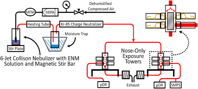
ENM-laden aerosol was generated from a suspension of NPs in water (Optima LC/MS grade, Fisher Scientific, Pittsburgh, PA) using a 6-jet Collison nebulizer (Mesa Labs, Butler NJ) (Fig. 1). Particle suspensions were sonicated in a cup horn sonicator (QSonica, Newtown CT) for 5 min. Table 2 lists the concentrations of NP suspensions that were used in the nebulizer. To prevent particle settling during nebulization, a magnetic stir bar was placed into the nebulizer and the nebulizer was positioned on a stir plate. Dehumidified and HEPA-filtered air was supplied to the nebulizer at 1.4 bar. Generated particle-laden aerosol from the nebulizer passed through a brass drying column heated to 110 °C and a particle neutralizer (Model 3012, TSI Inc., Shoreview, MN) prior to entering two nose-only inExpose towers. Mass exposure concentration of generated aerosol was determined using a photometric method with the use of an aerosol photometer (pDR, personal DataRAM™ pDR-1500, ThermoFisher Scientific, Waltham, MA), and a gravimetric method with the use of glass fiber filters (37 mm in diameter, Whatman, Middlesex, UK) located on the exhaust side of the pDR. An internal pump within the pDR sampled the air directly from one of the nose-only ports at 2 L/min. The filters were changed every 1 or 2 h. A stainless steel sampler holding 47-mm glass fiber filter was placed on the bottom of each nose-only tower to minimize particulate release into the hood. All filters were pre- and post-weighed in a dedicated climate-controlled room using a six-place microbalance (XP26, Mettler Toledo, Mettler-Toledo, Inc., Columbus, OH). A scanning mobility particle sizer (SMPS, Model 3938, TSI Inc.), with an aerosol neutralizer (Model 3088, TSI Inc.) and a standard differential mobility analyzer (Model 3081 L, TSI, Inc.), was used to determine the particle size distribution of generated aerosol sampled from the nose-only port on the inExpose tower. Geometric mean (GM) mobility diameter and geometric standard deviation (GSD) of aerosol sizes in individual exposures were obtained from software supplied by the vendor (Table 2).
Fig. 1.
Schematic of nanoparticle aerosol generation and nose-only exposure system used in in vivo acute studies. ENM suspensions were placed into a 6-jet Collison nebulizer supplied with dehumidified and HEPA-filtered air at 20 psi. Nebulizer-generated aerosol (indicated by red lines) flowed through a heating tube and moisture trap and then passed through a 85Kr charge neutralizer and subsequently entered each nose-only exposure tower at 6 L/min. Size distribution of the ENM aerosol within the exposure towers was evaluated using a SMPS. Real-time ENM aerosol concentration was monitored in each tower using an aerosol photometer (pDR). Time-weighted average ENM aerosol concentrations were determined gravimetrically from pDR filters. The entire system was housed in a chemical hood.
2.5.1. Estimated dose
The estimated deposited doses in the pulmonary region for each ENM used in these studies ranged between 3 and 5 μg/mouse (4–7 ng/cm2) and are reported in Table 3. The dose calculation assumed a minute volume of 25 mL/min. The pulmonary deposition fraction for each ENM was calculated using the computational Multiple-Path Particle Dosimetry (MPPD) model (software version 3.04, available at: https://www.ara.com/products/multiple-path-particle-dosimetry-model-mppd-v-304) (Asgharian et al., 2014).
Table 3.
Estimated exposure doses for acute nose-only exposure.
| ENM | Deposition fraction in pulmonary region | Estimated dosea |
|||
|---|---|---|---|---|---|
| By mass mg/kg | By animal μg/mouse | By lung mass μg/gc | By lung surfaceb ng/cm2 | ||
| ZnO | 0.21 | 0.23 | 4.7 | 39.0 | 6.9 |
| CuO | 0.22 | 0.25 | 4.9 | 41.1 | 7.2 |
| V2O5 | 0.20 | 0.25 | 4.9 | 41.1 | 7.2 |
| WO3 | 0.14 | 0.14 | 2.8 | 22.5 | 4.1 |
Assumes 165 breaths/min, tidal volume of 0.15 mL, and 20 g/mouse body mass.
680 cm2 surface area.
Wet weight of whole lungs used in this estimation was 120 mg.
2.6. End-point toxicity measurements in vivo
Pulmonary inflammatory responses at two time points were assessed in bronchoalveolar lavage (BAL) fluid: 4 h time point (n = 5) and 24 h time point (n = 6) in both sexes. Animals were euthanized, heart exsanguination was performed and the right lobes of the lungs were lavaged 3 times with 1 mL of 0.9% sterile sodium chloride solution (Baxter, Deerfield, IL). Collected BAL fluid was centrifuged at 800g for 5 min at 4 °C; supernatants were frozen at −80 °C for later determination of total protein and cytokines/chemokines. Cell pellets were resuspended in 200 μL Hank’s balanced salt solution (Life Technologies, Grand Island, NY). Cell viability (with PI stianing) and total cell counts were performed using MFS cassettes (MXC020) and an automated cell counter (Moxi GO II, Orflo Technologies, LLC, Ketchum, ID). The total cell counts and viability measurements were performed at two size ranges: 9.5 and 21.5 μm (representing mainly the diameter of monocytes/macrophages and large lymphocytes) and 6.5–23 μm (including neutrophils and eosinophils). For determination of differential cell counts (number of macrophages, neutrophils, lymphocytes, and eosinophils) cells were fixed on glass microscope slides with fetal calf serum (Cytospin 4, Thermo Shandon, Thermo Scientific, Waltham, MA) and stained using Protocol® HEMA 3 stain set (Fisher Diagnostics, Pittsburgh, PA). A total of 400 cells were evaluated to differentiate the cells by type using an optical microscope (Olympus, Center Valley, PA).
The number of samples evaluated for the cytokine and total protein concentrations in BAL fluid, as well as histopathology evaluation for each experimental group and time point was determined based on the inflammatory responses seen in the BAL fluid. We evaluated 10 lung tissues for each time point for CuO and ZnO and 2 lung tissues for each time point for V2O5 and WO3, representing both male and female mice. For statistical comparison of cytokine concentrations between experimental groups and shams, all values measured in shams were grouped together.
Cytokines/chemokines in BAL fluid supernatants (MIP-1a, KC, IL-10, IL-12p40, IL-6, MCP-1, G-CSF) were determined using a multiplex magnetic bead assay (Milliplex MAP kit, selected cytokines from MCYTOMAG-70K (EMD Millipore Corporation) and read on Luminex 100, Bio-Rad Laboratories, Inc., Hercules, CA). Total protein was determined using Quick Start Bradford Dye Reagent (Bio-Rad Laboratories).
After BAL collection, right lobes of the lungs were first perfused with 10% buffered formalin (Fisher Scientific, Kalamazoo, MI) through cannulated trachea and then stored in 10% buffered formalin until further processing. Tissues were then paraffin-embedded, sectioned (5-μm thickness) and stained with hematoxylin and eosin (H&E). Lung tissues were evaluated for key histopathologic changes including increased and activated number of macrophages and granulocytes, perivascular cuffing (infiltration of leukocytes around vessels), alveolar injury (evidenced by sloughing/cellular debris in alveolar lumen), reactive pneumocytes (hyperplastic/proliferative cells), necrosis (presence of cellular debris or overt necrosis of airway epithelia) and edema (eosinophilic fluid within airspaces). All lung tissues were evaluated by the board-certified veterinary pathologist using the post-examination masking method and following principles of reproducible tissue scoring (Meyerholz and Beck, 2018; Meyerholz et al., 2018).
2.7. In vivo dosimetry analyses using microwave assisted acid digestion and ICP-MS
To determine lung burden of each metal after inhalation exposure, n = 5 female mice per each ENM, per each time point (4 and 24 h) were exposed to generated nanoparticulate aerosol, as described above. Control animals were exposed to HEPA-filtered laboratory air. The exposure concentrations in these dosimetry assessment studies are listed in Table 2. Left lobes of lung tissues (harvested from mice without BAL) were freeze dried (FreeZone, Freeze Dry Systems, Labconco Co., Kansas City, MO) for 18 h and weighed using an analytical balance (Mettler Toledo, Mo. XPR206CDR). EPA Method 3051 was modified for digesting tissues from animals exposed to CuO, V2O5, and ZnO. Briefly, the entire left lobe (~8 mg) of lung tissue was weighed and transferred to a pre-weighed Teflon digestion vessel containing 9 mL of concentrated HNO3 and 1 mL of 30% H2O2. Samples were then acid digested using a Milestone ETHOS UP (Sorisole, Italy) microwave digestion system with a ramp set to 210 °C for 20 min with a hold at 210 °C for 15 min. Samples were then diluted with 40 mL of deionized water. The digestion vessel mass was recorded again post digestion to obtain a final solution mass. Samples were then diluted 10-fold in 2% HNO3 with 10 μg/L of 89Y and 209Bi as internal standards.
Studies have shown that tungsten chemistry, and solubility is complex, it can undergo polymerization in acidic conditions with HNO3 and form precipitates, thus yielding measurements of aqueous which are a gross underestimation (Sun and Bostick, 2015; Bostick et al., 2018). To overcome this issue, a method by Griggs et al. (2009) was adapted for acid digestion of tissue samples from mice exposed to WO3 carried out as described above but with 6 mL of HNO3, 2 mL of H3PO4, and 10 mL of 30% H2O2. To determine the digestion efficiency, each ENMs (~0.5 mg) was digested using both EPA method 3051 and the Griggs et al. method. Percent recoveries were found to be 100%, 70%, 84%, and 92% for CuO, ZnO, V2O5, and WO3, respectively. Further information on digestion efficiencies including a comparison between the two digestion methods is presented in supplementary materials (Supplementary Table 4). The concentrations of trace metals in lung tissue samples are expressed as μg/g of tissue (dry weight). All reagents were of trace metal grade or better. Digested samples were analyzed with inductively coupled plasma-mass spectrometry (ICP-MS, Agilent 7900, Santa Clara, CA) using helium gas in collision cell mode to remove isobaric interferences and increase sensitivity.
2.8. Statistical analyses
For the in vitro studies, the data were expressed as mean ± standard deviation. One-way analysis of variance (ANOVA) followed by Dunnett’s post hoc test in GraphPad Prism (GraphPad Software, San Diego, CA) was used for statistical analysis of ENM characterization and cytotoxicity data. All outcome measures obtained from in vivo studies after exposure to ENMs were compared to sham-exposed animals using one-way ANOVA (Sigma Plot v.11.0, Systat Software Inc., Point Richmond, CA) followed by multiple comparison versus control group (Dunn’s or Dunnett’s Method). If data were not normally distributed, Kruskal-Wallis ANOVA on Ranks was used. A t-test was used to test differences in outcomes between males and females. A p-value < 0.05 was considered statistically significant. Statistical probability (p values) in plots is expressed as follows: ****p < 0.0001, ***p < 0.001, **p < 0.01, and *p < 0.05. Data are expressed as mean ± standard error (SE) unless otherwise noted.
3. Results and discussion
3.1. ENM characterization
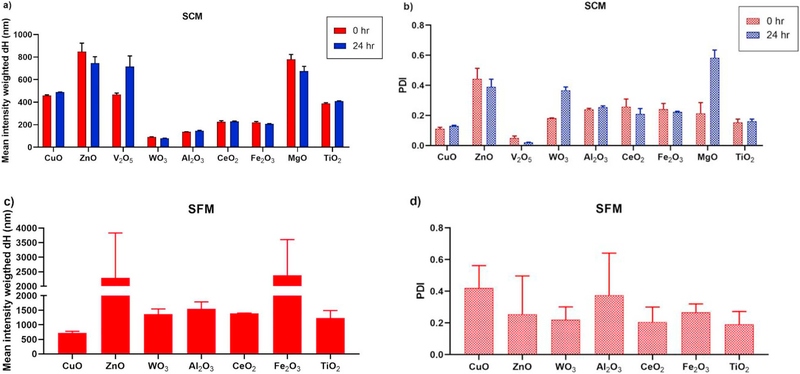
Hydrodynamic diameter (dH) of ENM was determined using dynamic light scattering (DLS) technique. Mean intensity-weighted dH of ENM in SCM at 0 and 24 h was provided from NHIR ERCC as shown in Fig. 2a. Most ENMs exhibited consistent mean dH at 0 and 24 h suggesting these ENM formed agglomerates can be stably suspended in the presence of serum protein for at least 24 h except V2O5. The agglomerate sizes of WO3, Al2O3, CeO2, and Fe2O3 in SCM were generally 100–200 nm exhibiting a smaller degree of agglomeration than the other 5 ENMs, CuO, ZnO, V2O5, MgO, and TiO2. These measured differences in aggregation may influence the delivered doses to cells for these materials, and therefore also affect the toxicity assessment.
Fig. 2.
(a) Mean intensity weighted hydrodynamic diameter (dH) of ENMs measured at 0 and 24 h incubation in SCM at 37 °C, (b) polydispersity index (PDI) of ENMs measured at 0 and 24 h incubation in SCM, (c) mean intensity weighted dH of ENMs for 0 h incubation, and (d) PDI of ENMs measured at 0 h incubation in SFM.
ENMs dispersed in SFM were measured for dH only at 0 h as we observed that, after 24 h incubation of ENMs in SFM, aggregates formed that were visible to the naked eye and were very large and sedimented too rapidly to be measured using the Zetasizer. ENMs dispersed in SCM consistently displayed less agglomeration or aggregation than the SFM (Fig. 2a, c). This observation is consistent with the literature - others have reported less aggregation of nanomaterials in the presence of serum (Liu et al., 2015; Anders et al., 2015; Wells et al., 2012; Tedja et al., 2012). We confirmed that the use of serum containing media in in vitro assays is superior considering physiological conditions and such obtained results agree more closely with in vivo results. Serum proteins adsorbed onto nanoparticle surfaces form protein corona which is a key factor in nanoparticle-nanoparticle interactions and directly impact the cellular uptake of nanoparticles. Deguchi et al. demonstrated that NPs can adsorb serum protein to the surface and form a steric layer that can prevent the aggregation induced by salts present in cell culture media (Deguchi et al., 2007). The hydrodynamic size for all ENMs in SFM was usually > 1000 nm with the one exception being CuO ENMs (718.5 ± 58.2 nm) (Fig. 2c). The V2O5 and MgO showed high degree of aggregation immediately (t = 0 h) after preparation, therefore, the hydrodynamic size and particle size distribution of these ENMs could not be determined.
3.2. In vitro cytotoxicity using MTS assay
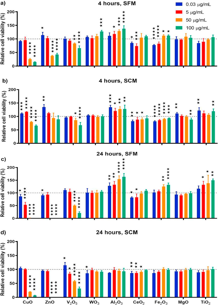
The cytotoxicity of ENMs to A549 cells was assessed using the colorimetric MTS assay for relative cell viability (Fig. 3). Materials that induced toxicity were defined as those that had significantly lower cell viability compared to the control group (cells that were incubated in either SFM or SCM and MTS reagent without any ENMs). The toxicity was assessed at 0.03, 5.0, 50, and 100 μg/mL of ENMs, which includes concentrations lower than what is typically reported in the literature (Pal et al., 2015; Wongrakpanich et al., 2016; Ahamed et al., 2010). For the present study it was imperative to investigate lower concentrations in vitro because the concentration of 0.03 μg/mL represents the epithelial surface-equivalent dose of the in vivo 4-h acute exposure at 3.5 mg/m3 (Jing et al., 2015).
Fig. 3.
Percentage relative cell viability of ENMs at the concentration of 0.03, 5, 50, and 100 μg/mL by MTS assay in (a) SFM for 4 h (b) SCM for 4 h (c) SFM for 24 h and (d) SCM for 24 h.
The results indicated that many of the metal oxide ENMs such as Al2O3, Fe2O3, CeO2, TiO2, MgO, and WO3 did not demonstrate significant cytotoxicity at any concentration tested. However, CuO, ZnO, and V2O5 ENMs exhibited significant cytotoxicity at the concentrations of 50 and 100 μg/mL at 4 and 24 h in SFM and SCM with the exception of the ZnO ENMs at 4 h in SCM (Fig. 3). Ahamed et al. reported that CuO ENMs reduced the relative cell viability of A549 cells after 24 h of exposure in a dose-dependent manner at 10–50 μg/mL (Ahamed et al., 2010). Wang et al. and Zhang et al. reported that ZnO ENMs decreased the relative viability of A549 cells after 24 h of exposure at 30 and 100 μg/mL (Zhang et al., 2017; Wang et al., 2018). It has also been reported that V2O5 ENMs induced cytotoxicity in A549 cells at much lower concentrations than observed in the current study, including 20, 30, and 40 μM (corresponding to 3.6, 5.5 and 7.3 μg/mL) (Karaboğa Arslan et al., 2017). Karlsson et al. investigated the cytotoxicity of different metal oxide NPs such as CuO, TiO2, ZnO, CuZnFe2O4, Fe3O4 and Fe2O3 using A549 (Karlsson et al., 2008). Their results showed that CuO NPs had the highest cytotoxicity due to DNA damage, and they were the only NPs in the study that promoted a significant increase in intracellular reactive oxygen species (ROS). ZnO NPs were the second most toxic, as they induced DNA damage, however, not to the same extent as CuO NPs (Karlsson et al., 2008). A similar trend was observed in the present study, in that CuO and ZnO were the most toxic metal oxide ENMs investigated, but the toxicity of CuO ENMs were slightly higher than ZnO based on the 4-h incubation results shown in Fig. 3. Therefore, the DNA damage and formation of intracellular ROS may explain the toxicity of the CuO ENMs investigated herein.
The lowest concentrations of 0.03 and 5 μg/mL of each of the ENMs did not exhibit cytotoxicity in either SFM or SCM at 4- and 24-h incubation, with the exception of 5 μg/mL of 50 nm CuO ENMs in SFM after 24 h incubation (Fig. 3). In general, when ENMs were incubated with A549 cells in SCM they were less cytotoxic compared to the same dose of ENMs in SFM. In particular, the relative cell viability of CuO was only 15% after a 4 h incubation in SFM whereas it was 65% after 4 h in SCM at the highest dose of 100 μg/mL. A similar trend was observed for CuO ENMs after 24 h incubation in which the highest dose resulted in a viability of almost zero in SFM whereas it was approximately 7% in SCM. ZnO and V2O5 ENMs exhibited a similar trend. Saikia et al. found that the existence of protein corona on silica NPs in SCM mitigated the cytotoxicity compared to SFM conditions because of the decrease in cell-mediated uptake of the NPs (Saikia et al., 2016). The surface energy of NPs was reduced after the formation of a protein corona which inhibits NP adhesion to the cell surface decreasing particle internalization (Lesniak et al., 2012; Lesniak et al., 2013).
The Al2O3, Fe2O3 and TiO2 ENMs induced increases in relative cell viability in a dose-dependent manner in SFM at 4 and 24 h (Fig. 3). This result might indicate an effect on cell proliferation of these materials, interference from ENM absorbance, or interactions between the dye and the ENMs. To address this concern, we tested the interference of ENMs with MTS reagent. The interference method is described in the Supplement. The result showed that only MgO 100 μg/mL caused a statistically significant increase in UV absorbance at 490 nm. In addition, V2O5 and CuO marginally, but not significantly, affected the readout/absorbance (Supplementary Fig. 1). In order to confirm the data obtained with MTS assay where cell viability (as opposed to interference) was measured, we performed an assay that did not rely on absorbance and instead directly assayed cell viability. Thus, viable cell counts were enumerated using propidium iodide (PI) staining (to stain dead cells) as an indicator of cytotoxicity as well as the effect of ENMs on cell proliferation.
3.3. In vitro cytotoxicity using viable cell count with PI staining
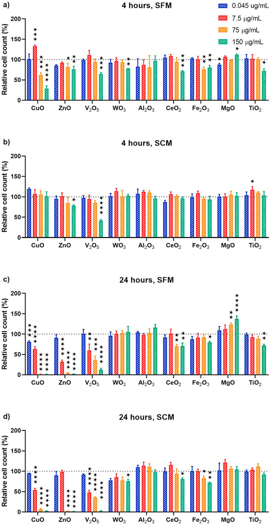
The viable cell count using PI staining was performed to assess cytotoxicity and cell proliferation effect to ensure no ENM interference. The toxicity for this test was assessed at 0.045, 7.5, 75, and 150 μg/mL ENMs in a 6-well plate with the same cell culture media condition and incubation time as the MTS assay. These concentrations are equivalent to those used in MTS assay base on ENM mass per surface area which are 0.009, 1.563, 15.625, and 31.25 μg/cm2.
Materials that exhibited cytotoxicity were defined as those that had significantly lower viable cell count compared to the control group (cells that were incubated in either SFM or SCM without any ENMs). The result indicated that CuO, ZnO, and V2O5 exhibited the cytotoxicity in a dose dependent manner at 24 h in SFM and SCM and these 3 ENMs were more toxic than other 6 metal oxide ENMs (Fig. 4). This toxicity finding was similar to the result from MTS assay. However, the significant increases in % relative viable cell count compared to the control groups were found at a concentration of 7.5 μg/mL CuO at 4 h in SFM and the concentrations of 75 and 150 μg/mL MgO at 24 h in SFM. This indicated the cell proliferation effect on both ENMs occurred at specific concentration and only in SFM condition. CuO exhibited the cell proliferation effect only at a concentration of 7.5 μg/mL at 4 h in SFM can be explained as the copper was an essential element to promote cell growth and maintain healthy cells at the specific concentration. Typically, cell culture media does not contain elemental copper, but the copper was in sera that was used to supplement the media. The amount of copper sufficient for cell growth is 50 to 100 ng/mL which is contained in 5 to 10% sera (Millipore Sigma, 2020). This explanation supports that adding specific amount of CuO to the cells in SFM could increase cell proliferation. D’Mello et al. showed that inclusion of copper in chitosan based scaffolds enhanced bone tissue regeneration (D’Mello et al., 2015). MgO in SFM had greater opportunity to react with water molecule and form magnesium hydroxide (MgO + H2O ➔ Mg(OH)2) than in SCM since there is no serum protein binding at the surface of MgO to reduce the reaction between MgO and water (ERCC). Mg2+ can be dissociated from Mg(OH)2 and stimulate cell proliferation as Mg promoted DNA and protein synthesis (Xing et al., 2018; Wolf and Cittadini, 1999).
Fig. 4.
Percentage of relative viable cell count of ENMs at the concentration of 0.045, 7.5, 75, and 150 μg/mL using viable cell count with PI staining in (a) SFM for 4 h (b) SCM for 4 h (c) SFM for 24 h and (d) SCM for 24 h.
There were no significant increases in % relative viable cell count on Al2O3, Fe2O3, and TiO2 in SFM at 4 and 24 h as we found from MTS assay result. Therefore, we can conclude that these 3 ENMs had no proliferation effect and the finding from MTS assay could result from the artifactual interference from ENM absorbance.
3.4. Delivered-to-cell dose of ENMs
Nanomaterial dosimetry provides meaningful and important information for in vitro cytotoxicity screening as the dose delivered to cells represents a more accurate measure of ENM dose than the nominal administered dose (DeLoid et al., 2015). The reason is because ENMs generally form agglomerates when dispersed in any liquid and these agglomerates have a specific sedimentation rate as a function of exposure time. The particle size distribution and effective density of these agglomerates affects the sedimentation rate of ENMs as shown in Eq. (1)
| (1) |
where g is acceleration due to gravity (m s−2), ρEV is effective density of particle (kg m−3), ρmedia is media density (kg m−3), and η is the media dynamic viscosity (kg m−1 s−1). Thus, the sedimentation velocity is proportional to the difference between the effective densities of particle and the densities of suspending media and the square of the suspended particle diameter (DeLoid et al., 2017a; DeLoid et al., 2015). Based on this equation, the agglomeration size influences sedimentation rate and therefore the dose delivered to the cell surface.
The mean volume-weighted dH, effective density, and delivered-to-cell doses of ENM in SCM were provided from NHIR ERCC as shown in Table 1 (we did not validate these data). The delivered-to-cell doses data would allow dosimetry-based toxicity assessment as described by DeLoid et al. (2017a, 2017b). The order from largest to smallest agglomeration size exhibited as follows: MgO, ZnO, TiO2, V2O5, CuO, Fe2O3, CeO2, Al2O3, and WO3. The first 4 largest ENMs also showed the highest mass of ENM delivered to cells. Similarly, the smallest agglomerate, WO3 had the smallest mass ENMs delivered to the cells. This finding was reasonable according to the previous explanation from Eq. (1).
The mass of ENM delivered to the cells in SFM is expected to be greater than in SCM since the agglomerate size for most ENMs in SFM was greater than in SCM. This explains our viable cell count cytotoxicity data for CuO and ZnO at 4 h incubation showing that ENMs in SFM exhibited more toxicity than in SCM.
The % relative viable cell count for CuO, ZnO, and V2O5 at 24 h incubation in SCM was plotted against the dosimetry dose based concentration (Supplementary Fig. 2) that was calculated by multiplying the mean fraction of deposited ENMs for 24 h and the nominal concentration. This figure showed that CuO induced the highest cytotoxicity.
3.5. Characterization of generated ENM aerosol for in vivo exposures
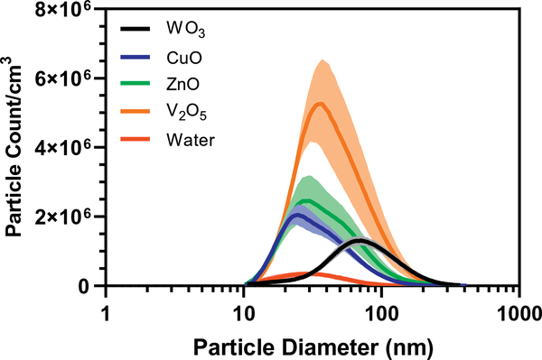
The conditions for the generation of nanoaerosol and exposure of mice in our nose-only system were meticulously controlled. The mass concentration of aerosol was held to our target of 3.5 mg/m3 by controlling the pressure, flow rate and mass of suspended nanomaterial in the Collison nebulizer. The exposure concentration was selected based on our (Adamcakova-Dodd et al., 2014; Adamcakova-Dodd et al., 2015; Kim et al., 2011) and other’s (Gosens et al., 2016) previous studies investigating various ENMs, as well as considering relevance to potential human exposures. The exact mass of each ENMs used in the nebulizer for the aerosol generation is reported in Table 2. Higher masses of nanomaterials were used for V2O5 and CuO aerosol generation (1 mg/mL), followed by ZnO and WO3 (0.3 and 0.4 mg/mL, respectively). We were able to achieve very similar exposure concentrations for all materials (Table 2), all were in the range between 3.24 and 4.15 mg/m3. The smallest geometric mean mobility diameter (GM) of generated particles was measured for CuO (33.26 nm), followed by ZnO (37.54 nm) and V2O5 (43.03 nm). The largest GM was generated for WO3 aerosol (77.61 nm) as shown in Table 2. Comparing the GM of the aerosol size distribution with the primary ENM particle sizes reported in Supplementary Table 1, the smallest primary sized particles, WO3 (20 nm) generated the highest agglomeration/aggregation of particles. Peak particle counts were all similar with the highest counts for V2O5 (Table 2, Fig. 5). We also measured the particle size distribution of particulates/contaminants that are always present in water (Fig. 5). The particle size distribution of all ENMs reported in Fig. 5 contains a small fraction of particles derived from the water (Knight and Petrucci, 2003). Since pure, analytical grade water was used as a vehicle for the generation of ENM aerosol the contribution of contaminant particle counts to the total measured was relatively insignificant.
Fig. 5.
Particle size distribution of ENM aerosols generated for nose-only exposures. Measurement was performed using SMPS sampling from the nose-only port of the exposure system.
3.6. In vivo pulmonary responses after acute exposures
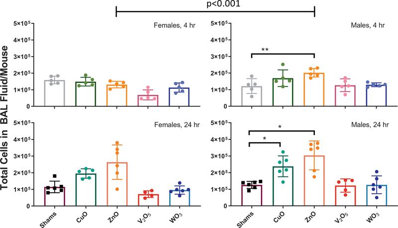
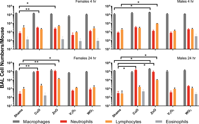
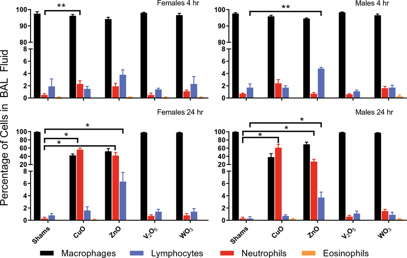
The inflammatory potency of ENMs after 4 h exposure was assessed by the evaluation of cells obtained by BAL, cell viability, total protein, cytokine concentrations and histopathology evaluation of lung tissues. There was a significantly higher recruitment of cells into the lungs in ZnO-exposed male mice at the 4 h time point (Fig. 6) in comparison to shams. At the 24 h time point, the total number of cells increased in both males and females in CuO- and ZnO-exposed mice compared to shams, however this increase was shown to be significant only in males due to a higher variability in females. There was a significant increase in the number of total cells in male ZnO-exposed mice at 4 h time point compared to female counterparts, however there was no significant difference between males and females at 24 h. The number of neutrophils in BAL fluid was slightly increased in CuO- and ZnO-exposed mice, both males and females at the 4-h time point (Fig. 7); however this increase was statistically significant only for CuO-exposed females. We observed much higher neutrophilic recruitment at the 24-h time point for the same ENMs in both, males and females. The number of lymphocytes was also significantly increased compared to shams in ZnO-exposed mice in both, male and females. The proportion of neutrophils in BAL fluid in CuO-exposed mice was increased from about 2% at 4 h to 56 ± 4% in females and to 61 ± 9% in males at 24 h (Fig. 8). In ZnO-exposed mice, this increase was from about 2 ± 0.5% at 4 h to 42 ± 8% at 24 h in females and from 1 ± 0.2% to 27 ± 6% in males. The number of eosinophils found in BAL fluid was not significantly increased at any time point after exposure, sex or type of ENM.
Fig. 6.
Number of total cells in BAL fluid in females and males at 4 h and 24 h time point.
Fig. 7.
Number of macrophages, neutrophils, lymphocytes and eosinophils in BAL fluid in females and males at 4 h and 24 h time point.
Fig. 8.
Proportion of macrophages, neutrophils, lymphocytes and eosinophils in BAL fluid in females and males at 4 h and 24 h time point.
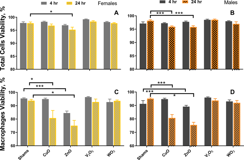
The viability of total cells that included the cells with the wider size range (6.5–23 μm) was decreased compared with sham-exposed mice after ZnO and CuO exposure at the 24 h time point in both, females and males (Fig. 9A and B). When we analyzed the viability of the cells with the cell diameter in the size range of mouse monocytes/macrophages (9.5 and 21.5 μm), we found a larger decrease in the viability compared to controls in ZnO and CuO-exposed mice at 24 h (Fig. 9C and D). The results of total protein determination support higher cell injury for ZnO-exposed animals at 24 h compared to shams or CuO-exposed mice (Supplementary Fig. 3).
Fig. 9.
BAL cell viability of total cells (A and B) and macrophages (C and D) in females and males at 4 h and 24 h time point.
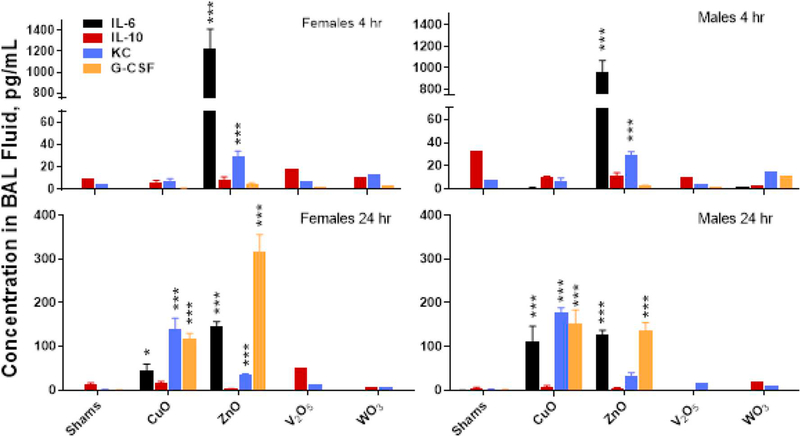
The concentrations of selected inflammatory cytokines/chemokines confirm the inflammatory responses after exposure to ZnO and CuO ENMs. However significant differences between CuO and ZnO ENMs in lavage cytokines were observed, indicating involvement different immunotoxicity pathways in inflammatory responses between these two ENMs (Fig. 10). The release of IL-6 in the lungs, the major mediator of the acute phase inflammation (Tanaka et al., 2014), was significantly higher in both females and males (1227 and 966 pg/mL, respectively) at the 4 h time point after ZnO exposure compared to controls or CuO ENM exposure (< 2.26 pg/mL [LLOD]). This increase was not observed after CuO exposure. At the 24 h time point after ZnO exposure, IL-6 concentrations decreased significantly in both females and males (146 and 126 pg/mL, respectively). Such a release of IL-6 is indicative of inflammation associated with lung tissue injury and increased total protein release at 24 h post exposure, as observed after ZnO exposure. Early release of IL-6 into circulation or BAL fluid was observed in workers after single exposure to ZnO fume after 2 h-exposure to 5 mg/m3 (Fine et al., 1997; Fine et al., 2000). Workers in that study, were exposed to freshly generated ZnO vapor condensed to ultrafine particles with the mass median diameter of 0.3 μm. Upregulation of IL-6 was also observed after acute (3-day) ZnO NP exposure of female ICR mice by inhalation (Rossner Jr et al., 2018). Acute exposure to CuO ENM caused higher release of keratinocyte-derived chemokine (KC) (neutrophil chemoattractant released by macrophages, neutrophils and epithelial cells) especially at 24 h post exposure in comparison to controls or ZnO exposure, even though both, CuO and ZnO caused a high number of neutrophils in BAL fluid. In agreement with the neutrophilic recruitment into BAL fluid 24 h post exposure, the concentration of KC was higher after CuO exposure than after ZnO exposure at the 24 h time point.
Fig. 10.
Concentration of cytokines in BAL fluid in females and males at 4 h and 24 h time point. Significant differences show difference in comparison with shams.
Most of the evaluated tissues had nominal or focal changes that were often in the lowest thresholds of detection. Supplementary Table 2 reports on the scores of the changes evaluated. A 5-point scale was used for the evaluation of potential pathologies. All of the tissues were scored either with the score of 0 (within the scope of normal), 1 (rare, but detectable change), or 2 (mild in distribution/severity). Increased numbers of macrophages and activated macrophages were found after CuO exposures in both females (average score of 1.8 ± 0.1) and males (1.2 ± 0.1). The most noticeable changes were represented by the presence of neutrophils around vessels (perivascular infiltration or aggregates) found for V2O5, in both time points post exposure and in females and males, however these changes were still scored as mild. None of the tissues were scored with moderate (score 3) or severe (score 4) in distribution/severity in any evaluated changes. Consistent with the results from BAL fluid, observed changes were more pronounced at the 24-h time point compared to the 4-h time point in all materials showing some inflammatory responses (CuO and ZnO). Supplementary Fig. 4 shows representative micrographs of lung tissues after V2O5 exposure (with a histology score 2 for perivascular infiltration of leukocytes around vessels at each time point for each sex), pointing out that all observed changes were minimal.
The effects of various types of ENMs after inhalation exposure differ by the duration and severity of the responses. The results of our studies confirmed the toxicity of CuO (Pettibone et al., 2008; Kim et al., 2011) and ZnO ENMs after inhalation exposure seen in many other inhalation studies (Chuang et al., 2014; Vandebriel and De Jong, 2012; Larsen et al., 2016), however the number of studies using only an acute protocol with shorter-term exposure for screening ENMs is limited (Gosens et al., 2016; Larsen et al., 2016). In our studies we also found that at least two time points post-acute exposure (e.g. 4 and 24 h) should be investigated to explore the toxicity of ENMs. Even though our in vitro screening assay did not point to an increased cytotoxicity of WO3, due to the lack of toxicological data after respiratory exposure, we thought to include this material in our in vivo acute screening protocol and compare the toxicity with other studied ENMs. None of the results of our WO3 inhalation study showed significant differences compared to controls. The recruitment of neutrophils into the lungs (Fig. 8) after WO3 exposure was < 2% in all experimental groups (ranging from 0.8% to 1.6%) and similar to sham-exposed mice (0.3% - 0.7%). In an inhalation study of WO3 NPs in golden Syrian hamsters (Prajapati et al., 2017), animals were exposed in a whole-body exposure chamber to low or high concentration (5 or 10 mg/m3, respectively) for 4 h/day for 4 or 8 days. An average geometric mean particle diameter of WO3 particulates produced in this study was 202 ± 49 nm. After inhalation exposure of WO3 particulate aerosol for 4 days, pulmonary inflammation (represented by an increase of neutrophils in BAL fluid) was significantly increased with the higher exposure concentration (10 mg/m3), but not with lower exposure concentration (5 mg/m3). In our studies, we did not see significant pulmonary inflammation or immunotoxicity after V2O5 ENM exposure, as has been described elsewhere (Worle-Knirsch et al., 2007; Kulkarni et al., 2014). Also, our in vitro screening studies showed that these materials lowered the relative cell viability. However most of the studies of V2O5 NPs were conducted in vitro or in vivo with repeated exposures (Kulkarni et al., 2014; Karaboğa Arslan et al., 2017). The toxicity of bulk vanadium pentoxide after inhalation exposures has been studied (Dill et al., 2004; Cooper, 2007; NTP toxicology and carcinogensis studies of vanadium pentoxide (CAS No. 1314–62-1) in F344/N rats and B6C3F1 mice (inhalation), 2002). The differences observed between in vitro and in vivo results in our screening studies might be due to short half-life of V2O5 after inhalation (Conklin et al., 1982). One hour after intratracheal instillation of 0.5 μCi 48V in V2O5 form (0.3 mg/kg, 40 μg/rat), 40% of the total 48V had been cleared, by day 3, 90% had been cleared (Conklin et al., 1982). Faster elimination of V2O5 has been reported in mice than rats (Dill et al., 2004). Such rapid clearance of V2O5 after acute exposure (which was not verified for nanoparticulate form of V2O5) might be responsible for the lack of toxicity present after acute inhalation exposure. The absence of clearance mechanisms in in vitro studies especially at submersed conditions (which is one of the limitations of such studies) must always be considered when comparing in vitro and in vivo screening methods. In our in vivo studies, retained doses of each particular ENM were determined using ICP-MS as reported in Table 4. We found higher clearance of W and V from the lungs than Cu at the 24 h time point post exposure. The lung burden of W decreased 3-fold in 24 h (from 58.6 μg/g at 4 h to 19.8 μg/g at 24 h). More than half of the V was cleared from the lungs 24 h post exposure (32 μg/g at 4 h and 18 μg/g at 24 h). Our estimations of deposited doses in the lungs using MPPD modeling (Table 3) agreed closely with our values measured by ICP-MS (Table 4) for all materials except ZnO. Concentration of Zn in the lungs after 4 h exposure was not significantly different from sham-exposed animals. We have seen similar results in our previous studies after sub-acute or sub-chronic inhalation exposure to ZnO nanomaterials (Adamcakova-Dodd et al., 2014).
Table 4.
Concentration of selected isotopes in lung tissues of exposed and control animals at 4 and 24 h post exposure, determined by ICP-MS. Data were adjusted for digestion efficiency.
| Investigated ENM | Measured isotope | Concentration, μg/g (dry wt) Mean ± SE |
||
|---|---|---|---|---|
| Shams (n = 5) | 4 h (n = 5) | 24 h (n = 5) | ||
| CuO | 63Cu | 12.1 ± 0.8 | 60.0 ± 1.6*** | 50.9 ± 2.1*** |
| ZnO | 64Zn | 196.1 ± 6.0 | 181.1 ± 20.9 | 116.7 ± 1.1 |
| V2O5 | 51V | 0.3 ± 0.0 | 31.8 ± 2.6*** | 18.2 ± 0.8* |
| WO3 | 183W | 0.2 ± 0.0 | 58.6 ± 4.8*** | 19.8 ± 2.1*** |
p < 0.05
p < 0.001 significantly higher from shams.
We hypothesize that rapid dissolution of ZnO NPs in lung fluids is a possible mechanism for the clearance of ENMs. To test this, we compared experimental data (observed) to theoretical dissolution data (modeled) for selected materials. Dissolution of ENMs was modeled using Visual MINTEQ (Gustafsson, 2011). Dissolution modeling was done for selected ENMs such as CuO (tenorite), ZnO (zincite) and V2O5 from our in vivo studies. Literature on tungsten is limited and ionic equilibrium data for WO3 was not available - this prevented us from modeling its dissolution. We simulated two physiologically relevant chemical conditions which include artificial lysosomal fluid (ALF, pH 4.5) and Gamble’s solution (pH 7.4). Components used to build these two simulations were those used for the measured dissolution experiments (Stopford et al., 2003; Midander et al., 2007; Moss, 1979). Results from our model (Supplemental Table 5) suggest that in ALF, we expect to see 100% of the CuO and ZnO dissolved while there will be very little dissolution of V2O5. This finding might serve to explain the loss of macrophage viability at both 4 h and 24 h for ZnO and CuO not seen for V2O5 resulting from rapid increase in metal ion concentration in the lysosome from rapid dissolution of the metal oxides. Interestingly, the opposite trend is observed for Gamble’s solution where we expect to see 100% dissolution of V2O5 but very little for CuO and ZnO.
Results from our dissolution modeling is supported by dissolution experiment results previously published by our group which also showed 100% of ZnO dissolved within 24 h in ALF, but < 1% in Gamble’s solution after 2 wks (Adamcakova-Dodd et al., 2014). Similarly, in our previous studies, CuO ENMs dissolved more in ALF than in Gamble’s solution (pH 7.4) (Pettibone et al., 2008). Based on our previous studies (Adamcakova-Dodd et al., 2014; Pettibone et al., 2008), the dissolution rates of ZnO and CuO were similar, however we observed lower clearance for Cu (Supplemental Table 5). V2O5 (fine powder, geometric mean diameter 0.31 μm) was found to be 8 times more soluble in Gamble’s solution than in a pure water (Toya et al., 2001). Based on our results from in vitro screening as well as considering known toxicity of bulk V2O5, a subacute or chronic inhalation exposure study is warranted to further assess the toxicity of this ENM. Determination of the dissolution characteristics of all studied materials in various simulated physiological conditions is a critical step towards explaining their higher or lower clearance and subsequent toxicity. This novel modeling approach might be a rapid, cost effective in silico tool for screening large quantities of ENMs in multiple simulated physiochemical conditions in addition to the lung, such as gastric fluid having pH < 2 would certainly change the dissolution kinetics of ENMs. This tool and possibly in conjunction with machine learning techniques also opens up the possibility of looking at dissolution of mixtures of ENMs leading to more environmentally relevant toxicity assessments. Further experimental studies are warranted to confirm and improve this newly emerging modeling approach.
A slight increase in the recruitment of total cells into the lungs at the 4 h time point was observed in males compared to females after exposure to ZnO ENMs (Fig. 6, p < 0.001). Conversely the concentration of IL-6 in BAL fluid also at the 4 h time point was higher in females (1227 ± 188 pg/mL) than males (966 ± 102 pg/mL), but not significantly (Fig. 10), and concentration of G-CSF in BAL fluid at 24 h time point was significantly higher (Fig. 10, p < 0.01) in females (319 ± 37 pg/mL) than males (138 ± 16 pg/mL). None of the other materials studied showed sex differences.
4. Conclusion
We aimed to establish a toxicity screening method for inhaled ENMs using in vitro and in vivo acute protocols. The in vitro assessments were conducted with the intention of a rapid screening of ENMs for their potential toxicities while the in vivo assessment was conducted to confirm or rebut the in vitro results in a model more relevant to human exposure. The results of a viability assay performed after exposure of A549 cells at submersed conditions were in the agreement with our in vivo results after acute nose-only inhalation exposure of C57Bl/6 mice for CuO, ZnO, and WO3 ENMs. However, the V2O5 ENMs showed a dose-dependent toxicity in vitro that was not observed in vivo. From all studied ENMs: ZnO, CuO, CeO2, Fe2O3, WO3, V2O5, TiO2, Al2O3 and MgO, the highest toxicity was shown for CuO and ZnO ENMs. The assessment of cell viability after in vitro exposure and lung inflammation after acute inhalation exposure in vivo were shown to be sensitive endpoints to predict toxicity after a short-term exposure. Two time points (4 and 24 h) after acute (one-time) inhalation exposure were necessary to observe inflammation from studied ENMs.
Supplementary Material
Acknowledgements
Research reported in this publication was supported by the National Institute of Environmental Health Sciences of the National Institutes of Health under Award Number [NIH grant # U01ES027252] as part of the Nanotechnology Health Implications Research (NHIR) Consortium which focuses on comprehensive evaluation of interactions between ENMs and biological systems. The research was conducted in laboratory facilities supported by the Environmental Health Sciences Research Center funded by NIH P30 ES005605. The content is solely the responsibility of the authors and does not necessarily represent the official views of the National Institutes of Health.
The engineered nanomaterials used in the research presented in this publication have been procured/developed, characterized, and provided by the Engineered Nanomaterials Resource and Coordination Core established at Harvard T. H. Chan School of Public Health (NIH grant # U24ES026946) as part of the NHIR Consortium.
The research was conducted in laboratory facilities supported by the Environmental Health Sciences Research Center funded by NIH P30 ES005605. B.E.G. was supported by the Alfred P. Sloan Foundation Minority PhD Scholarship, National GEM Consortium Associate Fellowship, The University of Iowa Graduate College Dean’s Fellowship. A.K.S. was supported by the Lyle & Sharon Bighley Endowed Professorship. We thank Dr. Xuefang Jing for her help with necropsies.
Footnotes
Declaration of competing interest
The authors declare that they have no known competing financial interests or personal relationships that could have appeared to influence the work reported in this paper.
Appendix A. Supplementary data
Supplementary data to this article can be found online at https://doi.org/10.1016/j.impact.2020.100214.
References
- Adamcakova-Dodd A, Stebounova LV, Kim JS, Vorrink SU, Ault AP, O’Shaughnessy PT, Grassian VH, Thorne PS, 2014. Toxicity assessment of zinc oxide nanoparticles using sub-acute and sub-chronic murine inhalation models. Particle and Fibre Toxicology. 11 (1), 15 10.1186/1743-8977-11-15. [DOI] [PMC free article] [PubMed] [Google Scholar]
- Adamcakova-Dodd A, Monick MM, Powers LS, Gibson-Corley KN, Thorne PS, 2015. Effects of prenatal inhalation exposure to copper nanoparticles on murine dams and offspring. Particle and Fibre Toxicology. 12 (1), 30 10.1186/s12989-015-0105-5. [DOI] [PMC free article] [PubMed] [Google Scholar]
- Ahamed M, Siddiqui MA, Akhtar MJ, Ahmad I, Pant AB, Alhadlaq HA, 2010. Genotoxic potential of copper oxide nanoparticles in human lung epithelial cells. Biochem. Biophys. Res. Commun. 396 (2), 578–583. 10.1016/j.bbrc.2010.04.156 (Epub 2010/05/08, PubMed PMID: 20447378). [DOI] [PubMed] [Google Scholar]
- Ahamed M, Akhtar MJ, Alhadlaq HA, Alrokayan SA, 2015. Assessment of the lung toxicity of copper oxide nanoparticles: current status. Nanomedicine 10, 2365+. [DOI] [PubMed] [Google Scholar]
- Anders CB, Chess JJ, Wingett DG, Punnoose A, 2015. Serum proteins enhance dispersion stability and influence the cytotoxicity and dosimetry of ZnO nanoparticles in suspension and adherent cancer cell models. Nanoscale Res. Lett. 10 (1), 448 10.1186/s11671-015-1158-y. [DOI] [PMC free article] [PubMed] [Google Scholar]
- Andreescu S, Ornatska M, Erlichman JS, Estevez A, Leiter JC, 2012. Biomedical applications of metal oxide nanoparticles In: Matijević E (Ed.), Fine Particles in Medicine and Pharmacy. Springer US, Boston, MA, pp. 57–100. [Google Scholar]
- Asgharian B, Price OT, Oldham M, Chen LC, Saunders EL, Gordon T, Mikheev VB, Minard KR, Teeguarden JG, 2014. Computational modeling of nanoscale and microscale particle deposition, retention and dosimetry in the mouse respiratory tract. Inhalation toxicology 26 (14), 829–842. 10.3109/08958378.2014.935535 (Epub 2014/11/07, PubMed PMID: 25373829; PMCID: PMC4668803). [DOI] [PMC free article] [PubMed] [Google Scholar]
- Bakand S, Winder C, Khalil C, Hayes A, 2005. Toxicity assessment of industrial chemicals and airborne contaminants: transition from in vivo to in vitro test methods: a review. Inhal. Toxicol. 17 (13), 775–787. 10.1080/08958370500225240 (Epub 2005/10/01, PubMed PMID: 16195213). [DOI] [PubMed] [Google Scholar]
- Beltran-Huarac J, Zhang Z, Pyrgiotakis G, DeLoid G, Vaze N, Demokritou P, 2018. Development of reference metal and metal oxide engineered nanomaterials for nanotoxicology research using high throughput and precision flame spray synthesis approaches. NanoImpact. 10, 26–37. 10.1016/j.impact.2017.11.007. [DOI] [PMC free article] [PubMed] [Google Scholar]
- Bostick BC, Sun J, Landis JD, Clausen JL, 2018. Tungsten speciation and solubility in munitions-impacted soils. Environmental science & technology. 52 (3), 1045–1053. 10.1021/acs.est.7b05406. [DOI] [PubMed] [Google Scholar]
- Chavali MS, Nikolova MP, 2019. Metal oxide nanoparticles and their applications in nanotechnology. SN Applied Sciences. 1 (6), 607 10.1007/s42452-019-0592-3. [DOI] [Google Scholar]
- Chuang HC, Juan HT, Chang CN, Yan YH, Yuan TH, Wang JS, Chen HC, Hwang YH, Lee CH, Cheng TJ, 2014. Cardiopulmonary toxicity of pulmonary exposure to occupationally relevant zinc oxide nanoparticles. Nanotoxicology. 8 (6), 593–604. 10.3109/17435390.2013.809809 (Epub 2013/06/07, PubMed PMID: 23738974). [DOI] [PubMed] [Google Scholar]
- Cohen JM, Beltran-Huarac J, Pyrgiotakis G, Demokritou P, 2018. Effective delivery of sonication energy to fast settling and agglomerating nanomaterial suspensions for cellular studies: implications for stability, particle kinetics, dosimetry and toxicity. NanoImpact. 10, 81–86. 10.1016/j.impact.2017.12.002. [DOI] [PMC free article] [PubMed] [Google Scholar]
- Conklin AW, Skinner CS, Felten TL, Sanders CL, 1982. Clearance and distribution of intratracheally instilled 48Vanadium compounds in the rat. Toxicology letters 11 (1), 199–203. 10.1016/0378-4274(82)90128-X. [DOI] [PubMed] [Google Scholar]
- Cooper RG, 2007. Vanadium pentoxide inhalation. Indian J Occup Environ Med. 11 (3), 97–102. 10.4103/0019-5278.38457 (PubMed PMID: 21957373). [DOI] [PMC free article] [PubMed] [Google Scholar]
- Deguchi S, Yamazaki T, Mukai S, Usami R, Horikoshi K, 2007. Stabilization of C60 nanoparticles by protein adsorption and its implications for toxicity studies. Chem. Res. Toxicol. 20 (6), 854–858. 10.1021/tx6003198. [DOI] [PubMed] [Google Scholar]
- DeLoid GM, Cohen JM, Pyrgiotakis G, Pirela SV, Pal A, Liu J, Srebric J, Demokritou P, 2015. Advanced computational modeling for in vitro nanomaterial dosimetry. Particle and Fibre Toxicology. 12 (1), 32 10.1186/s12989-015-0109-1. [DOI] [PMC free article] [PubMed] [Google Scholar]
- DeLoid GM, Cohen JM, Pyrgiotakis G, Demokritou P, 2017a. Preparation, characterization, and in vitro dosimetry of dispersed, engineered nanomaterials. Nature protocols 12 (2), 355–371. 10.1038/nprot.2016.172 (Epub 2017/01/20, PubMed PMID: 28102836; PMCID: PMC5857388). [DOI] [PMC free article] [PubMed] [Google Scholar]
- DeLoid GM, Wang Y, Kapronezai K, Lorente LR, Zhang R, Pyrgiotakis G, Konduru NV, Ericsson M, White JC, De La Torre-Roche R, Xiao H, McClements DJ, Demokritou P, 2017b. An integrated methodology for assessing the impact of food matrix and gastrointestinal effects on the biokinetics and cellular toxicity of ingested engineered nanomaterials. Particle and Fibre Toxicology. 14 (1), 40 10.1186/s12989-017-0221-5. [DOI] [PMC free article] [PubMed] [Google Scholar]
- Dill JA, Lee KM, Mellinger KH, Bates DJ, Burka LT, Roycroft JH, 2004. Lung deposition and clearance of inhaled vanadium pentoxide in chronically exposed F344 rats and B6C3F1 mice. Toxicological sciences: an official journal of the Society of Toxicology. 77 (1), 6–18. 10.1093/toxsci/kfh005 (Epub 2003/11/06, PubMed PMID: 14600283). [DOI] [PubMed] [Google Scholar]
- D’Mello S, Elangovan S, Hong L, Ross RD, Sumner DR, Salem AK, 2015. Incorporation of copper into chitosan scaffolds promotes bone regeneration in rat calvarial defects. Journal of biomedical materials research Part B, Applied biomaterials 103 (5), 1044–1049. 10.1002/jbm.b.33290 (Epub 2014/09/18, PubMed PMID: 25230382; PMCID: PMC4363304). [DOI] [PMC free article] [PubMed] [Google Scholar]
- Fine JM, Gordon T, Chen LC, Kinney P, Falcone G, Beckett WS, 1997. Metal fume fever: characterization of clinical and plasma IL-6 responses in controlled human exposures to zinc oxide fume at and below the threshold limit value. J. Occup. Environ. Med. 39 (8), 722–726 (Epub 1997/08/01, PubMed PMID: 9273875). [DOI] [PubMed] [Google Scholar]
- Fine JM, Gordon T, Chen LC, Kinney P, Falcone G, Sparer J, Beckett WS, 2000. Characterization of clinical tolerance to inhaled zinc oxide in naive subjects and sheet metal workers. J. Occup. Environ. Med. 42 (11), 1085–1091. 10.1097/00043764-200011000-00010 (PubMed PMID: 11094787). [DOI] [PubMed] [Google Scholar]
- Golbamaki N, Rasulev B, Cassano A, Marchese Robinson RL, Benfenati E, Leszczynski J, Cronin MTD, 2015. Genotoxicity of metal oxide nanomaterials: review of recent data and discussion of possible mechanisms. Nanoscale. 7 (6), 2154–2198. 10.1039/C4NR06670G. [DOI] [PubMed] [Google Scholar]
- Gosens I, Cassee FR, Zanella M, Manodori L, Brunelli A, Costa AL, Bokkers BGH, de Jong WH, Brown D, Hristozov D, Stone V, 2016. Organ burden and pulmonary toxicity of nano-sized copper (II) oxide particles after short-term inhalation exposure. Nanotoxicology. 10 (8), 1084–1095. 10.3109/17435390.2016.1172678 (Epub 2016/05/02, PubMed PMID: 27132941). [DOI] [PMC free article] [PubMed] [Google Scholar]
- Grassian VH, O’Shaughnessy PT, Adamcakova-Dodd A, Pettibone JM, Thorne PS, 2007. Inhalation exposure study of titanium dioxide nanoparticles with a primary particle size of 2 to 5 nm. Environmental health perspectives 115 (3), 397–402. 10.1289/ehp.9469 (Epub 2007/04/14, PubMed PMID: 17431489; PMCID: PMC1849915). [DOI] [PMC free article] [PubMed] [Google Scholar]
- Griggs CS, Larson S, Nestler C, Wynter M, 2009. Coupling of oxygen and pH requirements for effective microwave-assisted digestion of soils for tungsten analysis. Land Contamination & Reclamation. 17, 121–128. 10.2462/09670513.927. [DOI] [Google Scholar]
- Gröhn AJ, Pratsinis SE, Sánchez-Ferrer A, Mezzenga R, Wegner K, 2014. Scale-up of nanoparticle synthesis by flame spray pyrolysis: the high-temperature particle residence time. Ind. Eng. Chem. Res. 53 (26), 10734–10742. 10.1021/ie501709s. [DOI] [Google Scholar]
- Gustafsson JP, 2011. Visual MINTEQ 3.0 User Guide. KTH, Department of Land and Water Recources, Stockholm, Sweden. [Google Scholar]
- Holder AL, Goth-Goldstein R, Lucas D, Koshland CP, 2012. Particle-induced artifacts in the MTT and LDH viability assays. Chem Res Toxicol. 25 (9), 1885–1892. 10.1021/tx3001708 (Epub 2012/07/18, PubMed PMID: 22799765; PMCID: PMC3446248). [DOI] [PMC free article] [PubMed] [Google Scholar]
- Jing X, Park JH, Peters TM, Thorne PS, 2015. Toxicity of copper oxide nanoparticles in lung epithelial cells exposed at the air–liquid interface compared with in vivo assessment. Toxicol. in Vitro 29 (3), 502–511. 10.1016/j.tiv.2014.12.023. [DOI] [PMC free article] [PubMed] [Google Scholar]
- Karaboğa Arslan A, Öztürk E, Yerer M, 2017. Monitoring the Effectiveness and Time Dependency of Vanadium Pentoxide Cytotoxicity on A549 and Beas-2b Cell Lines. pp. 5. [Google Scholar]
- Karlsson HL, Cronholm P, Gustafsson J, Möller L, 2008. Copper oxide nanoparticles are highly toxic: a comparison between metal oxide nanoparticles and carbon nanotubes. Chem. Res. Toxicol. 21 (9), 1726–1732. 10.1021/tx800064j. [DOI] [PubMed] [Google Scholar]
- Kim JS, Adamcakova-Dodd A, O’Shaughnessy PT, Grassian VH, Thorne PS, 2011. Effects of copper nanoparticle exposure on host defense in a murine pulmonary infection model. Part Fibre Toxicol. 8, 29 10.1186/1743-8977-8-29 (Epub 2011/09/29, PubMed PMID: 21943386; PMCID: PMC3193802). [DOI] [PMC free article] [PubMed] [Google Scholar]
- Knight M, Petrucci GA, 2003. Study of residual particle concentrations generated by the ultrasonic nebulization of deionized water stored in different container types. Anal. Chem. 75 (17), 4486–4492. 10.1021/ac034355n. [DOI] [PubMed] [Google Scholar]
- Kulkarni A, Kumar GS, Kaur J, Tikoo K, 2014. A comparative study of the toxicological aspects of vanadium pentoxide and vanadium oxide nanoparticles. Inhal. Toxicol. 26 (13), 772–788. 10.3109/08958378.2014.960106 (Epub 2014/10/10, PubMed PMID: 25296879). [DOI] [PubMed] [Google Scholar]
- Lai X, Zhao H, Zhang Y, Guo K, Xu Y, Chen S, Zhang J, 2018. Intranasal delivery of copper oxide nanoparticles induces pulmonary toxicity and fibrosis in C57BL/6 mice. Sci. Rep. 8 (1), 4499 10.1038/s41598-018-22556-7. [DOI] [PMC free article] [PubMed] [Google Scholar]
- Larsen ST, Jackson P, Poulsen SS, Levin M, Jensen KA, Wallin H, Nielsen GD, Koponen IK, 2016. Airway irritation, inflammation, and toxicity in mice following inhalation of metal oxide nanoparticles. Nanotoxicology. 10 (9), 1254–1262. 10.1080/17435390.2016.1202350 (Epub 2016/07/18, PubMed PMID: 27323801). [DOI] [PMC free article] [PubMed] [Google Scholar]
- Lee JY, Wang H, Pyrgiotakis G, DeLoid GM, Zhang Z, Beltran-Huarac J, Demokritou P, Zhong W, 2018. Analysis of lipid adsorption on nanoparticles by nanoflow liquid chromatography-tandem mass spectrometry. Anal. Bioanal. Chem. 410 (24), 6155–6164. 10.1007/s00216-018-1145-0. [DOI] [PMC free article] [PubMed] [Google Scholar]
- Lesniak A, Fenaroli F, Monopoli MP, Aberg C, Dawson KA, Salvati A, 2012. Effects of the presence or absence of a protein corona on silica nanoparticle uptake and impact on cells. ACS Nano 6 (7), 5845–5857. 10.1021/nn300223w (Epub 2012/06/23, PubMed PMID: 22721453). [DOI] [PubMed] [Google Scholar]
- Lesniak A, Salvati A, Santos-Martinez MJ, Radomski MW, Dawson KA, Aberg C, 2013. Nanoparticle adhesion to the cell membrane and its effect on nanoparticle uptake efficiency. J. Am. Chem. Soc. 135 (4), 1438–1444. 10.1021/ja309812z (Epub 2013/01/11, PubMed PMID: 23301582). [DOI] [PubMed] [Google Scholar]
- Liu R, Liu HH, Ji Z, Chang CH, Xia T, Nel AE, Cohen Y, 2015. Evaluation of toxicity ranking for metal oxide nanoparticles via an in vitro dosimetry model. ACS Nano 9 (9), 9303–9313. 10.1021/acsnano.5b04420. [DOI] [PubMed] [Google Scholar]
- Louro H, Saruga A, Santos J, Pinhao M, Silva MJ, 2019. Biological impact of metal nanomaterials in relation to their physicochemical characteristics. Toxicology in vitro: an international journal published in association with BIBRA. 56, 172–183. 10.1016/j.tiv.2019.01.018 (Epub 2019/02/02, PubMed PMID: 30707927). [DOI] [PubMed] [Google Scholar]
- Meyerholz DK, Beck AP, 2018. Principles and approaches for reproducible scoring of tissue stains in research. Lab. Investig. 98 (7), 844–855. 10.1038/s41374-018-0057-0. [DOI] [PubMed] [Google Scholar]
- Meyerholz DK, Sieren JC, Beck AP, Flaherty HA, 2018. Approaches to evaluate lung inflammation in translational research. Veterinary pathology 55 (1), 42–52. 10.1177/0300985817726117 (Epub 2017/08/16, PubMed PMID: 28812529; PMCID: PMC5600706). [DOI] [PMC free article] [PubMed] [Google Scholar]
- Midander K, Pan J, Wallinder IO, Heim K, Leygraf C, 2007. Nickel release from nickel particles in artificial sweat. Contact Dermatitis 56 (6), 325–330. 10.1111/j.1600-0536.2007.01115.x. [DOI] [PubMed] [Google Scholar]
- Millipore Sigma, Jan 21, 2020. Copper in cell culture 2020. Available from: https://www.sigmaaldrich.com/life-science/cell-culture/learning-center/media-expert/copper.html.
- Moss OR, 1979. Simulants of lung interstitial fluid. Health Phys. 36 (3), 447–448 (PubMed PMID: 489300). [PubMed] [Google Scholar]
- Navrotsky A, 2001. Thermochemistry of nanomaterials. Rev. Mineral. Geochem. 44 (1), 73–103. 10.2138/rmg.2001.44.03. [DOI] [Google Scholar]
- NTP toxicology and carcinogensis studies of vanadium pentoxide (CAS No. 1314–62-1) in F344/N rats and B6C3F1 mice (inhalation). In: National Toxicology Program Technical Report Series; 507. pp. 1–343 (Epub 2003/01/21. PubMed PMID: 12533744). [PubMed] [Google Scholar]
- Pal AK, Bello D, Cohen J, Demokritou P, 2015. Implications of in vitro dosimetry on toxicological ranking of low aspect ratio engineered nanomaterials. Nanotoxicology 9 (7), 871–885. 10.3109/17435390.2014.986670 (Epub 2015/02/13, PubMed PMID: 25672815; PMCID: PMC4782604). [DOI] [PMC free article] [PubMed] [Google Scholar]
- Pettibone JM, Adamcakova-Dodd A, Thorne PS, O’Shaughnessy PT, Weydert JA, Grassian VH, 2008. Inflammatory response of mice following inhalation exposure to iron and copper nanoparticles. Nanotoxicology. 2 (4), 189–204. 10.1080/17435390802398291. [DOI] [Google Scholar]
- Prajapati MV, Adebolu OO, Morrow BM, Cerreta JM, 2017. Original research: evaluation of pulmonary response to inhaled tungsten (IV) oxide nanoparticles in golden Syrian hamsters. Experimental biology and medicine (Maywood, NJ) 242 (1), 29–44. 10.1177/1535370216665173 (Epub 2016/08/19, PubMed PMID: 27534980; PMCID: PMC5206983). [DOI] [PMC free article] [PubMed] [Google Scholar]
- Rossner P Jr., Vrbova K, Strapacova S, Rossnerova A, Ambroz A, Brzicova T, Libalova H, Javorkova E, Kulich P, Vecera Z, Mikuska P, Coufalik P, Krumal K, Capka L, Docekal B, Moravec P, Sery O, Misek I, Fictum P, Fiser K, Machala M, Topinka J, 2018. Inhalation of ZnO nanoparticles: splice junction expression and alternative splicing in mice. Toxicol. Sci. 168 (1), 190–200. 10.1093/toxsci/kfy288. [DOI] [PMC free article] [PubMed] [Google Scholar]
- Saikia J, Yazdimamaghani M, Hadipour Moghaddam SP, Ghandehari H, 2016. Differential protein adsorption and cellular uptake of silica nanoparticles based on size and porosity. ACS Appl. Mater. Interfaces 8 (50), 34820–34832 (Epub 2016/12/06, PubMed PMID: 27998138). [DOI] [PMC free article] [PubMed] [Google Scholar]
- Schwotzer D, Ernst H, Schaudien D, Kock H, Pohlmann G, Dasenbrock C, Creutzenberg O, 2017. Effects from a 90-day inhalation toxicity study with cerium oxide and barium sulfate nanoparticles in rats. Part Fibre Toxicol. 14 (1), 23 10.1186/s12989-017-0204-6 (Epub 2017/07/14, PubMed PMID: 28701164; PMCID: PMC5508701). [DOI] [PMC free article] [PubMed] [Google Scholar]
- Stopford W, Turner J, Cappellini D, Brock T, 2003. Bioaccessibility testing of cobalt compounds. J. Environ. Monit. 5 (4), 675–680. 10.1039/B302257A. [DOI] [PubMed] [Google Scholar]
- Stueckle TA, Davidson DC, Derk R, Kornberg TG, Schwegler-Berry D, Pirela SV, Deloid G, Demokritou P, Luanpitpong S, Rojanasakul Y, Wang L, 2017. Evaluation of tumorigenic potential of CeO2 and Fe2O3 engineered nanoparticles by a human cell in vitro screening model. NanoImpact 6, 39–54. 10.1016/j.impact.2016.11.001. [DOI] [PMC free article] [PubMed] [Google Scholar]
- Sun J, Bostick BC, 2015. Effects of tungstate polymerization on tungsten(VI) adsorption on ferrihydrite. Chem. Geol. 417, 21–31. 10.1016/j.chemgeo.2015.09.015. [DOI] [Google Scholar]
- Tanaka T, Narazaki M, Kishimoto T, 2014. IL-6 in inflammation, immunity, and disease. Cold Spring Harbor perspectives in biology 6 (10), a016295 10.1101/cshperspect.a016295 (Epub 2014/09/06, PubMed PMID: 25190079; PMCID: PMC4176007). [DOI] [PMC free article] [PubMed] [Google Scholar]
- Tedja R, Lim M, Amal R, Marquis C, 2012. Effects of serum adsorption on cellular uptake profile and consequent impact of titanium dioxide nanoparticles on human lung cell lines. ACS Nano 6 (5), 4083–4093. 10.1021/nn3004845. [DOI] [PubMed] [Google Scholar]
- Teoh WY, Amal R, Mädler L, 2010. Flame spray pyrolysis: an enabling technology for nanoparticles design and fabrication. Nanoscale. 2 (8), 1324–1347. 10.1039/C0NR00017E. [DOI] [PubMed] [Google Scholar]
- Toya T, Fukuda K, Takaya M, Arito H, 2001. Lung lesions induced by intratracheal instillation of vanadium pentoxide powder in rats. Ind. Health 39 (1), 8–15. 10.2486/indhealth.39.8 (Epub 2001/02/24, PubMed PMID: 11212290). [DOI] [PubMed] [Google Scholar]
- Vandebriel RJ, De Jong WH, 2012. A review of mammalian toxicity of ZnO nanoparticles. Nanotechnology, science and applications 5, 61–71. 10.2147/nsa.S23932 (Epub 2012/01/01, PubMed PMID: 24198497; PMCID: PMC3781722). [DOI] [PMC free article] [PubMed] [Google Scholar]
- Wang B, Zhang J, Chen C, Xu G, Qin X, Hong Y, Bose DD, Qiu F, Zou Z, 2018. The size of zinc oxide nanoparticles controls its toxicity through impairing autophagic flux in A549 lung epithelial cells. Toxicol. Lett. 285, 51–59. 10.1016/j.toxlet.2017.12.025 (Epub 2018/01/01, PubMed PMID: 29289694). [DOI] [PubMed] [Google Scholar]
- Wells MA, Abid A, Kennedy IM, Barakat AI, 2012. Serum proteins prevent aggregation of Fe2O3 and ZnO nanoparticles. Nanotoxicology 6, 837–846. 10.3109/17435390.2011.625131 (Epub 2011/12/14, PubMed PMID: 22149273; PMCID: PMC3963816). [DOI] [PMC free article] [PubMed] [Google Scholar]
- Wiemann M, Vennemann A, Sauer UG, Wiench K, Ma-Hock L, Landsiedel R, 2016. An in vitro alveolar macrophage assay for predicting the short-term inhalation toxicity of nanomaterials. Journal of Nanobiotechnology. 14 (1), 16 10.1186/s12951-016-0164-2. [DOI] [PMC free article] [PubMed] [Google Scholar]
- Wolf FI, Cittadini A, 1999. Magnesium in cell proliferation and differentiation. Frontiers in bioscience: a journal and virtual library. 4, D607–D617. 10.2741/wolf (Epub 1999/08/03, PubMed PMID: 10430554). [DOI] [PubMed] [Google Scholar]
- Wongrakpanich A, Mudunkotuwa IA, Geary SM, Morris AS, Mapuskar KA, Spitz DR, Grassian VH, Salem AK, 2016. Size-dependent cytotoxicity of copper oxide nanoparticles in lung epithelial cells. Environ Sci Nano. 3 (2), 365–374. 10.1039/C5EN00271K (Epub 2016/02/24, PubMed PMID: 27347420). [DOI] [PMC free article] [PubMed] [Google Scholar]
- Worle-Knirsch JM, Kern K, Schleh C, Adelhelm C, Feldmann C, Krug HF, 2007. Nanoparticulate vanadium oxide potentiated vanadium toxicity in human lung cells. Environmental science & technology. 41 (1), 331–336. 10.1021/es061140x (Epub 2007/02/03, PubMed PMID: 17265967). [DOI] [PubMed] [Google Scholar]
- Xing Z, Bai L, Ma Y, Wang D, Li M, 2018. Mechanism of magnesium oxide hydration based on the multi-rate model. Materials (Basel, Switzerland) 11 (10). 10.3390/ma11101835 (Epub 2018/09/29, PubMed PMID: 30261674; PMCID: PMC6212817). [DOI] [PMC free article] [PubMed] [Google Scholar]
- Zhang J, Qin X, Wang B, Xu G, Qin Z, Wang J, Wu L, Ju X, Bose DD, Qiu F, Zhou H, Zou Z, 2017. Zinc oxide nanoparticles harness autophagy to induce cell death in lung epithelial cells. Cell death & disease 8 (7), e2954 10.1038/cddis.2017.337 (Epub 2017/07/28, PubMed PMID: 28749469; PMCID: PMC5550878). [DOI] [PMC free article] [PubMed] [Google Scholar]
- Zhang Z, Zhang R, Xiao H, Bhattacharya K, Bitounis D, Demokritou P, McClements DJ, 2019. Development of a standardized food model for studying the impact of food matrix effects on the gastrointestinal fate and toxicity of ingested nanomaterials. NanoImpact 13, 13–25. 10.1016/j.impact.2018.11.002. [DOI] [PMC free article] [PubMed] [Google Scholar]
- Zimmerman JF, Ardoña HAM, Pyrgiotakis G, Dong J, Moudgil B, Demokritou P, Parker KK, 2019. Scatter enhanced phase contrast microscopy for discriminating mechanisms of active nanoparticle transport in living cells. Nano Lett. 19 (2), 793–804. 10.1021/acs.nanolett.8b03903. [DOI] [PMC free article] [PubMed] [Google Scholar]
Associated Data
This section collects any data citations, data availability statements, or supplementary materials included in this article.


