
Figure 1. SPEG protein levels are decreased in patients with pAF.
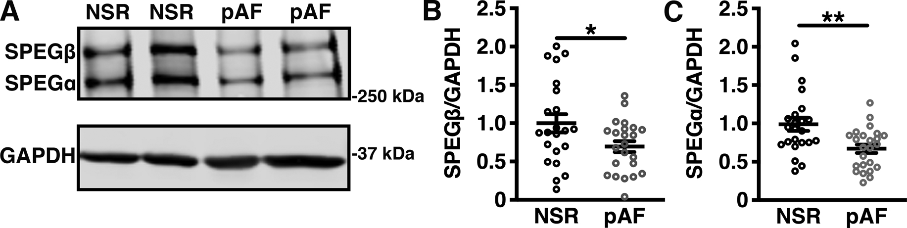
Representative western blots (A) and bar graph quantifications of (B) SPEGβ and (C) SPEGα protein levels relative to GAPDH loading control in right atrial appendage biopsies from patients in paroxysmal AF (pAF; n=23) or normal sinus rhythm (NSR; n=22). Significance determined using Student’s t-test. *P<0.05. **P<0.01.