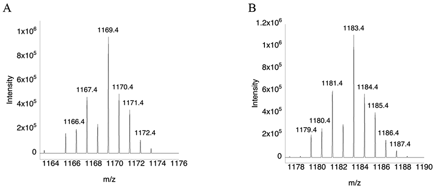
Figure 3: Mass spectra of the C-terminus redox center peptide for the WT and (αMe)Sec-containing mutant.

(A) Mass spectrum of peptide SGLEPTVTGCUG that results from a trypsin digest. (B) Mass spectrum of peptide SGLEPTVTGC(αMe)UG that results from a trypsin digest. The mass peaks differ by 14 daltons due to the presence of the αMe group. The sample were not reduced and alkylated prior to MS analysis, so Cys/Sec residues exist in the oxidized form as a selenosulfide bridge.
