Abstract
The current global severe acute respiratory syndrome coronavirus 2 (SARS-CoV-2) pandemic has magnified the risk to healthcare providers when inititiating airway management, and safe tracheal intubation has become of paramount importance. Mitigation of risk to frontline providers requires airway management to be an orchestrated exercise based on training and purposeful simulation. Role allocation and closed-loop communication form the foundation of this exercise. We describe a methodical, 10-step approach from decision-making and meticulous drug and equipment choices to donning of personal protective equipment, and procedural concerns. This bundled approach will help reduce unplanned actions, which in turn may reduce the risk of aerosol transmission during airway management in resource-limited settings.
BACKGROUND
The emergence of the severe acute respiratory syndrome coronavirus 2 (SARS-CoV-2), or COVID-19, pandemic has brought infections that are transmitted via droplet and aerosol under the spotlight.1 Infections such as influenza A subtype H1N1, Nipah virus infection, Ebola virus disease, and multidrug-resistant tuberculosis are equally contagious and pose a significant risk to healthcare professionals, especially those involved in airway management.2,3 Herin, we describe a step-by-step approach to endotracheal intubation of critically ill patients with suspected or confirmed COVID-19 and other airborne diseases with the goal of limiting the risk of exposure to healthcare providers.
CONCEPTS
Rapid sequence intubation (RSI) and invasive mechanical ventilation are preferred. Non-invasive positive pressure ventilation (NIPPV) increases the risk of aerosol generation; NIPPV has been associated wth increased risk of healthcare worker infection and hence should be avoided.4–6
-
The care area is divided as follows:
Hot zone: A three-meter [9.85 feet] radius around the patient
Warm zone: The area between hot and cold zone where decontamination takes place
Cold zone: The outermost noncontaminated area.
The intubation team members are described in Table 1.
Encourage closed-loop communication.
Table 1.
Role Allocation and Personnel Details of intubation team.
| S. No | Personnel | Stationed in | Responsibility |
|---|---|---|---|
| 1 | Team leader | Hot Zone (3-meter radius) | Performs tracheal intubation |
| 2 | Registered respiratory therapist | Hot Zone | Oversees airway and ventilator equipment |
| 3 | Registered nurse | Hot Zone | Ensures IV access and administers IV medications |
| 4 | Infection control nurse | Warm Zone | Oversees procedure and protocols |
IV, intravenous; TL, Team Leader; RRT, Registered Respiratory Therapist; RN, Registered Nurse; ICN, Infection Control Nurse.
Population Health Research Capsule.
What do we already know about this issue?
Several protocols for intubation during the COVID-19 pandemic aim at reducing transmissibility of infections by using sophisticated equipment.
What was the research question?
How can we reduce the risk of aerosol hazards from infectious diseases transmitted during intubation?
What was the major finding of the study?
Execution of intubation in suspected aerosol-transmitted infections can be performed systematically in low-resource settings.
How does this improve population health?
The protocol is aimed at safeguarding healthcare professionals against aerosol hazards while performing airway interventions.
STEPS OF MIST (Modified Intubating Sequence for Transmissibility) BUNDLE
-
Pre-assessment phase and pre-briefing phase – Cold Zone
Step a. Review patient clinical data to determine appropriateness of endotracheal intubation and mechanical ventilation for the patient.
Step b. Team leader (TL) debriefs the intubation plan to the team to avoid unplanned and unarticulated actions.
Step c. Infection control nurse (ICN) alerts team to any breach of protocol or infection control practice.
-
Preparatory phase – Cold Zone
Step a. Use continous positive airway pressure mode with non-invasive ventilation (NIV) mask for preoxygenation. The registered respiratory therapist (RRT) assembles the ventilator with its circuit including preparation of the NIV mask with a viral filter and checks for possible leaks and disconnections. Ventilator settings: pressure support 0 centimeters of water, positive end expiratory pressure as per the requirement, and fraction of inspired oxygen to 100%. Deselect the apnea setting.
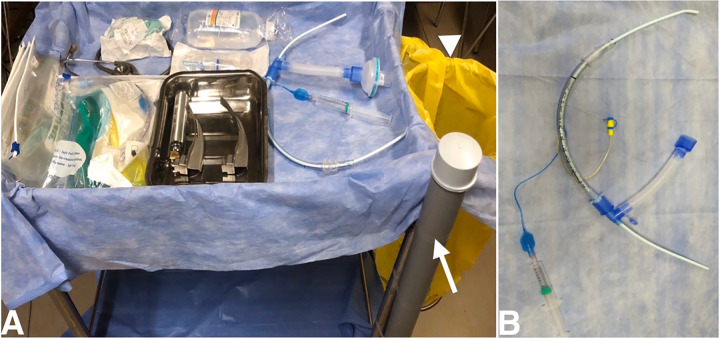
Step b. Review the equipment required for intubation (Figure 1A) (Table 2); the registered nurse (RN) loads pre-calculated doses of RSI medications (Table 3).
Step c. The assembly (Figure 1B) of the endotracheal tube (ETT) should be preset with a catheter mount containing a viral filter, and an inflation syringe with an intubating bougie.
-
Preoxygenation Phase: Hot Zone
Step a: The RRT ensures wall-mounted suction unit is properly connected. A Yankauer suction connected to the wall-mounted suction unit should be available, but its usage should be judicious. The suction tip, if used, should be disposed of in a Ziploc bag.
Step b: TL at the head of the bed places the NIV mask with the viral filter onto the patient and ensures proper sealing to avoid leak. The RRT “starts” the ventilator and preoxygenates until adequate oxygen saturation is attained. Avoid bag-valve mask for preoxygenation. Meticulous preoxygenation should be done for 3–5 minutes.
Step c: RN ensures patent intravenous access, assesses the vitals periodically, and communicates them to the TL.
-
Peri Induction phase: Hot Zone
Step a: RN administers the pre-calculated dose of the induction agent followed by the paralytic agent to the patient.
Step b: Approrpiate patient positioning should be performed to maximise safe apnoea time.
-
Peri-intubation Phase: Hot Zone
Step a: The RRT sets the ventilator on standby mode b after adequate paralysis and oxygen saturation is achieved.
Step b: TL subsequently removes the NIV mask, which is disconnected from the ventilator by the RRT and placed in a Ziploc bag.
Step c: TL performs laryngoscopy. During this time, the RRT is required to change the settings of the ventilator to “assist control mode ventilation.”. Once the vocal cords are visualized, the RRT hands over the intubating unit to the TL who should then pass the bougie between the cords under direct visualization. Video laryngoscope is a preferred choice for intubation of such patients, if available.
Step d: The RRT assists in guiding the ET over the bougie and should subsequently inflate the cuff with the pre-filled air syringe.
Step e: The RRT then removes the bougie and disposes of it in the preset disposition system (Figure 1A).
Step f: The RRT then proceeds to connect the ET to the ventilator and convert the ventilator from standby to its preset settings. Simultaneously, the TL removes the laryngoscope and places it in a Ziploc bag.
Step g: The RN confirms the position of the tube with five-point auscultation, following which the stethoscope should be disposed of in the Ziploc bag. End-tidal carbon dioxide confirmation is advised, if available.
-
Post-intubation Phase: Hot Zone
Step a: Continue ventilation and monitor hemodynamics. Initiate early sedation and analgesia.
Step b: Ensure all soiled equipment has been disposed of appropriately into the yellow bag for decontamination (Figure 1A).8
Step c: The order of doffing and decontamination is TL, followed by the RN, and then the RRT who should be separately overviewed and monitored by the ICN.
Figure 1.
A. List of equipment required in intubation trolley. PVC pipe sealed at one end (white arrow) filled with 1% sodium hypochlorite solution is used for discarding the soiled bougie and yellow bag (arrow head) for discarding the soiled wastes. B. Intubation unit.
Table 2.
List of equipment required in intubation trolley.
| Equipment | Numbers |
|---|---|
| Intubation unit (Figure 1B) | 1 |
| Macintosh laryngoscope with size 3 and 4 blades in a sterile tray | 1 each |
| iGel | 2 |
| Stethoscope | 1 |
| Cuffed ETT, size 7 | 1 |
| ETT fixator | 1 |
| IV fluid with infusion set | 1 |
| Ziploc bags | 5 |
ETT, endotracheal tube; IV, intravenous.
Table 3.
List of drugs used in Intubation.
| Drugs | Dose |
|---|---|
| Inducing agent | |
| Inj. etomidate or | 0.3mg/kg IV |
| Inj. ketamine | 1–2mg/kg IV |
| Paralytic agent | |
| Inj. rocuronium | 1–1.2mg/kg IV |
Inj, injection; mg, milligram; kg, kilogram; IV, intravenous.
CONCLUSION
This sequence should guide healthcare professionals to minimize aerosol and droplet transmission during intubation and expedite better patient care. This approach does not involve significant resource intensification and can be done in resource-limited settings.
Supplementary Information
Footnotes
Section Editor: Gabriel Wardi, MD
Full text available through open access at http://escholarship.org/uc/uciem_westjem
Disclaimer: Due to the rapidly evolving nature of this outbreak, and in the interests of rapid dissemination of reliable, actionable information, this paper went through expedited peer review. Additionally, information should be considered current only at the time of publication and may evolve as the science develops.
Conflicts of Interest: By the WestJEM article submission agreement, all authors are required to disclose all affiliations, funding sources and financial or management relationships that could be perceived as potential sources of bias. No author has professional or financial relationships with any companies that are relevant to this study. There are no conflicts of interest or sources of funding to declare.
REFERENCES
- 1.Van Doremalen N, Bushmaker T, Morris DH, et al. Aerosol and surface stability of SARS-CoV-2 as compared with SARS-CoV-1. N Engl J Med. 2020;382:1564–7. doi: 10.1056/NEJMc2004973. [DOI] [PMC free article] [PubMed] [Google Scholar]
- 2.Li Q, Guan X, Wu P, Wang X, et al. Early transmission dynamics in Wuhan, China, of novel coronavirus-infected pneumonia. N Engl J Med. 2020;382:1199–1207. doi: 10.1056/NEJMoa2001316. [DOI] [PMC free article] [PubMed] [Google Scholar]
- 3.Tang JW, Tambyah PA, Hui DS. Emergence of a novel coronavirus causing respiratory illness from Wuhan, China. J Infect. 2020;80(3):350–71. doi: 10.1016/j.jinf.2020.01.014. [DOI] [PMC free article] [PubMed] [Google Scholar]
- 4.Peng PW, Ho PL, Hota SS. Outbreak of a new coronavirus: what anaesthetists should know. Br J Anaesth. 2020;124(5):497–501. doi: 10.1016/j.bja.2020.02.008. [DOI] [PMC free article] [PubMed] [Google Scholar]
- 5.Cheung JC, Ho LT, Cheng JV, Cham EY, et al. Staff safety during emergency airway management for COVID-19 in Hong Kong. Lancet Respir Med. 2020;8(4):e19. doi: 10.1016/S2213-2600(20)30084-9. [DOI] [PMC free article] [PubMed] [Google Scholar]
- 6.Detsky ME, Jivraj N, Adhikari NK, et al. Will this patient be difficult to intubate? The rational clinical examination systematic review. JAMA. 2019;321(5):493–503. doi: 10.1001/jama.2018.21413. [DOI] [PubMed] [Google Scholar]
- 7.World Health Organization. Clinical management of severe acute respiratory infection (SARI) when COVID-19 disease is suspected - Interium Guidance. [Accessed March 29, 2020]. Available at: https://www.who.int/publications-detail/clinical-management-of-severe-acute-respiratory-infection-when-novel-coronavirus-(ncov)-infection-is-suspected.
- 8.World Health Organization. Safe management of wastes from health-care activities: a summary. [Accessed March 29, 2020]. Available at: https://www.who.int/water_sanitation_health/publications/wastemanag/en/
Associated Data
This section collects any data citations, data availability statements, or supplementary materials included in this article.


