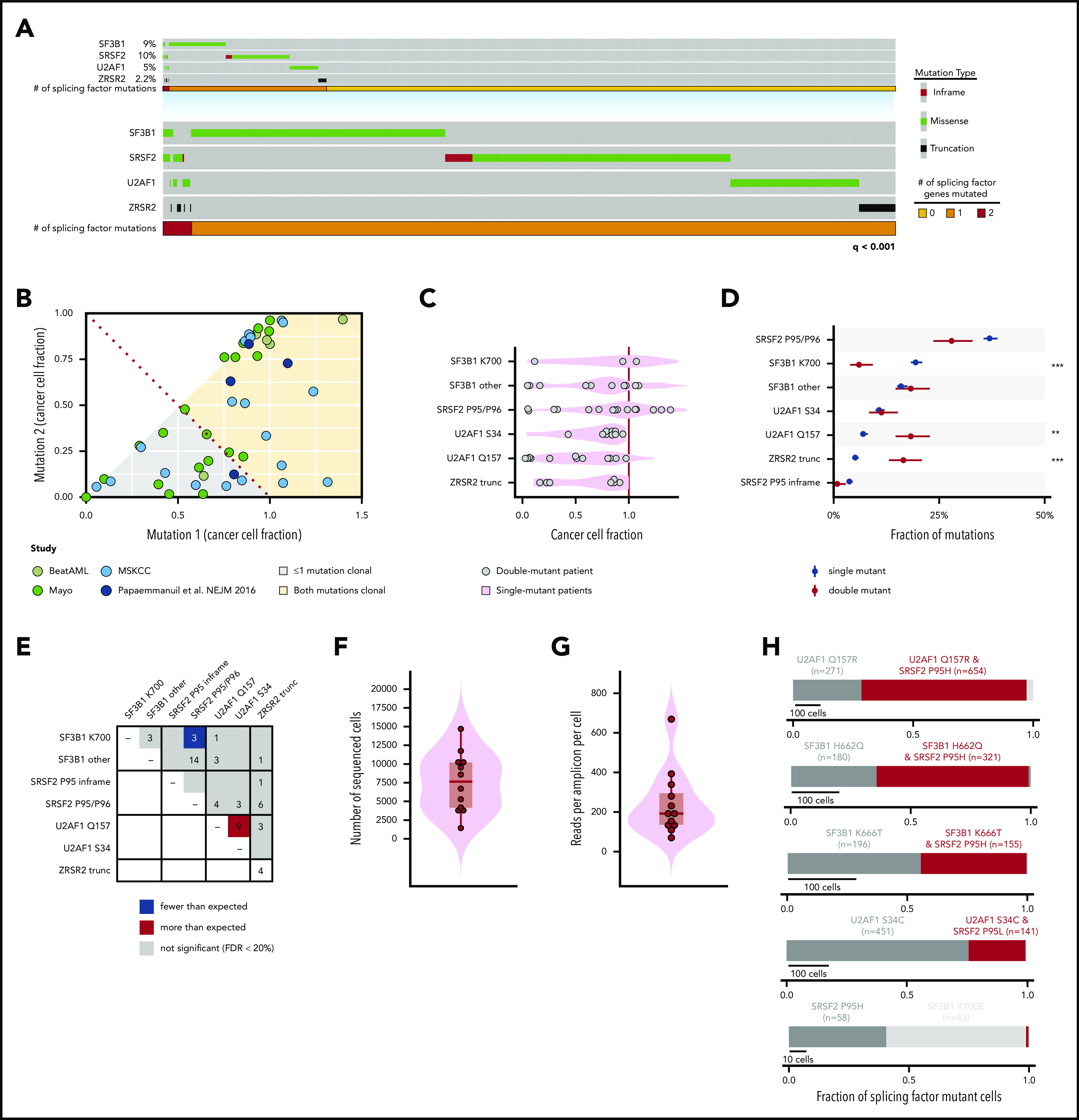
Figure 1.

Genetic features of patients harboring 2 concomitant mutations in RNA splicing factors at the bulk and single-cell level. (A) Oncoprint of hotspot mutations in SF3B1, SRSF2, an U2AF1, as well as clearly deleterious mutations (nonsense or frameshift mutations) in ZRSR2 across 4231 patients. Each column represents 1 patient. The number of patients with 0, 1, or 2 splicing factor mutations is shown in yellow, orange, and red, respectively. Overall, mutations in each gene exhibited strong mutual exclusivity (q < .001; Fisher’s exact test). (B) CCF of each mutant splicing factor from genomic DNA sequencing of a cohort of 58 dual mutant splicing factor samples, including those from a single study.16 (C) CCF of mutations at SF3B1K700, other residues of SF3B1 (SF3B1 other), SRSF2P95/P96, U2AF1S34, and U2AF1Q157 as well as ZRSR2 truncation mutations (ZRSR2 trunc). (D) Percentage of patients with single or double splicing factor mutations (in black and red, respectively) with mutations in SRSF2, SF3B1, U2AF1, and ZRSR2. Error bars: 1 standard deviation, based on a binomial distribution. **P < .005; ***P < .0005 (Fisher’s exact test). (E) Plot describing the number of patients with coexisting mutant alleles in splicing factors. The expected number was based on the fraction of samples with exactly 2 mutations under the assumption of no mutual exclusivity and using a Poisson distribution. The distribution of the number of (F) total sequenced cells per patient and (G) reads per amplicon per cell from single-cell genomic DNA sequencing. Each point represents a sample from a unique patient. (H) Fraction of mutated cells with 1 or 2 mutations in RNA splicing factors within each patient with a dual splicing factor mutation. Red bar denotes fraction of individual cells where 2 splicing factor mutations were identified within the same cell. The number of cells containing each mutation is indicated.
