Abstract
Pancreatic cancer remains a lethal type of cancer with poor prognosis. Molecular classification enables in-depth, precise prognostic assessment. This study is aimed at identifying a robust and simple mRNA signature to predict the overall survival (OS) of pancreatic cancer (PC) patients. Differentially expressed genes (DEGs) between 45 paired pancreatic tumor samples and adjacent healthy tissues were selected. For risk determination, a LASSO Cox regression model with DEGs was used to generate the OS-associated risk score formula for the training cohort containing 177 PC patients. Another five independent datasets were used as the testing cohort to determine the predictive efficiency for further validation. In total, 441 DEGs were selected after considering the enrichment of classical pathways, such as EMT, cell cycle, cell adhesion, and PI3K-AKT. A five-gene signature for risk discrimination was established with high efficacy using LASSO Cox regression in the training group. External validation showed that patients identified by the gene expression signature to be in the high-risk group had poorer prognosis compared with the low-risk patients. Further investigation identified the differential epigenetic modification patterns of the five genes, which indicated their roles in tumor progression and their effect on therapy. In conclusion, we constructed a robust five-gene expression signature that could predict the OS of PC patients, offering a new insight for risk discrimination in daily clinical practice.
1. Introduction
Although great improvements have been achieved in detection and treatment of many types of highly malignant tumors, such as lung and breast cancers, the overall survival (OS) and prognosis of pancreatic cancer (PC) remain poor, with a five-year survival rate of only around 8% [1]. Surgical resection offers the only chance for long-term survival, since PC is naturally resistant to chemo- and radiotherapy. Only about 20% of patients have the opportunity to receive surgical resection, and the median OS is only around 24 months [2]. In daily clinical practice, TNM staging formulated by AJCC is used to determine the course of the treatment. However, even two patients at the same TNM stage may have totally different prognoses [3]. This means that clinical histopathological classification has inherent limitations in predicting the prognosis for PC patients, and thus, identification of new biomarkers for prognostic assessment is urgently required [4].
With the development of next-generation sequencing, genetic markers have been pursued for cancer classification, and these have come to play an important role in the assessment of prognosis and the best course of treatment. The term “molecular subtypes” refers to tumors with similar morphology but with very different clinical features. Although molecular subtyping is a highly complex system, it is the major diagnostic and prognostic strategy in clinical practice [5]. For instance, breast cancer is divided into different subtypes based on the markers ER, PR, and HER2, and each subtype is associated with different treatment modalities and overall survival. A similar pattern has been determined for EGFR and ALK in non-small-cell lung adenocarcinoma [6, 7]. However, the much-needed investigation of PC subgroups is still in its infancy. Scholars previously attempted to classify PC according to the expression patterns of single genes based on studies which showed the relevance of these genes to OS. However, despite the promising progress in the laboratory, no significant improvement has been achieved in the clinic.
Transcriptomic sequencing has provided new opportunities and already helped make some achievements, in PC classification. On the one hand, Collisson et al. classified PC into three subtypes based on a 62-mRNA gene expression signature and named them classical, quasimesenchymal, and exocrine-like tumors. These three groups differed in the survival time of the patients and sensitivity to chemotherapeutics [8]. On the other hand, Moffitt et al. divided PC into two subtypes, basal-like and stromal, of which the former one had a worse prognostic outcome. Stromal subtypes were further divided into two groups, “normal” and “activated,” which immensely differed from each other in terms of their prognosis [9]. Based on these two studies, Bailey et al. used RNA-seq of 96 genes and 232 microarray data and identified ten key signaling pathways in PC. They then accordingly divided PC into four subtypes—squamous, immunogenic, pancreatic progenitor, and ADEX [10]. Wartenberg et al. classified PC into three subtypes according to their immune status: immune-rich, immune-exhausted, and immune-escape. These three groups greatly differed from each other in their prognostic outcomes [11].
Molecular subtype classification based on gene expression signatures can be used for prognostic and therapeutic assessment, including surgery and chemotherapy options, even potentially during early stages of the disease. In this respect, there has already been some progress in other tumor types. For instance, Li et al. established a four-miRNA signature for predicting trastuzumab's effect on HER2-positive breast cancer patients [12]. Zhou et al. established a seven-miRNA detection system for early diagnosis of hepatocarcinoma, and this system is currently in use for diagnostic assessment in clinics [13]. However, a similar approach has not been comprehensively pursued for PC, and there have been only a few studies. Taking this into consideration, we used meta-analysis on pooled datasets, totaling 875 PC patients' samples. The entire dataset included transcriptomic sequencing data, survival information, and epigenetic background. Using this, we determined a multigene expression signature that predicts the OS and analyzed the mechanism underlying this pattern for the development of potential therapies in the future.
2. Materials and Methods
2.1. Data Availability
The raw gene expression data and corresponding clinical information of pancreatic patients were downloaded from the Gene Expression Omnibus (GEO, https://www.ncbi.nlm.nih.gov/gds/). The processed TCGA data was derived from UCSC-Xena (https://xenabrowser.net/). These samples had been profiled using whole-genome DNA microarray (Affymetrix or Agilent) and RNA-seq (Illumina). The datasets contained 875 patient data, including 555 patients with available survival data. The dataset of TCGA was used as the training cohort after removing the patients that lacked survival data information. The dataset of GSE28735 was processed to obtain the differentially expressed genes between pancreatic cancer and adjacent normal tissues. The datasets of GSE21501, GSE57495, GSE62165, GSE62452, GSE79668, and Bailey et al., 2016 served as the independent validation cohorts. The information about all the datasets is shown in Table 1 and Supplementary Table 3.
Table 1.
Characterization of the included datasets.
| Datasets | Study | Platform | Cases | Description |
|---|---|---|---|---|
| GSE28735 | Zhang et al. 2012 | Affymetrix HG-1.0 ST | 90 | 45 pairs of tumor with adjacent healthy tissues |
| GSE21501 | Stratford et al. 2010 | Agilent-014859 WHG 4X44K | 132 | 102 patients with survival data |
| GSE57495 | Chen et al. 2015 | Affymetrix | 63 | 63 patients with survival data |
| GSE79668 | Kirby et al. 2016 | RNAseq, Illumina Hiseq2000 | 51 | 51 patients with survival data |
| Bailey_ICGC_PACA_AU | Bailey et al. 2016 | RNAseq Illumina | 96 | 96 patients with survival data |
| GSE62165 | Janky et al. 2016 | Affymetrix HG-U219 | 131 | 118 surgically resected PDAC and 13 healthy tissues |
| GSE62452 | Yang et al. 2014 | Affymetrix HG-1.0 ST | 130 | 66 patients with survival data; 69 tumors + 61 nontumors |
| TCGA | TCGA_PAAD_UCSC_Xena | RNA Illumina v3 | 182 | 177 patients with survival and clinical data |
2.2. Normalization and Annotation of GEO Data and TCGA Data
First, we normalized each DNA microarray-based dataset using the Robust Multichip Average (RMA) method for the raw Affymetrix data derived from GEO. Then, we mapped hybridization probes across the different technological platforms with the corresponding SOFT-formatted family files in R. When multiple probes were mapped to the same gene symbol, we calculated the average expression of the genes in the dataset. For the data from the Agilent; TCGA; Bailey et al., 2016; GSE62165; and GSE79668 datasets, we used the available normalized data. The datasets above were log2-transformed.
2.3. Selection of the Differentially Expressed Genes and Construction of LASSO Cox Regression Model
The GSE28735 dataset consisted of 45 pairs of pancreatic tumor and adjacent healthy tissues. We used these datasets to identify the differentially expressed genes. The genes with significant expression differences were defined based on the following parameters: FDR < 0.05, ∣log2fc | >1. Having identified 441 differentially expressed transcripts, we sought to establish an association with patient outcomes. Then, we merged the differently expressed gene list derived from the GSE28735 dataset with the TCGA (n = 177) dataset to generate the training cohort. Next, the logical regression analysis with Least Absolute Shrinkage and Selection Operator (LASSO) was applied to select the gene expression signature [14], which is a selection method that handles the high-dimensional regression variables with no prior feature selection step by shrinking all regression coefficients toward zero and thus forcing many regression variables to be exactly zero. The penalty regularization parameter lambda was chosen via 10-fold cross-validation cv. glmnet, which is implemented in the R package glmnet [15]. The lambda was finalized using the lambda.min = 0.2215, which is the value of lambda giving the minimum mean cross-validated error. Finally, we obtained five-gene expression signatures and corresponding coefficients.
2.4. Establishment of the Risk Score Formula
Based on the expression levels of the five genes, a formula was constructed to calculate an OS risk score for each patient as follows:
| (1) |
In our risk score formula, N (N = 5) is the number of genes, Exp is the expression value of each gene, and Coefficient is their corresponding coefficient from the LASSO Cox regression. In this case, we would be able to generate a risk score for each patient, from which patients could be divided into high- and low-risk score groups with an optimal cutoff score determined by X-tile plots [16] based on the association with OS.
2.5. Receiver Operating Characteristic (ROC) Curve Analysis
Based on the LASSO Cox regression, a group of four genes were selected; ROC was employed to demonstrate the sensitivity and specificity of different variables by risk score. The prognosis performance was evaluated using a time-dependent receiver operating characteristic (ROC) curve analysis [17]. In order to evaluate the predictive accuracy and robustness of our prognostic model, AUC at 1 year, 2 years, and 3 years was calculated in the training and different validation cohorts according to the five-gene expression signature. The spanning parameter of the NNE approach was span =0.25 × nobs−0.20, which was performed in R package survivalROC [17].
2.6. Overall and Stratified Survival Analysis
According to the risk score formula, we divided each patient into high- or low-risk groups with the optimal cutoff value derived from the training cohort. The Kaplan-Meier method was used to assess the difference in the survival rates of high- and low-risk patients. Then, univariate and multivariate Cox regression survival analysis was performed to evaluate the various clinicopathological features, such as age, gender, tumor stage, and grades. Moreover, to further explore the impact of clinical pathological features on the value of risk score, stratified survival analysis related to age at the time of diagnosis (>60 or ≤60), gender (male or female), AJCC stage (I/IIA or IIB/III/IV), T stage (T1/T2 or T3/T4), N stage (N+ or N-), and histological grade (G1/G2 or G3/G4) was conducted. A P value < 0.05 according to the log-rank test was considered significant. The hazard ratio (HR) and 95% confidence interval (CI) were calculated. All of these statistical analyses were performed in R or corresponding R packages survival and survminer.
2.7. Pathway Enrichment Analysis
The enrichment analysis was performed to predict the biological processes and KEGG pathways of the DEGs in an online tool—Metascape [18]. The GSEA was shown to predict the hallmarks of the tumor and healthy pancreas enrichment [19]. Both DEGs and GSEA input data were derived from GSE28735.
2.8. Epigenetic Modification Analysis
DNA methylation data is from TCGA-PAAD Illumina 450K methylation microarray. The histone modification ChIP-seq data (H3K4me3, H3K27ac) were derived from GEO and ENCODE [20] (data accession: GSM3376452, GSM2466034, GSM1574235, ENCSR520BIM, GSM2700597, ENCSR-596PFU, GSM945261, GSM2286771, GSM1574256, ENCSR876DCP, and ENCSR554RQQ). The Cistrome Data Browser [21], which has one pipeline to process all containing ChIP-seq data, was used to link WashU Browser online visualization.
3. Results
3.1. Preparation of Clinical Pancreatic Disease Datasets and Construction of the Workflow
A total of 875 datasets covering 555 patients with available survival data were included in this study (Table 1). The dataset of GSE28735 was used for the selection of differentially expressed genes (DEGs) and functional enrichment analysis. For this, we used the TCGA-PAAD dataset, which contained 177 PC patients with detailed survival data, combined with the selected DEGs to construct a gene expression signature prognostic risk score model based on the LASSO Cox regression. The GSE21501 (102 patients), Bailey et al., 2016 (96 patients), GSE57495 (63 patients), GSE62165 (131 patients), GSE62452 (66 patients), and GSE79668 (51 patients) cohorts were used for further validation. The workflow of the experimental strategy is shown in Supplementary Figure 1.
3.2. Selection of DEGs between Pancreatic Cancer and Adjacent Normal Tissues
In order to select specific genes associated with pancreatic tumorigenesis, we first used the GSE28735 dataset, which contained the gene expression information of 45 pairs of pancreatic cancer and adjacent normal tissues. The supervised analysis compared the expression profiles of the 45 pairs of pancreatic cancer and adjacent normal tissues using the paired t-test in R. A false discovery rate (FDR) was applied to correct for the multiple testing hypothesis, and the significant genes were selected by the following threshold: FDR < 0.05, ∣log2 (fold change) | >1. The results indicated that 441 genes were differentially expressed between the two groups (Figure 1(a)). Of these, 238 upregulated genes were involved in cell adhesion and cell cycle by Gene Ontology (GO) analysis, while 203 genes related to secretion function were downregulated compared to the tumor with normal tissues. Further analysis by gene set enrichment analysis (GSEA) showed that the p53, cell cycle, cell adhesion junction, PI3K-AKT-mTORC, Notch, TGFβ, epithelial-mesenchymal transition (EMT), and other cancer-related signaling pathways were enriched in the tumors, which was in accordance with previous studies (Figures 1(c)–1(e) and Supplementary Figure 2A-L).
Figure 1.
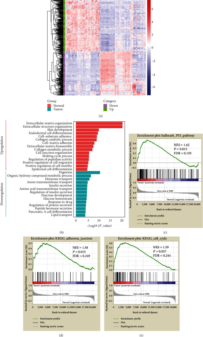
Identification of the genes differentially expressed between tumor and normal tissues. (a) Hierarchical clustering of DEGs in 45 paired PC (in green) and adjacent normal tissue samples (in red). Each row represents an individual differentially expressed gene, and each column represents an individual sample. Pseudocolors indicate relative expression levels from low to high on a log2 scale from -8 to 8; (b) GO biological process analysis for the upregulated and downregulated DEGs in Metascape online tool; (c–e) GSEA of the expression profile of the tumor samples in comparison to that of normal tissues from the GSE28735 dataset.
3.3. Establishment of a Five-Gene Expression Signature in Pancreatic Cancer
To identify the mRNAs associated with OS in PC patients, we downloaded the transcriptome data from TCGA, which contained 177 patients with detailed survival information, for further investigation. The DEGs were merged with TCGA transcriptome data to form the training dataset. We observed collinearity among the DEGs (Figure 2(a)) in the training cohort, which would prejudice the results of traditional Cox regression analysis. Therefore, the LASSO Cox regression model selects the prognostic mRNAs to predict the survival-associated genes (Figures 2(b) and 2(c)). Finally, five genes out of 441 DEGs were selected: CHGA, COL17A1, ITGB6, LAMC2, and S100P (Table 2). Among these, COL17A1, ITGB6, LAMC2, and S100P were upregulated in tumor tissues, whereas only CHGA was downregulated. The five-gene expression levels were further validated in an independent cohort (GSE62165), which contains 118 neoplastic and 13 normal tissues. There was a significant differential expression in pancreatic cancer patients compared to normal ones (P < 0.001), suggesting that they might be a potential biomarker signature for pancreatic patients (Figures 3(a)–3(e)). Next, in order to explore the interaction network of the identified gene signature, we generated a protein-protein interaction network (PPI) from DEGs using the STRING online tool. Clustered by MCODE algorithm [22] in Cytoscape [23], two modules with the five-gene signature were selected (Figure 4); each module protein might form a large complex to regulate some biological process. The GO and KEGG analysis showed that the Cluster 1 genes were significantly enriched in the cell adhesion and ECM-receptor interaction signal pathways, while the Cluster 2 genes participated in the pancreatic secretion process (Supplementary Figure 3).
Figure 2.
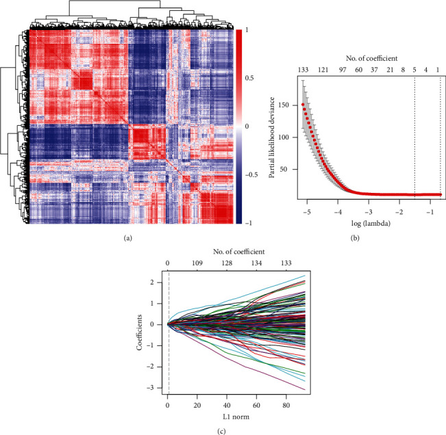
Establishment of a gene expression signature for overall survival prediction in the training cohort. (a) Hierarchical clustering shows the collinear expression of the DEGs. A correlation matrix heatmap of DEGs in the training cohort, in which each cell represents the Pearson correlation between the row and column of DEGs, the heatmap bar color along with the change of correlation coefficient from -1 to 1; (b, c) selection of the OS-associated genes by the LASSO Cox regression model. LASSO coefficient profiles of 441 differentially expressed associated genes. Each curve corresponds to a gene; the vertical line is drawn at the value lambda = 0.2215 chosen by 10-fold cross-validation.
Table 2.
Characterization of the five candidate genes in hg 19 genome.
| Gene symbol | Description | Expression status | Coordinate | P valuea | χ 2 b | Coefficientc |
|---|---|---|---|---|---|---|
| CHGA | Chromogranin A | Down tumor/normal | Chr14:93389445-93401638 | <0.0001 | 18.35 | -0.0481 |
| COL17A1 | Collagen type XVII alpha 1 chain | Up tumor/normal | Chr10:105791046-105845638 | <0.0001 | 21.05 | 0.0402 |
| ITGB6 | Integrin subunit beta 6 | Up tumor/normal | Chr2:160956182-161110349 | <0.0001 | 16.13 | 0.0697 |
| LAMC2 | Laminin subunit gamma 2 | Up tumor/normal | Chr1:183147952-183214262 | 0.00015 | 14.44 | 0.0021 |
| S100P | S100 calcium binding protein P | Up tumor/normal | Chr4:6695566-6698897 | 0.0023 | 9.256 | 0.0063 |
aDerived from the univariate Cox proportional hazards regression analysis in the training cohort (log-rank test). bDerived from the univariate Cox proportional hazards regression analysis in the training cohort (Chi2 test). cDerived from the LASSO Cox regression analysis coefficients in the training cohort.
Figure 3.
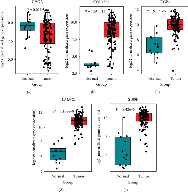
The expression of five genes in PC and normal tissues. (a) CHGA; (b) COL17A1; (c) ITGB6; (d) LAMC2; (e) S100P. P values were calculated by a t-test on the log2 RMA normalization of the expression data.
Figure 4.
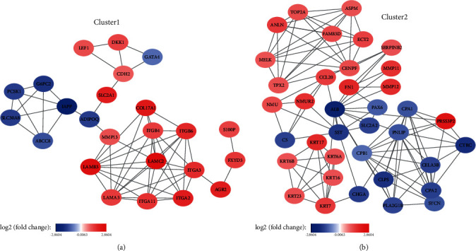
Five genes of protein-protein interaction (PPI) networks clustered by MCODE algorithm. (a) Cluster 1 module contains COL17A1, ITGB6, LAMC2, and S100P protein; (b) Cluster 2 module contains CHGA protein. The color of a node in the PPI network reflects the log2 (fold change) value of the gene expression.
3.4. Construction of Prognostic Risk Score Model for Long-Term Survival Prediction
Based on the expression levels of these five genes, the following risk score formula was generated for further evaluation from the TCGA training cohort: Risk score = −0.0481∗CHGA + 0.0402∗COL17A1 + 0.0697∗ITGB6 + 0.0021∗LAMC2 + 0.0063∗S100P. Using this formula, PC patients in the training cohort were divided into high- and low-risk score subgroups according to the optimal selected cutoff score (0.46) calculated by X-tile plots [16] based on their association with the OS (Supplementary Figure 4). Figure 5(a) indicates the division of PC patients into high- and low-risk groups by this formula, and Figure 5(b) shows the expression patterns of the five genes from low- to high-risk score groups. The results indicated that the distribution of mortality in the high-risk group was significantly higher than that in the low-risk group (67.2% vs. 42.7%, P < 0.0001, Figures 5(c) and 5(d)). In addition, Kaplan-Meier analysis indicated that patients with high- and low-risk scores had a median OS time of 15.6 months and 24.6 months, respectively (HR = 2.46, 95%CI = 1.62‐3.73, P < 0.0001, Figure 5(e)), and the median disease-free survival time was 18.5 vs. 40.3 months (HR = 1.73, 95%CI = 1‐2.98, P = 0.045, Figure 5(f)). To know about the prognostic efficiency of our model with the survival time, we performed the time-dependent ROC curve analysis. The ROC 1-, 2-, and 3-year survival predicted by the risk score is depicted, with AUCs of 0.654 (1-year), 0.615 (2-year), and 0.651 (3-year), respectively (Supplementary Figure 5A). These results imply that the five-gene expression signature has relatively high sensitivity and specificity in predicting the OS of PC patients.
Figure 5.
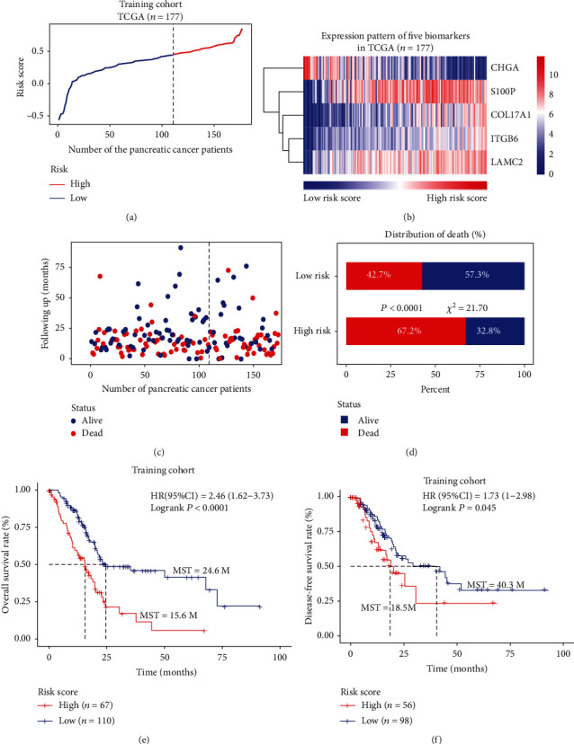
The five-gene signature-based risk score in the prognosis of survival in the training cohort. (a) Risk score distribution of the final five-gene expression signature in high- and low-risk groups; (b) the expression profiles of the five-gene signatures from low- to high-risk score; (c) the vital survival status of patients in the high- and low-risk cohorts; (d) distribution of mortality rate in high- and low-risk score groups. (e) Kaplan-Meier analysis with log-rank test for overall survival of the PC patients in the high- and low-risk score groups; (f) Kaplan-Meier analysis with log-rank test for disease-free survival of the PC patients in the high- and low-risk score groups.
3.5. External Validation of the Five-Gene Prognostic Signature with Different PC Datasets
To further validate the efficiency of this five-gene expression signature, we applied the formula and cutoff to five external independent validation datasets. Patients of each cohort were then divided into high- or low-risk subgroups. In the GSE21501 cohort, the five-gene signature expression pattern and OS analysis were similar to those in the training cohort (P < 0.001, HR = 2.61, 95%CI = 1.47‐4.62, Figures 6(a) and 6(b)). In the Bailey et al., 2016 cohort, the OS of patients discriminated by the gene expression signature was not statistically significant between the high- and low-risk groups (P = 0.064, HR = 2.62, 95%CI = 0.94‐7.25, Figures 6(c) and 6(d)). These hazard ratio values indicate that the gene expression signature could still be a potential risk factor in this cohort. Three other GEO datasets were also independently validated; the overall median survival time of high- and low-risk groups in the GSE57495 cohort was 16.2 months and 31.6 months, respectively (HR = 1.86, 95%CI = 1.0‐3.43, P = 0.047, Figures 6(e) and 6(f)). In the GSE62452 cohort, he median survival time of high- and low-risk groups was 13.8 months and 45.9 months (HR = 3.24, 95%CI = 1.46‐7.18, P = 0.003, Figures 6(g) and 6(h)), and in the GSE79668 cohort, they were 16.5 months and 96.9 months, respectively (HR = 4.44, 95%CI = 1.83‐10.73, P = 0.0009, Figures 6(i) and 6(j)). Time-dependent ROC was conducted in validation dataset analysis, showing a robust model constructed from our five-gene signature (Supplementary Figure 5B-E). The above results indicate the high predictive efficiency of this five-gene expression signature in PC patients.
Figure 6.
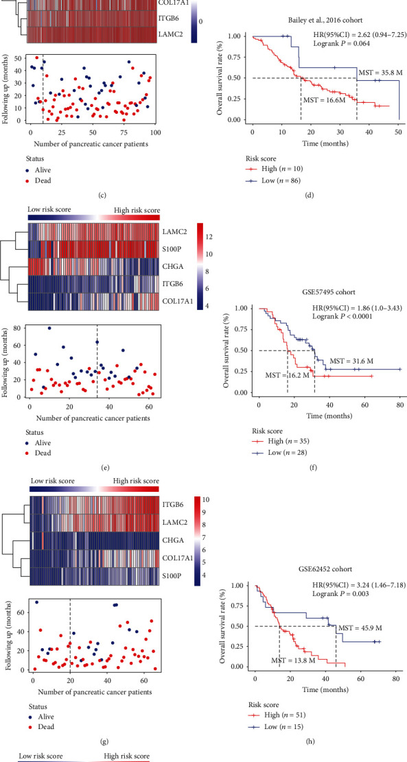
Performance assessment of the five-gene expression signature model in another five external independent validation datasets. (a, c, e, g, i) The heatmap and distribution of the five-gene expression profiles from low- to high-risk scores for the five external independent validation cohorts. (b, d, f, h, j) Kaplan-Meier overall survival analysis with log-rank test for the PC patients in high- and low-risk groups in five external independent validation cohorts.
3.6. Univariate and Multivariate Analysis Combined with Stratified Survival Analysis
Univariate and multivariate survival analysis was performed on the five-gene signature and clinicopathological features for OS. We found that the five-gene signature was an independent prognostic factor of PC patients between the training and external independent cohorts (Table 3, Supplementary Table 1). The univariate analysis showed that the AJCC, T, and N stages and histological grade had relatively significant impacts on prognosis. Therefore, we performed stratified survival analysis by the individual clinicopathological features to evaluate the prognostic values of our risk score model in the training cohort and external independent datasets. According to the results of stratified analysis (Supplementary Table 2), we concluded that this signature pattern could be further used to discriminate those patients in the relatively late-stage AJCC IIB-IV stages (Figure 7), T3/T4 tumors (Figures 8(a) and 8(b)), lymph node metastasis (Figures 8(c) and 8(d)), and lower-grade tumor G1 and G2 tumors (Figures 8(e) and 8(f)). This observation indicates that the five-gene expression signature also could be applied in clinicopathological subgroups, which, to some extent, indicated the reliability and general applicability of our risk score model.
Table 3.
Univariate and multivariate Cox regression analysis of five-gene signature and clinicopathological characteristics with overall survival in the training and another two external validation datasets.
| Variable | Training (TCGA) cohort | GSE21501 cohort | GSE79668 cohort | ||||||||||||
|---|---|---|---|---|---|---|---|---|---|---|---|---|---|---|---|
| N | Univariate | Multivariate | N | Univariate | Multivariate | N | Univariate | Multivariate | |||||||
| P | HR (95% CI) | P | HR (95% CI) | P | HR (95% CI) | P | HR (95% CI) | P | HR (95% CI) | P | HR (95% CI) | ||||
| AJCC stage | |||||||||||||||
| I-IIA vs. IIB-IV | 48/125 | 0.012 | 1.93 (1.15-3.24) | 0.68 | 1.37 (0.30-6.27) | 26/71 | 0.044 | 1.83 (1.0-3.29) | 0.059 | 1.83 (0.97-3.42) | 13/38 | 0.25 | 1.52 (0.74-3.1) | 0.98 | 0.97 (0.11-8.86) |
| T stage | |||||||||||||||
| T1/T2 vs. T3/T4 | 31/144 | 0.03 | 2.02 (1.07-3.81) | 0.87 | 1.06 (0.51-2.23) | 18/80 | 0.85 | 0.94 (0.51-1.74) | 0.24 | 0.68 (0.36-1.30) | 15/36 | 0.05 | 1.97 (0.97-3.98) | 0.36 | 1.48 (0.63-3.49) |
| N stage | |||||||||||||||
| N0 vs. N+ | 50/118 | 0.003 | 2.16 (1.28-3.65) | 0.58 | 1.48 (0.36-6.00) | 28/73 | 0.035 | 1.83 (1.04-3.22) | 0.087 | 1.74 (0.92-3.28) | 14/37 | 0.29 | 1.44 (0.72-2.87) | 0.96 | 0.97 (0.12-7.69) |
| Grade | |||||||||||||||
| G1/G2 vs. G3/G4 | 125/50 | 0.05 | 1.54 (0.99-2.37) | 0.53 | 1.17 (0.72-1.89) | ||||||||||
| Gender | |||||||||||||||
| Female vs. male | 80/97 | 0.31 | 1.24 (0.82-1.86) | 0.51 | 0.86 (0.55-1.35) | 19/32 | 0.501 | 1.24 (0.66-2.30) | 0.87 | 1.05 (0.55-2.01) | |||||
| Age (years) | |||||||||||||||
| >60 vs. ≤60 | 58/119 | 0.12 | 1.42 (0.90-2.24) | 0.13 | 1.49 (0.89-2.51) | 18/33 | 0.35 | 1.34 (0.72-2.49) | 0.61 | 1.19 (0.61-2.31) | |||||
| Signature | |||||||||||||||
| High vs. low risk | 67/110 | <0.0001 | 2.46 (1.62-3.73) | 0.002 | 2.05 (1.29-3.24) | 27/75 | <0.001 | 2.61 (1.47-4.62) | 0.005 | 2.31 (1.28-4.18) | 38/13 | 0.0009 | 4.44 (1.83-10.73) | 0.0044 | 3.84 (1.52-72) |
Figure 7.
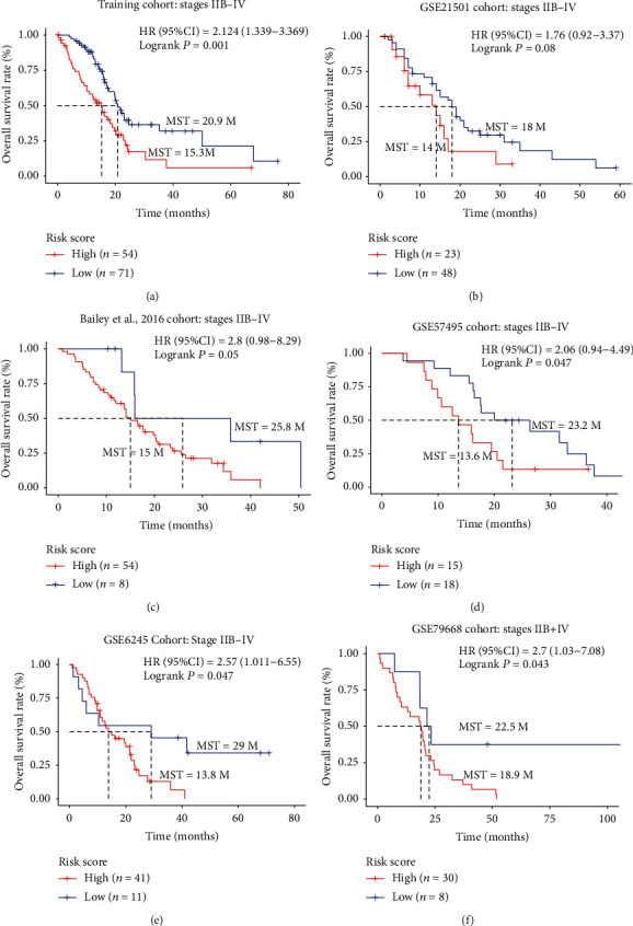
The association between five-gene signature and overall survival in patients with stages IIB-IV. The Kaplan-Meier survival curve of the training cohort (a) and the five external independent validation cohorts (b–f).
Figure 8.
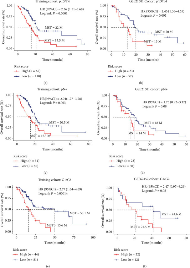
The five-gene signature was associated with prognosis in patients with advanced-stage cancer. (a, b) Kaplan-Meier analysis of the OS of patients with T3/4 stage in the training cohort and GSE21501 cohort; (c, d) Kaplan-Meier analysis of the OS of patients with lymph node metastasis in the training cohort and GSE21501 cohort; (e, f) Kaplan-Meier analysis of the OS of patients with grade 1/2 stage in the training cohort and GSE62452 cohort.
3.7. Epigenetic Regulation of the “Five Genes” in PC
In order to clarify the mechanism underlying the expression pattern of these five genes, and given that epigenetic modifications are highly related to tumorigenesis, we examined their epigenetic regulation by comparing promoter DNA methylation and histone modification markers of the five genes in pancreatic tumor and healthy cells. DNA methylation analysis showed that COL17A1, LAMC2, and S100P gene promoter methylation was downregulated (Figures 9(b), 9(d), and 9(e)), while CHGA gene promoter methylation was significantly upregulated (Figure 9(a)), which was in accordance with the gene expression pattern (Figure 3). Although methylation of the ITGB6 promoter was not obviously different (Figure 9(c)), the activated histone markers, H3K27ac and H3K4me3, were significantly upregulated in the ITGB6 promoter in the tumor cells (Figure 10(c)). Likewise, the promoter regions of the other upregulated genes, S100P, COL17A1, and LAMC2, were also associated with the activated chromatin state (Figures 10(b), 10(d), and 10(e)), while the downregulated gene CHGA lacked activated histone modification (Figure 10(a)). These observations presumably elucidate that the differential expression of these genes between healthy and PC cells was coregulated by multiple epigenetic factors.
Figure 9.
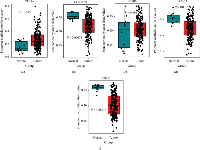
Promoter methylation status of five genes between PC and normal tissues in TCGA Illumina 450K methylation dataset: (a) CHGA; (b) COL17A1; (c) ITGB6; (d) LAMC2; (e) S100P.
Figure 10.
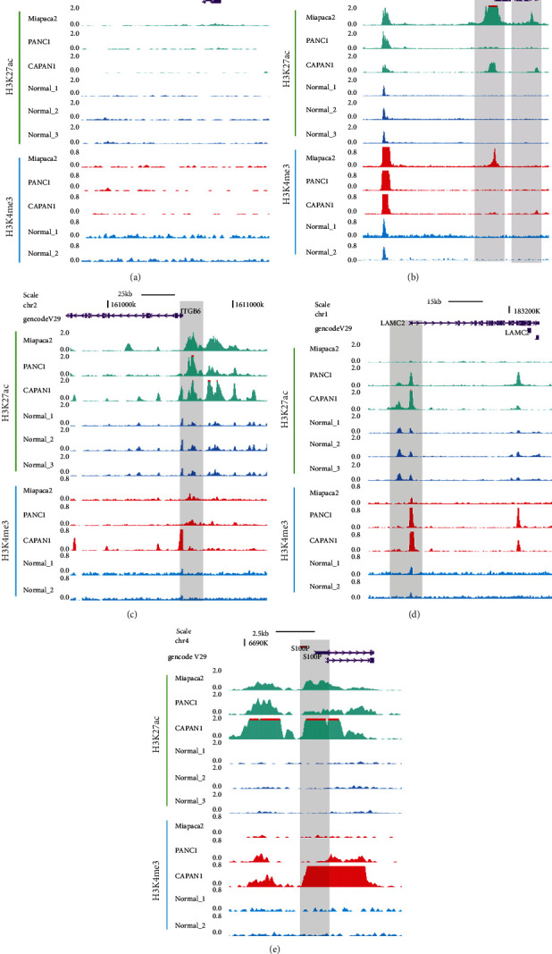
WashU Epigenome browser view of activated (H3K27ac and H3K4me3) histone modification tracks showing the promoter region around CHGA (a), COL17A1 (b), ITGB6 (c), LAMC2 (d), and S100P (e) in pancreatic cancer cell line (Miapaca2, PANC1, and CAPAN1) and normal tissue cells.
4. Discussion
Clinical histopathological parameters, such as TNM stage and the level of tumor differentiation, are currently used for prognostic prediction of PC patients. However, this system has obvious limitations due to the lack of understanding of tumor heterogeneity. Genetic molecular subtyping of PC is only in its infancy, but current progress has already shown its potential value to discriminate patients into different subtypes, related to very different OS and therapeutic response. Along the same line, based on the development of cancer genomics, use of gene expression signatures for clinical prediction has also made great progress. Haider et al. established a 36-gene expression signature for prognosis with satisfactory results [24]. Klett et al. reported a 17-gene subset that could be applied for prognostic evaluation and early diagnosis and could discriminate pancreatic cancer from nontumor tissues, pancreatic precursor lesions, and pancreatitis [25]. Currently, blood-based CA 19-9 is widely used for diagnosis and prognosis in PC patients; however, due to its low sensitivity and specificity, clinical prediction is not satisfactory. Furthermore, there are CA 19-9-negative patients due to limited Lewis antigen [26].
Genetic sequencing offers a new approach to precision medicine. Moreover, current targeted cancer therapy has essentially been established on the results of studies about gene detection [27–29]. We therefore attempted to construct a gene expression signature for prognostic assessment. This study was built on a clinical problem: some early-stage PC patients do not show a favorable survival rate even in comparison to the OS of late-stage PC patients who underwent resection. This observation indicates that histopathological classification is not sufficient for the prognostic and therapeutic assessment. We hypothesized that molecular differences between the samples categorized into the same groups by traditional approaches might be the underlying reason. Thus, prognostic assessment using a gene expression signature could allow patients to avoid unnecessary or even detrimental treatment modalities, such as operations, or chemotherapy. The ideal prognostic model should have high prediction efficiency with as few genes as possible, to increase clinical practicality. Therefore, we first selected PC-associated genes by screening the differentially expressed genes between 45 pairs of PC and adjacent healthy tissues. Next, 177 PC patients with recorded survival information were used as the training cohort to construct the prognostic model by LASSO Cox regression. Finally, a five-gene expression signature with a risk score equation was constructed. In order to test the efficiency of this signature, several other datasets were used as the validation cohorts. The results were also highly positive given that this signature correlated with DFS and discriminated patients in several different cohorts into high- and low-risk groups, who also had different prognoses. These results indicated that the prognosis of some patients in the high-risk groups was poor, and these patients even belonged to an early-stage category, such as IIA. In lymph node-positive, T3, and T4 PC patients, the low-risk groups still had a relatively better prognosis compared to that of the high-risk groups. These results were congruent with our hypothesis that traditional histopathological and blood-based CA 19-9 approaches were insufficient for prognostic evaluation compared with the genetic classifiers when tumor heterogeneity is taken into consideration.
Although this five-gene expression signature was tested successful in different cohorts which contained hundreds of PC patients, the potential mechanism affecting the expression of these genes was still unclear. CHGA is a member of the chromogranin/secretogranin family of neuroendocrine secretory proteins found in the secretory vesicles of neurons and endocrine cells. It is involved in pancreatic beta cell secretion, negative regulation of insulin, and hormone secretion (Figure 1(b)). In recent years, some previous works have revealed CHGA as a novel biomarker for PC [30–32]. ITGB6 and LAMC2 had been reported to be associated with activation of the EMT, cell adhesion, TGFβ, PI3K-AKT, and MAPK pathways [33–39], which are all involved in PC tumorigenesis. Additionally, these pathways were also enriched in our study (Figure 1 and Supplementary Figure 2). COL17A1 is a transmembrane protein, which mediates cell adhesion and extracellular matrix organization. It is underexpressed in breast cancer and overexpressed in cervical and other epithelial cancers. The COL17A1 promoter methylation status accurately predicts both the direction of misexpression and the increasingly invasive nature of epithelial cancers [40]. Our work also implied that COL17A1 was overexpressed and its promoter displayed aberrant DNA methylation in PC compared to that in adjacent healthy tissues (Figures 3(b) and 9(b)). S100P had been revealed to be related to increased cancer cell invasion and metastasis in PC [41, 42], and Matsunaga et al. had found S100P presence in the duodenal fluid to be a useful diagnostic marker for pancreatic ductal adenocarcinoma [43]. Next, we looked into the involvement of epigenetic modifications. Without altering the genetic sequence, epigenetic modification can regulate gene expression at the transcriptional and posttranscriptional levels, which has become the main target for cancer therapy [44]. We found that H3K27ac and H3K4me3 modification was significantly different between pancreatic cell lines and normal pancreatic tissues (Figures 10(b)–10(e)), and these epigenetic differences can cause differences in the expression levels of these genes in tumor and adjacent healthy tissues. Furthermore, epigenetic inhibition is currently a potential cancer therapy, and some inhibitors have already been approved for some types of cancer, such as vorinostat in T-cell lymphoma and bortezomib in melanoma [45, 46]. Although some of these five prognosis-associated genes had previously been used for therapeutic purposes, their epigenetic regulations were unknown. Our results could offer new insight for identifying new therapeutic targets.
There are still some limitations to our study. Firstly, clinical parameters such as gender, age, medical history, (neo)adjuvant chemotherapy, or radiotherapy were not always complete; thus, we could not evaluate the relationship between the gene expression signature with these parameters in all the datasets. Furthermore, the input for our study was derived from public databases, and hence, our study is retrospective. Validation with a prospective study is needed. We are currently in the process of evaluating this gene expression signature in blood, urine, and saliva samples in order to clarify whether this signature can be used for early detection through these routes. Simultaneously, we are working on the identification of a gene expression signature to be used in patients undergoing chemotherapy.
5. Conclusion
Taken together, we established a novel model for robust biomarker identification for PC. Subsequent analysis and review of previous works revealed the diagnostic and prognostic influence of the five-gene signature on PC. In the future, we believe that therapeutic targeting of specific genes will be an effective method.
Acknowledgments
We are grateful for the experimental support of the Uli Schwarz public laboratory platform in PICB, SIBS, and CAS. We thank Dr. Yujie Chen for her assistance in this study. We also thank Prof. Wei Huang for his organization of a biomedical database at Dalian Medical University. This work was mainly funded by the National Key Research and Development Program of China (No. 2016YFA0100703) and National Nature Science Foundation of China (No.31771431) through Gang Wei. This study was also funded by the National Nature Science Foundation of China (No.81902382), Scientific Research Starting Foundation for PhD, and Liaoning Province of Science and Technology Hall (No.2019-BS-077) through Jiangning Gu and Outstanding Doctor Training Program through Haifeng Luo.
Contributor Information
Guang Tan, Email: tanguangdl@yeah.net.
Haifeng Luo, Email: luohaifeng1975@163.com.
Gang Wei, Email: weigang@picb.ac.cn.
Data Availability
The datasets supporting this study can be found in the GEO (https://www.ncbi.nlm.nih.gov/gds/) and the UCSC-Xena browser (http://xenabrowser.net/datapages/) repository.
Conflicts of Interest
The authors declare that they have no conflicts of interest.
Authors' Contributions
Zhiqiang Zhang and Jiangning Gu are co-first authors and have contributed equally to this work.
Supplementary Materials
Supplementary Table 1: univariate and multivariate Cox analysis in external three independent datasets. Supplementary Table 2: stratified survival analysis of clinical clinicopathological characteristics of training and validation cohorts. Supplementary Table 3: statistics information of the datasets included in this study. Supplementary Figure 1: the workflow of construction and evaluation of our prognostic model. Supplementary Figure 2: GSEA of the hallmarks and KEGG pathway between PC and adjacent normal tissues. A-G: the hallmarks enrichment of PC; H-L: KEGG pathway enrichment in PDAC. Supplementary Figure 3: GO term and KEGG pathway analysis for five genes related to protein network annotation. A: Cluster 1 contains COL17A1, ITGB6, LAMC2, and S100P; B: Cluster 2 contains CHGA. The barplot is derived from the Metascape online tool. Supplementary Figure 4: X-tile plots of the five selected gene expression signatures associated with the overall survival of the patients in the training cohort and with the LASSO risk values. Supplementary Figure 5: evaluation of gene expression signature-based risk score and robustness.
References
- 1.Siegel R. L., Miller K. D., Jemal A. Cancer statistics, 2018. CA: A Cancer Journal for Clinicians. 2018;68(1):7–30. doi: 10.3322/caac.21442. [DOI] [PubMed] [Google Scholar]
- 2.McGuigan A., Kelly P., Turkington R. C., Jones C., Coleman H. G., McCain R. S. Pancreatic cancer: a review of clinical diagnosis, epidemiology, treatment and outcomes. World Journal of Gastroenterology. 2018;24(43):4846–4861. doi: 10.3748/wjg.v24.i43.4846. [DOI] [PMC free article] [PubMed] [Google Scholar]
- 3.Chun Y. S., Pawlik T. M., Vauthey J. N. 8th Edition of the AJCC Cancer Staging Manual: pancreas and hepatobiliary cancers. Annals of Surgical Oncology. 2018;25(4):845–847. doi: 10.1245/s10434-017-6025-x. [DOI] [PubMed] [Google Scholar]
- 4.Loosen S. H., Neumann U. P., Trautwein C., Roderburg C., Luedde T. Current and future biomarkers for pancreatic adenocarcinoma. Tumor Biology. 2017;39(6) doi: 10.1177/1010428317692231. [DOI] [PubMed] [Google Scholar]
- 5.Collisson E. A., Bailey P., Chang D. K., Biankin A. V. Molecular subtypes of pancreatic cancer. Nature Reviews Gastroenterology & Hepatology. 2019;16(4):207–220. doi: 10.1038/s41575-019-0109-y. [DOI] [PubMed] [Google Scholar]
- 6.Fallahpour S., Navaneelan T., De P., Borgo A. Breast cancer survival by molecular subtype: a population-based analysis of cancer registry data. CMAJ Open. 2017;5(3):E734–E739. doi: 10.9778/cmajo.20170030. [DOI] [PMC free article] [PubMed] [Google Scholar]
- 7.Lindeman N. I., Cagle P. T., Beasley M. B., et al. Molecular testing guideline for selection of lung cancer patients for EGFR and ALK tyrosine kinase inhibitors: guideline from the College of American Pathologists, International Association for the Study of Lung Cancer, and Association for Molecular Pathology. The Journal of Molecular Diagnostics. 2013;15(4):415–453. doi: 10.1016/j.jmoldx.2013.03.001. [DOI] [PubMed] [Google Scholar]
- 8.Collisson E. A., Sadanandam A., Olson P., et al. Subtypes of pancreatic ductal adenocarcinoma and their differing responses to therapy. Nature Medicine. 2011;17(4):500–503. doi: 10.1038/nm.2344. [DOI] [PMC free article] [PubMed] [Google Scholar]
- 9.Moffitt R. A., Marayati R., Flate E. L., et al. Virtual microdissection identifies distinct tumor- and stroma-specific subtypes of pancreatic ductal adenocarcinoma. Nature Genetics. 2015;47(10):1168–1178. doi: 10.1038/ng.3398. [DOI] [PMC free article] [PubMed] [Google Scholar]
- 10.Bailey P., Initiative A. P. C. G., Chang D. K., et al. Genomic analyses identify molecular subtypes of pancreatic cancer. Nature. 2016;531(7592):47–52. doi: 10.1038/nature16965. [DOI] [PubMed] [Google Scholar]
- 11.Wartenberg M., Cibin S., Zlobec I., et al. Integrated genomic and immunophenotypic classification of pancreatic cancer reveals three distinct subtypes with prognostic/predictive significance. Clinical Cancer Research. 2018;24(18):4444–4454. doi: 10.1158/1078-0432.CCR-17-3401. [DOI] [PubMed] [Google Scholar]
- 12.Li H., Liu J., Chen J., et al. A serum microRNA signature predicts trastuzumab benefit in HER2-positive metastatic breast cancer patients. Nature Communications. 2018;9(1):p. 1614. doi: 10.1038/s41467-018-03537-w. [DOI] [PMC free article] [PubMed] [Google Scholar]
- 13.Zhou J., Yu L., Gao X., et al. Plasma microRNA panel to diagnose hepatitis B virus-related hepatocellular carcinoma. Journal of Clinical Oncology. 2011;29(36):4781–4788. doi: 10.1200/JCO.2011.38.2697. [DOI] [PubMed] [Google Scholar]
- 14.Tibshirani R. The lasso method for variable selection in the Cox model. Statistics in Medicine. 1997;16(4):385–395. doi: 10.1002/(SICI)1097-0258(19970228)16:4<385::AID-SIM380>3.0.CO;2-3. [DOI] [PubMed] [Google Scholar]
- 15.Friedman J., Hastie T., Tibshirani R. Regularization paths for generalized linear models via coordinate descent. Journal of Statistical Software. 2010;33(1):1–22. [PMC free article] [PubMed] [Google Scholar]
- 16.Camp R. L., Dolled-Filhart M., Rimm D. L. X-tile: a new bio-informatics tool for biomarker assessment and outcome-based cut-point optimization. Clinical Cancer Research. 2004;10(21):7252–7259. doi: 10.1158/1078-0432.CCR-04-0713. [DOI] [PubMed] [Google Scholar]
- 17.Heagerty P. J., Lumley T., Pepe M. S. Time-dependent ROC curves for censored survival data and a diagnostic marker. Biometrics. 2000;56(2):337–344. doi: 10.1111/j.0006-341X.2000.00337.x. [DOI] [PubMed] [Google Scholar]
- 18.Zhou Y., Zhou B., Pache L., et al. Metascape provides a biologist-oriented resource for the analysis of systems-level datasets. Nature Communications. 2019;10(1):p. 1523. doi: 10.1038/s41467-019-09234-6. [DOI] [PMC free article] [PubMed] [Google Scholar]
- 19.Subramanian A., Tamayo P., Mootha V. K., et al. Gene set enrichment analysis: a knowledge-based approach for interpreting genome-wide expression profiles. Proceedings of the National Academy of Sciences of the United States of America. 2005;102(43):15545–15550. doi: 10.1073/pnas.0506580102. [DOI] [PMC free article] [PubMed] [Google Scholar]
- 20.Maher B. ENCODE: the human encyclopaedia. Nature. 2012;489(7414):46–48. doi: 10.1038/489046a. [DOI] [PubMed] [Google Scholar]
- 21.Liu T., Ortiz J. A., Taing L., et al. Cistrome: an integrative platform for transcriptional regulation studies. Genome Biology. 2011;12(8, article R83) doi: 10.1186/gb-2011-12-8-r83. [DOI] [PMC free article] [PubMed] [Google Scholar]
- 22.Bader G. D., Hogue C. W. An automated method for finding molecular complexes in large protein interaction networks. BMC Bioinformatics. 2003;4(1) doi: 10.1186/1471-2105-4-2. [DOI] [PMC free article] [PubMed] [Google Scholar]
- 23.Shannon P., Markiel A., Ozier O., et al. Cytoscape: a software environment for integrated models of biomolecular interaction networks. Genome Research. 2003;13(11):2498–2504. doi: 10.1101/gr.1239303. [DOI] [PMC free article] [PubMed] [Google Scholar]
- 24.Haider S., Wang J., Nagano A., et al. A multi-gene signature predicts outcome in patients with pancreatic ductal adenocarcinoma. Genome Medicine. 2014;6(12) doi: 10.1186/s13073-014-0105-3. [DOI] [PMC free article] [PubMed] [Google Scholar]
- 25.Klett H., Fuellgraf H., Levit-Zerdoun E., et al. Identification and validation of a diagnostic and prognostic multi-gene biomarker panel for pancreatic ductal adenocarcinoma. Frontiers in Genetics. 2018;9 doi: 10.3389/fgene.2018.00108. [DOI] [Google Scholar]
- 26.Llop E., Guerrero P. E., Duran A., et al. Glycoprotein biomarkers for the detection of pancreatic ductal adenocarcinoma. World Journal of Gastroenterology. 2018;24(24):2537–2554. doi: 10.3748/wjg.v24.i24.2537. [DOI] [PMC free article] [PubMed] [Google Scholar]
- 27.Campos C. D. M., Jackson J. M., Witek M. A., Soper S. A. Molecular profiling of liquid biopsy samples for precision medicine. Cancer Journal. 2018;24(2):93–103. doi: 10.1097/PPO.0000000000000311. [DOI] [PMC free article] [PubMed] [Google Scholar]
- 28.Yoshihara K., Tsunoda T., Shigemizu D., et al. High-risk ovarian cancer based on 126-gene expression signature is uniquely characterized by downregulation of antigen presentation pathway. Clinical Cancer Research. 2012;18(5):1374–1385. doi: 10.1158/1078-0432.CCR-11-2725. [DOI] [PubMed] [Google Scholar]
- 29.Zhang H., Liu X., Liu M., et al. Gene detection: an essential process to precision medicine. Biosensors & Bioelectronics. 2018;99:625–636. doi: 10.1016/j.bios.2017.08.033. [DOI] [PubMed] [Google Scholar]
- 30.Corsello A., di Filippo L., Massironi S., et al. Vasostatin-1: a novel circulating biomarker for ileal and pancreatic neuroendocrine neoplasms. PLoS One. 2018;13(5, article e0196858) doi: 10.1371/journal.pone.0196858. [DOI] [PMC free article] [PubMed] [Google Scholar]
- 31.Mirkin K. A., Hollenbeak C. S., Wong J. Impact of chromogranin a, differentiation, and mitoses in nonfunctional pancreatic neuroendocrine tumors ≤ 2 cm. The Journal of Surgical Research. 2017;211:206–214. doi: 10.1016/j.jss.2016.12.033. [DOI] [PubMed] [Google Scholar]
- 32.Miki M., Ito T., Hijioka M., Kawabe K., Jensen R. T. Utility of serum chromogranin B compared with chromogranin A as a biomarker in Japanese patients with pancreatic neuroendocrine tumors. Neuroendocrinology. 2017;105:p. 157. doi: 10.1093/jjco/hyx032. [DOI] [PMC free article] [PubMed] [Google Scholar]
- 33.Cao D., Qi Z., Pang Y., et al. Retinoic acid-related orphan receptor C regulates proliferation, glycolysis, and chemoresistance via the PD-L1/ITGB6/STAT3 signaling axis in bladder cancer. Cancer Research. 2019;79(10):2604–2618. doi: 10.1158/0008-5472.CAN-18-3842. [DOI] [PubMed] [Google Scholar]
- 34.Niu J., Dorahy D. J., Gu X., et al. Integrin expression in colon cancer cells is regulated by the cytoplasmic domain of the ?6 integrin subunit. International Journal of Cancer. 2002;99(4):529–537. doi: 10.1002/ijc.10397. [DOI] [PubMed] [Google Scholar]
- 35.Takahashi S., Hasebe T., Oda T., et al. Cytoplasmic expression of laminin gamma 2 chain correlates with postoperative hepatic metastasis and poor prognosis in patients with pancreatic ductal adenocarcinoma. Cancer. 2002;94(6):1894–1901. doi: 10.1002/cncr.10395. [DOI] [PubMed] [Google Scholar]
- 36.Kosanam H., Prassas I., Chrystoja C. C., et al. Laminin, gamma 2 (LAMC2): a promising new putative pancreatic cancer biomarker identified by proteomic analysis of pancreatic adenocarcinoma tissues. Molecular & Cellular Proteomics. 2013;12(10):2820–2832. doi: 10.1074/mcp.M112.023507. [DOI] [PMC free article] [PubMed] [Google Scholar]
- 37.Katayama M., Sanzen N., Funakoshi A., Sekiguchi K. Laminin gamma2-chain fragment in the circulation: a prognostic indicator of epithelial tumor invasion. Cancer Research. 2003;63(1):222–229. [PubMed] [Google Scholar]
- 38.Katayama M., Funakoshi A., Sumii T., Sanzen N., Sekiguchi K. Laminin γ2-chain fragment circulating level increases in patients with metastatic pancreatic ductal cell adenocarcinomas. Cancer Letters. 2005;225(1):167–176. doi: 10.1016/j.canlet.2004.11.052. [DOI] [PubMed] [Google Scholar]
- 39.Long N. P., Jung K. H., Anh N. H., et al. An integrative data mining and omics-based translational model for the identification and validation of oncogenic biomarkers of pancreatic cancer. Cancers. 2019;11(2):p. 155. doi: 10.3390/cancers11020155. [DOI] [PMC free article] [PubMed] [Google Scholar]
- 40.Thangavelu P. U., Krenacs T., Dray E., Duijf P. H. G. In epithelial cancers, aberrant COL17A1 promoter methylation predicts its misexpression and increased invasion. Clinical Epigenetics. 2016;8(1) doi: 10.1186/s13148-016-0290-6. [DOI] [PMC free article] [PubMed] [Google Scholar]
- 41.Nakayama H., Ohuchida K., Yonenaga A., et al. S100P regulates the collective invasion of pancreatic cancer cells into the lymphatic endothelial monolayer. International Journal of Oncology. 2019;55(1):211–222. doi: 10.3892/ijo.2019.4812. [DOI] [PubMed] [Google Scholar]
- 42.Barry S., Chelala C., Lines K., et al. S100P is a metastasis-associated gene that facilitates transendothelial migration of pancreatic cancer cells. Clinical and Experimental Metastasis. 2013;30(3):251–264. doi: 10.1007/s10585-012-9532-y. [DOI] [PubMed] [Google Scholar]
- 43.Matsunaga T., Ohtsuka T., Asano K., et al. S100P in duodenal fluid is a useful diagnostic marker for pancreatic ductal adenocarcinoma. Pancreas. 2017;46(10):1288–1295. doi: 10.1097/MPA.0000000000000940. [DOI] [PubMed] [Google Scholar]
- 44.Jaenisch R., Bird A. Epigenetic regulation of gene expression: how the genome integrates intrinsic and environmental signals. Nature Genetics. 2003;33(S3):245–254. doi: 10.1038/ng1089. [DOI] [PubMed] [Google Scholar]
- 45.Kavanaugh S. A., White L. A., Kolesar J. M. Vorinostat: a novel therapy for the treatment of cutaneous T-cell lymphoma. American Journal of Health-System Pharmacy. 2010;67(10):793–797. doi: 10.2146/ajhp090247. [DOI] [PubMed] [Google Scholar]
- 46.Chen D., Frezza M., Schmitt S., Kanwar J., Dou Q. P. Bortezomib as the first proteasome inhibitor anticancer drug: current status and future perspectives. Current Cancer Drug Targets. 2011;11(3):239–253. doi: 10.2174/156800911794519752. [DOI] [PMC free article] [PubMed] [Google Scholar]
Associated Data
This section collects any data citations, data availability statements, or supplementary materials included in this article.
Supplementary Materials
Supplementary Table 1: univariate and multivariate Cox analysis in external three independent datasets. Supplementary Table 2: stratified survival analysis of clinical clinicopathological characteristics of training and validation cohorts. Supplementary Table 3: statistics information of the datasets included in this study. Supplementary Figure 1: the workflow of construction and evaluation of our prognostic model. Supplementary Figure 2: GSEA of the hallmarks and KEGG pathway between PC and adjacent normal tissues. A-G: the hallmarks enrichment of PC; H-L: KEGG pathway enrichment in PDAC. Supplementary Figure 3: GO term and KEGG pathway analysis for five genes related to protein network annotation. A: Cluster 1 contains COL17A1, ITGB6, LAMC2, and S100P; B: Cluster 2 contains CHGA. The barplot is derived from the Metascape online tool. Supplementary Figure 4: X-tile plots of the five selected gene expression signatures associated with the overall survival of the patients in the training cohort and with the LASSO risk values. Supplementary Figure 5: evaluation of gene expression signature-based risk score and robustness.
Data Availability Statement
The datasets supporting this study can be found in the GEO (https://www.ncbi.nlm.nih.gov/gds/) and the UCSC-Xena browser (http://xenabrowser.net/datapages/) repository.


