Abstract
Teaching point: A fibroepithelial polyp is an intraluminal, long, slender neoplasm that frequently occurs in the ureteropelvic junction and upper ureter.
Keywords: ureter, fibroepithelial polyp, computed tomography, urography
Case History
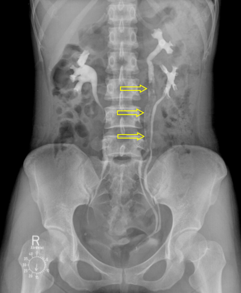
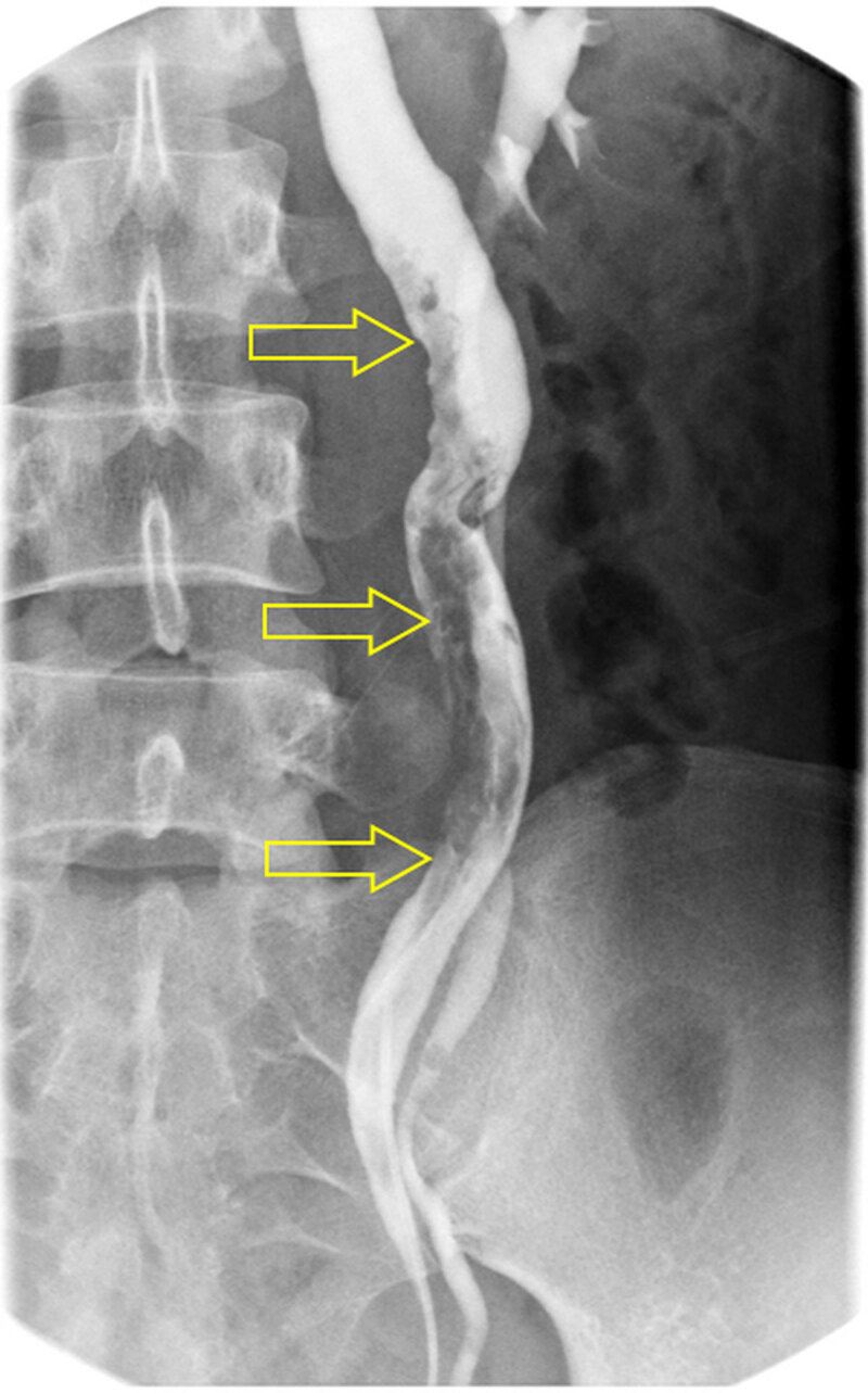
A 36-year-old female was admitted to our hospital with hematuria and left flank pain. Her past medical history was unremarkable. Urine analysis showed >100 leukocytes and 5–9 erythrocytes per high-power field. Intravenous urography (Figure 1) revealed incomplete duplication of the left ureter with a 9-cm-long filling defect (open arrows) in the upper moiety. Contrast-enhanced computed tomography (CT) was performed, and axial nephrographic phase CT images (Figure 2A and 2B) showed an enhancing mass (open arrow) in the left upper ureter, with ipsilateral mild hydronephrosis. Axial and coronal reformatted excretory phase CT images (Figure 2C–F) demonstrated an intraluminal mass (open arrow) filling the left upper ureter. Contrast material flowed around the mass (open arrow). Retrograde pyelography (Figure 3) confirmed along and irregular filling defect (open arrows) in the left ureter. The patient underwent left ureterostomy and excision of the mass, and was diagnosed with fibroepithelial polyp (FEP).
Figure 1.

Figure 2.

Figure 3.

Comment
Ureteral FEP is a rare benign tumor that consists of a fibrovascular stromal core covered by transitional epithelium. It is the most common benign, non-epithelial, ureteral neoplasm and predominantly presents in the third to the fifth decades of life. Although the exact etiology of FEP is unclear, congenital factors and chronic inflammation have been proposed. FEPs can develop in any part of the urinary tract, but most occur in the ureter, particularly in the ureteropelvic junction or upper ureter. The majority of patients with FEP demonstrate hematuria and/or flank pain. FEP is usually smaller than 5 cm, but a 14 cm polyp has been reported [1]. Most FEPs are solitary, but multiple or bilateral occurrences have been reported [1].
On imaging, FEP is frequently mistaken for urothelial carcinoma, which is the most common neoplasm in the urinary tract. Although the imaging features of FEP are variable, it usually manifests as a long, slender, and smooth ureteral filling defect on urography images. Compared to urothelial carcinomas, FEPs occur in younger patients. In addition, FEPs occasionally show mobility, unlike the fixed urothelial carcinomas. Nevertheless, it is difficult to pre-operatively differentiate FEP from urothelial carcinoma due to the rarity of FEP and the overlap in clinical presentations and radiological findings. The treatment of choice for FEP is complete excision [1].
Funding Statement
This work was supported by the Soonchunhyang University Research Fund and the National Research Foundation of Korea (NRF) grant funded by the Korea government (MSIT) (No. 2018R1C1B5085419).
Ethics and Consent
All procedures performed in studies involving human participants were in accordance with the ethical standards of the institutional and/or national research committee and with the 1964 Helsinki Declaration and its later amendments or comparable ethical standards. This article does not contain any studies with animals performed by any of the authors. Statement of informed consent was not applicable because the manuscript does not contain any patient data.
Funding Information
This work was supported by the Soonchunhyang University Research Fund and the National Research Foundation of Korea (NRF) grant funded by the Korea government (MSIT) (No. 2018R1C1B5085419).
Competing Interests
The authors have no competing interests to declare.
Author Information
On a title page separate from the manuscript.
References
- 1.Williams T, Wagner B, Corse W, Vestevich J. Fibroepithelial polyps of the urinary tract. Abdominal Imaging. 2002; 27(2): 217–21. DOI: 10.1007/s00261-001-0066-z [DOI] [PubMed] [Google Scholar]


