Abstract
Background
Computed Tomography (CT) is an invaluable imaging tool in the diagnostic workup of patients presenting with head trauma, stroke, brain tumour and epilepsy. The objective of this study was to document the common intracranial pathologies as revealed by CT in our setting and also determine if the indications for CT scan are justified so that patients are not exposed to radiation unnecessarily.
Materials and methods
This was a cross-sectional study carried out in Hansa Clinic Enugu, Enugu State, Nigeria. Demographic data and brain CT radiological reports with imaging findings and clinical indications for patients referred to this study centre from January, 2017 to January 2019 were retrieved from the CT reports' archives and reviewed retrospectively. Relevant information such age, gender, radiological CT findings and clinical indications were collected using structured proforma.
Results
A total of 300 patients' brain CT radiological reports were included in this study. The mean age of the patients was 41.25 ± 16.5 years with majority been within the age group of 31–40 years 92 (30.67%). Out of 300 cases, normal finding was highest 117 (39%) and the least was intracranial physiological calcification, which is 1 (0.33%). Headache is the most common clinical indication, 53 (17.67%) the least was unsteady Gait, which is 3 (1%). The Chi-square test revealed that there was statistically significance relationship between brain CT findings and clinical indications for the investigations (X2 = 285.60, p = 0.002).
Conclusion
The study showed that more males than females undergo brain CT scan with headache being the most common presenting complaint. The majority of findings of the brain CT scans in this study are normal despite, myriads of complaints necessitating the investigations. The study also revealed significant association between clinical indications and CT findings.
Keywords: brain computed tomography, common findings, exposure
1. Introduction
The invention of computed tomography (CT) in the 1970's significantly changed radiological imaging and medicine as a whole [1]. CT is essential in the medical imaging armamentarium used in the detection, prevention and screening for disease. It is a widely and readily available imaging modality in the developed world and in Nigeria. CT scanning is increasingly used both in research and in clinical medicine with improved image quality obtained especially with the newer helical scanners [2]–[4].
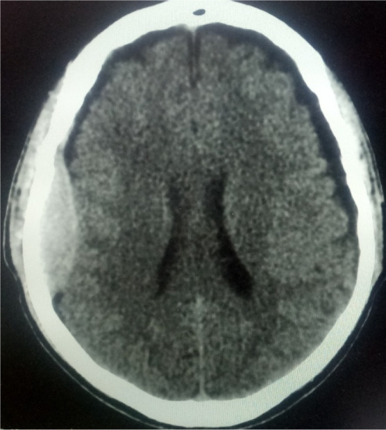
CT is an invaluable imaging tool in the diagnostic workup of patients presenting with head trauma, stroke, brain tumour and epilepsy. The common brain CT findings vary depending on the indications for the study and may include findings unrelated to the patients' complaints. According to Ijeh-Tarila et al. [5], in a study evaluating stroke patients, the common findings were ischemic infarct, cerebral haemorrhage, cerebral atrophy, cerebral abscess and brain tumour. In another study of patients with head trauma, Ogolodom et al. [6], documented intracerebral hematoma, skull fracture, contusion and extra-axial collection as the common findings. Other common brain CT imaging abnormalities include physiological intracranial calcification, cyst, aneurysm, white matter demyelinating disease, intraventricular haemorrhage, epidural haemorrhage (Figure 1), hydrocephalus, oedema and inflammatory disease. Despite patients' complaints, CT scan may reveal an absence of abnormal findings. Milad and Gamal [7] in a study, which evaluated the profile of head CT findings in 255 patients in their hospital reported 178 cases with no abnormal findings (64%) as the most common outcome.
Figure 1. Axial non-contrast brain CT showing acute epidural haemorrhage in the right parietal region.

CT scanners uses high doses of ionizing radiation, which may cause cancer. It is estimated that 0.4% of current cancer in the United States are due to CT scans performed in the past and this may increase to as high as 1.5–2% with 2007 rate of CT usage [8]. Patient exposure is more perilous in CT because, aside using ionizing radiation, the doses are usually higher than for radiographic or fluoroscopic procedures [9]. In addition, the introduction of multi-section CT scanners resulted in relatively large dose increase compared with doses from single-section scanners [10]. It is uncertain whether the cost and risk of brain CT are justified especially when considered vis-à-vis the indications for the study. Furthermore, CT often misses brain dysfunction following mild-to-moderate brain injury [11].
Usage of CT has increased dramatically over the past two decades in many countries [12]. The increased availability of CT scanners and use by especially Nigerian head injury patients has been reported [13]. The advances in technology of the CT scanners available in our environment in recent times have also increased the resolutions and abilities of radiologists in picking up many more subtle findings. To the best of our knowledge, no study has ever examined the common brain CT findings in low-resource setting like ours. The goal of this study was to document the common intracranial pathologies as revealed by CT in our setting and also determine if the indications for CT scan are justified so that patients are not exposed to radiation unnecessarily.
2. Materials and methods
This is a cross-sectional study carried out in the Radiology department of Hansa Clinic Enugu, Enugu State, Nigeria after obtaining ethical clearance from the study center. Demographic data and brain CT radiological reports with imaging findings and clinical indications for patients referred to this study center from January, 2017 to January 2019 were retrieved from the CT reports' archives and reviewed retrospectively. Relevant information such age, gender, radiological CT findings and clinical indications were collected using structured proforma. The obtained data were processed on SPSS version 21 and analyzed statistically using both descriptive statistics and inferential statistic (Ch-square test). The level of statistical significance was set at p < 0.05.
3. Results
A total of 300 patients' brain CT radiological reports and request forms were evaluated and included in this study. The mean age of the patients was 41.25 ± 16.5 years. Majority (N = 92) within the age of 31–40 years (30.67%), followed by (N = 73) within the age group of 41–50 years (24.33%) and the least (N = 13) were within the age group of <20 years (4.33%) (Table 1). The majority (N = 176) of the cases were males (58.7%), while (N = 124) were females (41.3%) (Table 1). With regards to the common brain CT findings, out of 300 cases, normal finding 39% (N = 117) was highest, followed by Ischemic infarct 20.67% (N = 62) and the least 0.33% (N = 1) was intracranial physiological Calcification. Out of 62 cases of ischemic infarcts, chronic type 61.29% (N = 38) was highest, while acute ischemic infarct was 38.71% (N = 24). Haemorrhage accounted for 14.33% (N = 43) of the total cases studied, with intracerebral haemorrhage 5.33% (N = 16) been highest, followed by (N = 11) subduaral haemorrhage (3.67%), and the least (N = 4) was extradural Haemorrhage (1.33%) (Table 2).
Table 1. Frequency and percentage of demographic variables.
| Frequency | Percentage (%) | |
| Age (years) | ||
| ≤20 years | 13 | 4.3 |
| 21–30 years | 37 | 12.3 |
| 31–40 years | 92 | 30.7 |
| 41–50 years | 73 | 24.3 |
| 51–60 years | 59 | 19.67 |
| ≥61 years | 26 | 8.67 |
| Total | 300 | 100.0 |
| Gender | ||
| Male | 176 | 58.7 |
| Female | 124 | 41.3 |
| Total | 300 | 100.0 |
Table 2. Frequency and percentage of common brain CT findings.
| CT findings | Frequency | Percent (%) |
| Cerebral Atrophy | 28 | 9.33 |
| Ischemic Infarct | 62 | 20.67 |
| Normal Brain CT | 117 | 39.00 |
| Fracture | 17 | 5.67 |
| Haemorrhage | ||
| Intracerebral haemorrhage | 16 | 5.33 |
| Intraventricular haemorrhage | 6 | 2.00 |
| Extraduaral haemorrhage | 4 | 1.33 |
| Subdural haemorrhage | 11 | 3.67 |
| Subarahnoid haemorrhage | 6 | 2.00 |
| Tumour | 10 | 3.33 |
| Hydrocephalus | 8 | 2.67 |
| Brain Oedema | 4 | 1.33 |
| Abscess | 3 | 1.00 |
| Metastasis | 2 | 0.67 |
| Meningitis | 5 | 1.67 |
| Intracranial physiological calcification | 1 | 0.33 |
| Total | 300 | 100 |
Headache is the most common clinical indication, 17.67% (N = 53), followed by seizure disorders 13.67% (N = 41), loss of consciousness 12.33% (N = 37) cerebrovascular disease (CVD) 11.33% (N = 34), RTA with head injury 9.33% (N = 28) , space occupying lesion, 6.67% (N = 20) and the least was unsteady Gait, which is 1% (N = 3) (Table 3). The Chi-square test revealed that there was statistically significance relationship between brain CT findings and clinical indications for the investigations (X2 = 285.60, p = 0.002).
Table 3. Common Clinical indication for Brain CT.
| Clinical Indications | Frequency | Percentage |
| Loss of Consciousness | 37 | 12.33 |
| Headache | 53 | 17.67 |
| Seizure disorder | 41 | 13.67 |
| Cerebrovascular disease | 34 | 11.33 |
| RTA with Head injury | 28 | 9.33 |
| RVD Encephalopathy | 9 | 3.00 |
| Meningitis | 15 | 5.00 |
| Cerebral Abscess | 11 | 3.67 |
| Intracranial Metastasis | 10 | 3.33 |
| Space occupying lesion | 20 | 6.67 |
| Proptosis | 6 | 2.00 |
| Facial Nerve Palsy | 4 | 1.33 |
| Paranasal Sinusitis | 13 | 4.33 |
| Unsteady Gait | 3 | 1.00 |
| Sinonasal mass | 4 | 1.33 |
| Hyperprollactinemia | 5 | 1.67 |
| Anosmia | 7 | 2.67 |
| Total | 300 | 100 |
RTA—Road Traffic Accident; RVD—Retroviral Disease.
4. Discussion
The demographic description of the cases included in our study revealed that majority of the cases were in their third, fourth, fifth and sixth decades of life with the peak value in the fourth decade of life, which accounted for over 30% of the total study population. Males were highest in number when compared with their female counterparts. This demographic finding is in agreement with the findings of similar studies conducted by Ohaegbulam et al. [14] in a medium-sized city (Enugu, Nigeria) and by Ogolodom et al. [6] in a large city (Port Harcourt, Nigeria). According to Ohaegbulam et al. [14], these categories of people are the most active and productive group in the society and appears to be more prone to both occupational and social risks, while Ogolodom et al. [6] in their study, attributed their finding to the fact that people of this age group commonly involved in the consumption of hard drugs, cultism, militancy activities and disobeying of traffic rules and regulations, especially the male population.
Normal brain CT finding was the most common outcome in our study, despite positive clinical indication to perform the CT scan. This finding is consistent with the findings of similar studies conducted by Haghighi et al. [15] in Taba Radiology centre in Shiraz and Khodapanahandeh and Hadizadeh [16] in Tehran. According to Haghighi et al. [15] finding, out of 167 patients data included in their study, 147 (88.02%) cases were normal findings, while the abnormal findings accounted for only 20 (11.98%) of the total study population. In Khodapanahandeh and Hadizadeh [16] study, CT scans and MRIs were performed for 108 and 11 patients respectively, those with normal findings accounted for 90% of the total case. The differences in the absolute values of the normal findings in our studies could be attributed to the different sample sizes and the nature of the different studies. Contrary to our finding, Ohaegbulam et al. [14] in their study, reported 80.1% of abnormal CT findings with the remaining 19.9% been unremarkable. The nature of our study and that of Ohaegbulam et al., greatly influenced the observed differences in our findings.
In our study, we found that the most common clinical indication for the brain CT investigations was headache. Headache is common complaints in clinical practice, though most of the patients who present with headache have no neurological abnormality on CT and MRI investigations [17]. This finding is consistent with the result of similar study carried out by Haghighi et al. [15]. In a retrospective study conducted by Haghighi et al., which evaluated the abnormal findings in brain CT scans among children in Taba Radiology centre in Shiraz, reported headache as the major complaint for the CT scan, 739 (60.80%). The discrepancies noted in our findings could be ascribed to the different nature and the sample size of the two studies.
It is also important to note that CT often misses mild-to-moderate traumatic brain injury (TBI). CT is widely available and remains a useful tool for visualizing fracture and bleeding, but it contributes little to understanding the pathology of common neurological indications for neuroimaging—acute ischaemic stroke and traumatic brain injury. CT particularly fails in the area of diagnostic imaging of TBI. This is in agreement with the findings of the studies conducted by Haydel et al. [18], Jacobs et al. [19] and Abu-Judeh et al. [20]. Haydel et al. found only 5–10% of CT scans were abnormal in over 4,000 patients with documented mild traumatic brain injury and they concluded that for the investigation of patients with minor head injury, the use of CT can be safely restricted to those who have certain clinical findings. Jacobs et al. documented positive findings of traumatic brain injury by SPECT scan in 126 cases which were negative on CT and MRI. In the study by Abu-Judeh et al., they observed that CT missed or underestimated injury from TBI when compared to the findings from SPECT scans.
In this present study, we found a statistical significant relationship between clinical indications and brain CT findings. This implies that there was justification for the CT requests, although normal findings was found to be highest, which is usually common when the chief complaint is headache as in the case of this study.
5. Conclusion
Computed tomography scan is a common imaging modality used to evaluate brain pathologies. The study showed that more males than females undergo brain CT scan with headache being the most common presenting complaint. The majority of findings of brain CT scans in this study is normal despite myriads of complaints necessitating the investigations. The study also revealed significant association between clinical indications and CT findings.
Footnotes
Conflict of interest: The authors declare no conflict of interest.
References
- 1.Looking back on the millennium in medicine. N Engl J Med. 2000;342:42–49. doi: 10.1056/NEJM200001063420108. [DOI] [PubMed] [Google Scholar]
- 2.Ogunseyinde AO, Obajimi MO, Ogundare SM. Radiological evaluation of head trauma by computer tomography in Ibadan, Nigeria. West Afr J Med. 1999;18:33–38. [PubMed] [Google Scholar]
- 3.Obajimi MO, Shokunbi MT, Malomo AA, et al. Computed tomography (CT) in civilian gunshot head injuries in Ibadan. West Afr J Med. 2004;23:58–61. doi: 10.4314/wajm.v23i1.28084. [DOI] [PubMed] [Google Scholar]
- 4.Arogundade RA. Brain tumour as a predisposing factor to head trauma: A review of two cases diagnosed by computed tomography. Niger Postgrad Med J. 2010;17:168–171. [PubMed] [Google Scholar]
- 5.Ijeh-Tarila KI, Alazigha N, Mbaba AN, et al. Brain computed tomography findings in stroke patients in port Harcourt: A retrospective hospital-based study. Am J Biomed Sci Res. 2020;8:280–284. [Google Scholar]
- 6.Ogolodom MP, David LK, Erondu OF, et al. Patterns of traumatic head injury among patients that underwent craniofacial computed tomography scan in Port Harcourt metropolis, Rivers state, Nigeria. Health Sci J. 2019;13:1–8. [Google Scholar]
- 7.Milad MH, Gamal AM. The profile of head CT scan in patients at Sebha medical center, Libya. Sudan J Med Sci. 2016;11:95–100. [Google Scholar]
- 8.Brenner DJ, Hall EJ. Computed Tomography—an increasing source of radiation exposure. N Engl J Med. 2007;357:2277–2284. doi: 10.1056/NEJMra072149. [DOI] [PubMed] [Google Scholar]
- 9.Amis ES, Jr, Butler PF, Applegate KE, et al. American College of Radiology white paper on radiation dose in medicine. J Am Coll Radiol. 2007;4:272–284. doi: 10.1016/j.jacr.2007.03.002. [DOI] [PubMed] [Google Scholar]
- 10.Tsalafoutas IA, Koukourakis GV. Patient dose considerations in computed tomography examinations. World J Radiol. 2010;2:262–268. doi: 10.4329/wjr.v2.i7.262. [DOI] [PMC free article] [PubMed] [Google Scholar]
- 11.Raji CA, Tarzwell R, Pavel D, et al. Clinical utility of SPECT Neuroimaging in the diagnosis and treatment of traumatic brain injury: A systematic review. PLoS One. 2014;9:e91088. doi: 10.1371/journal.pone.0091088. [DOI] [PMC free article] [PubMed] [Google Scholar]
- 12.Smith-Bindman R, Lipson J, Marcus R, et al. Radiation dose associated with common computed tomography examination and the associated lifetime attributed role of cancer. Arch Intern Med. 2009;169:2078–2086. doi: 10.1001/archinternmed.2009.427. [DOI] [PMC free article] [PubMed] [Google Scholar]
- 13.Ogbole GI, Adeleye AO, Owolabi MO, et al. Incidental cranial CT findings in head injury patients in a Nigerian tertiary hospital. J Emerg Trauma Shock. 2015;8:77–82. doi: 10.4103/0974-2700.155499. [DOI] [PMC free article] [PubMed] [Google Scholar]
- 14.Ohaegbulam SC, Mezue WC, Ndubuisi CA, et al. Cranial computed tomography scan findings in head trauma patients in Enugu, Nigeria. Surg Neurol Int. 2011;2:182. doi: 10.4103/2152-7806.91137. [DOI] [PMC free article] [PubMed] [Google Scholar]
- 15.Haghighi M, Baghery MH, Rashidi F, et al. Abnormal findings in brain CT scans among children. J Compr Pediatr. 2014;5:e13761. [Google Scholar]
- 16.Khodapanahandeh F, Hadizadeh H. Neuroimaging in children with first afebrile seizures to order or not order? Arch Iran Med. 2006;9:156–158. [PubMed] [Google Scholar]
- 17.Ogolodom MP, Mbaba AN, Abam R, et al. Magnetic resonance imaging findings in patients presenting with headache in Port Harcourt, Rivers State, Nigeria. J Biomed Sci. 2019;8:3. [Google Scholar]
- 18.Haydel MJ, Preston CA, Mills JT, et al. Indications for computed tomography in patients with minor head injury. N Engl J Med. 2000;342:100–105. doi: 10.1056/NEJM200007133430204. [DOI] [PubMed] [Google Scholar]
- 19.Jacob A, Put E, Ingels M, et al. Prospective evaluation of Technetium-99m-HMPAO SPECT in mild and moderate traumatic head injury. J Nucl Med. 1994;35:942–947. [PubMed] [Google Scholar]
- 20.Abu-Juden HH, Parker R, Aleksics S, et al. SPECT brain perfusion findings in mild or moderate traumatic brain injury. Nucl Med Rev. 2000;3:5–11. [PubMed] [Google Scholar]


