Abstract
Purpose
Patients and physicians are often pleased when uncorrected visual acuity (UCVA) on post-operative day 1 (POD1) after cataract surgery is 20/20. Unfortunately, this UCVA does not always last. This article aims to investigate the relationship between excellent uncorrected visual acuity on post-operative day 1 and final post-operative UCVA after uncomplicated cataract surgery.
Patients and Methods
The medical records of patients who had undergone uncomplicated cataract surgery between 2012 and 2017 were assessed. UCVA on POD1 and final UCVA were obtained for patients who had a final best-corrected visual acuity of 20/20 or better.
Results
Of 309 patients with UCVA of 20/20 on POD 1, 62.4% maintained 20/20 and 87.4% maintained 20/25 or better as their final uncorrected visual outcome. Of 204 patients with UCVA of 20/25 on POD 1, 44.1% achieved 20/20 and 69.6% maintained 20/25 or better as their final uncorrected visual outcome. Patients with 20/20 UCVA on POD1 were more likely to have a better final UCVA compared with those who were 20/25 on POD1. Of the 531 patients with UCVA of 20/25 or better on POD1, 20% had final UCVA worse than 20/25 with 4% losing more than 2 lines for their final UCVA.
Conclusion
The majority of patients with 20/20 UCVA on POD1 after cataract surgery maintained excellent UCVA as their final visual outcome. However, a significant percentage of these patients experienced a decrease in UCVA over the course of the postoperative period.
Keywords: phacoemulsification, cataract, pseudophakia, visual acuity, treatment outcome
Introduction
Cataract surgery is one of the mainstays of ophthalmic surgery. With advances in cataract surgery, demand has risen for improved refractive outcomes and decreased reliance on spectacle correction after surgery. To accommodate this demand, surgeons employ detailed preoperative planning, including robust biometry measurements and the use of increasingly advanced formulas for IOL calculations such as the SRK/T, Holladay 1 and 2, Barrett, Olsen, Hill-RBF, and the Ladas Super Formula.1–4 These and other formulas are continually being modified to refine refractive results based on factors such as axial length, corneal refractive power, and anterior chamber depth.
Technological advances have also led to improved efficiency and safety of cataract surgery and the potential for rapid visual recovery. Newer generation phacoemulsification machines allow efficient nuclear removal with less energy, and viscoelastics protect from early corneal edema.5 Patients are routinely achieving good visual acuities, even shortly after surgery. However, it can be challenging to manage patient expectations regarding visual outcomes even after surgery has been completed successfully. In particular, patients who achieve excellent uncorrected visual acuity (UCVA) on post-operative day 1 (POD1) may not maintain that level of UCVA. While numerous reports have been published correlating preoperative evaluation and IOL selection with visual outcomes, it is less clear how early post-operative UCVA is predictive of the final visual outcome.3,4 The purpose of this study is to analyze the relationship between excellent UCVA on post-operative day 1 (POD1) after uncomplicated cataract surgery and final post-operative UCVA. Such knowledge would help cataract surgeons guide patient expectations more precisely during the post-operative period.
Patients and Methods
This retrospective cohort study was approved by the Oregon Health & Science University (OHSU) Review Board. Patient consent to medical records review was waived by the OHSU IRB for minimal risk to patient confidentiality; de-identified data were extracted and analyzed to protect patient confidentiality in accordance with the Declaration of Helsinki. OHSU is an academic tertiary care center. Within the ophthalmology department, cataract surgery is performed by more than one division. Patients were included for the study if they had uncomplicated cataract surgery (CPT code 66984) performed within the Casey Eye Comprehensive Ophthalmology division between 2012 and 2017. The comprehensive division includes faculty who have completed residency/fellowship training, and residents who perform cataract surgery during their final year of residency training as primary surgeon.
While there were likely slight differences during surgery owing to surgeon preferences, the pre-surgical evaluation was standardized. Intraocular lens power was calculated using optical biometers (IOLMaster 500 and 700, Carl Zeiss Meditec) and formulas used were per surgeon preference. IOL selection, by institutional preference, was nearly exclusively monofocal SN60WF or toric variant SN6ATX Alcon lenses. Multifocal lenses were rarely used within our institution.
A POD1 visit was required; thus, patients seen for same-day post-operative visits were effectively excluded. Patients were included if they achieved excellent POD1 uncorrected visual acuity (20/25 or better). Vision was measured with standardized vision charts displayed on computer monitors. When determining vision, missing or additional letters were eliminated for ease of comparison. Thus, 20/20-2 or 20/25+2 were simplified to 20/20 and 20/25, respectively. Exclusion criteria included missing outcome data and a final post-operative best-corrected visual acuity of less than 20/20 in the operated eye.
The primary outcome measure was the final post-operative uncorrected visual acuity (defined as the last uncorrected visual acuity measurement between POD10 and POD60). Post-operative refractions and the best-corrected visual acuities (BCVA) were taken from this same time period, among other data including demographics, pre-operative spherical equivalent (SE), and intraocular pressure.
Results
A total of 5061 uncomplicated cataract surgeries were examined from the comprehensive service between 2012 and 2017. After excluding surgeries without a POD1 visit, and who did not achieve at least 20/20 as their final BCVA, 309 eyes achieved 20/20 UCVA on POD1 and 204 eyes achieved UCVA of 20/25 on POD1. Of these patients, only 0.97% (n=5) had multifocal or extended depth-of-focus lens implants, while the rest were monofocal lens variants.
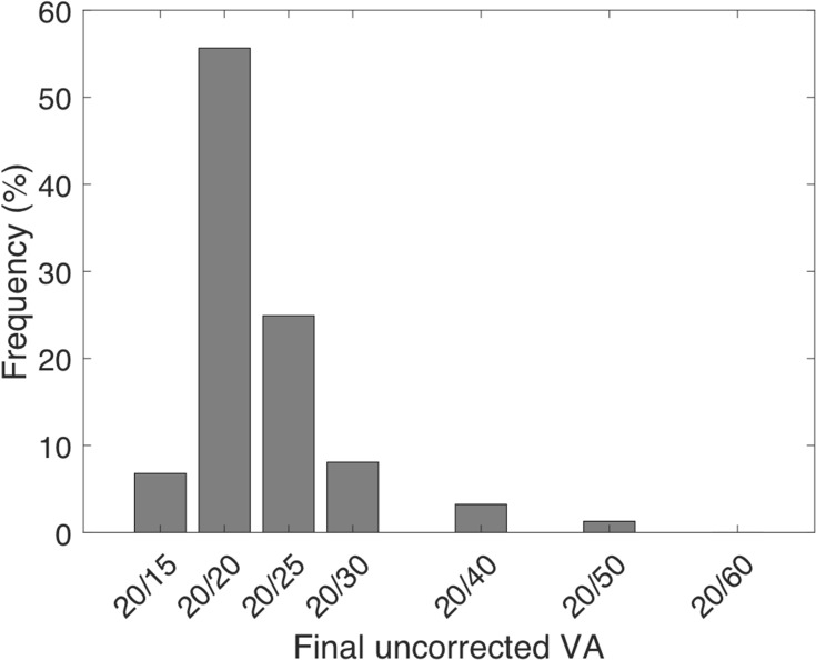
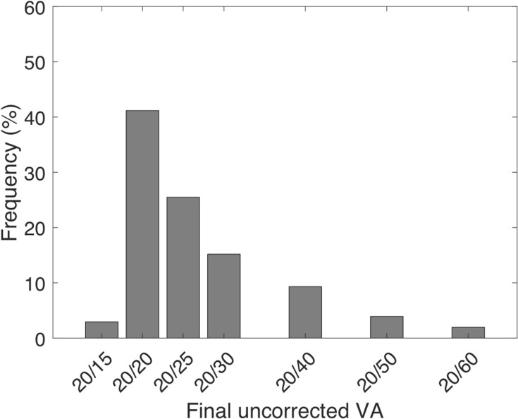
Figure 1 shows the distribution of final post-operative UCVA in patients with UCVA of 20/20 vision or better on POD1. These patients had a 62.4% chance of maintaining 20/20 UCVA or better and an 87.4% chance of maintaining 20/25 vision or better as their final UCVA. Figure 2 shows the distribution of final post-operative UCVA in patients with UCVA 20/25 vision on post-operative day one. These patients had a 44.1% chance of improving to an UCVA of 20/20 or better and a 69.6% chance of maintaining 20/25 vision or better as their final UCVA. The distribution of post-operative UCVA between these two groups of patients was significantly different using a Kolmogorov–Smirnov (KS) test (p<0.005, not shown).
Figure 1.
Distribution of final post-operative UCVA for all patients who achieved 20/20 on POD1 (n = 309).
Figure 2.
Distribution of final post-operative UCVA for all patients who achieved 20/25 on POD1 (n = 204).
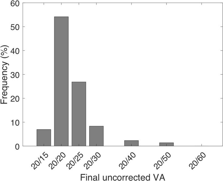
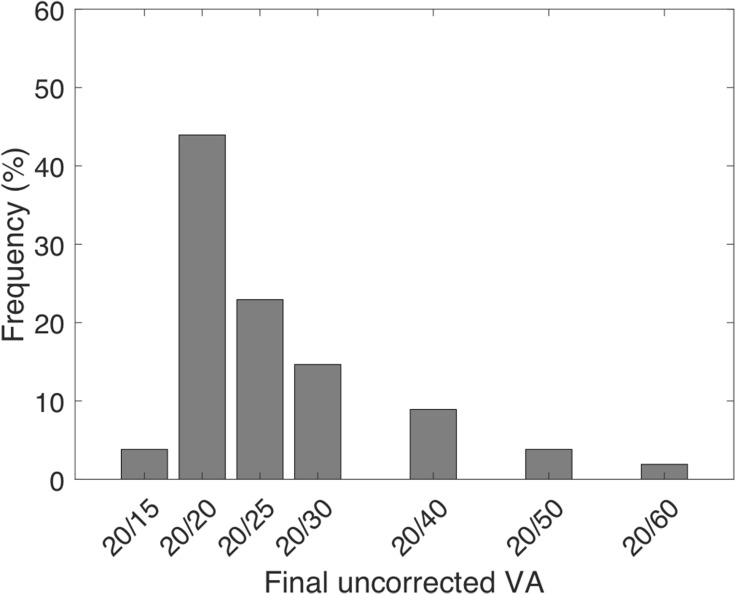
In order to understand the robustness of these distributions, we plotted similar outcomes after excluding patients who had been evaluated by subspecialty ophthalmology clinics at Casey Eye Institute in the year preceding or subsequent to cataract surgery at the comprehensive department. This was done to reduce the likelihood of a patient having any severe preexisting ocular pathology. This exclusion decreased the number of patients with POD1 UCVA of 20/20 or 20/25 by approximately 30% percent. KS testing revealed that the specialist-included distribution and the specialist-excluded distribution were not significantly different (compare Figures 1 to 3 and Figures 2 to 4).
Figure 3.
Distribution of final post-operative UCVA for patients who achieved 20/20 on POD1 after excluding patients who were seen in sub-specialty clinics (n = 216).
Figure 4.
Distribution of final post-operative UCVA for patients who achieved 20/25 on POD1 after excluding patients who were seen in sub-specialty clinics (n = 143).
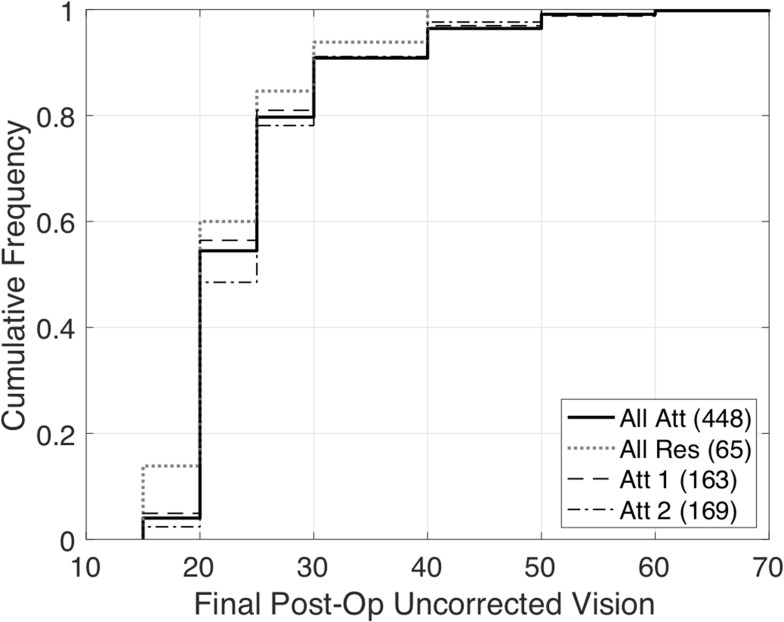
We also sought to understand if these distributions were surgeon-dependent. We created four different surgeon groups: attendings only, resident only, and two high-volume individual attendings. We found that the distributions of final post-operative UCVA in patients achieving either 20/20 or 20/25 UCVA on POD1 were not statistically different between these groups in pairwise KS testing (Figure 5).
Figure 5.
Cumulative distribution plots showing final post-operative UCVA among different surgeon groups. These distributions were not statistically different.
We observed some patients who lost more than two lines of UCVA after POD1 (see Figures 1 through 4). We termed these patients “outliers” and attempted to understand patient risk factors that might help predict this outcome. Of the 513 patients with uncorrected visual acuity of 20/25 or better on POD1, 31 (6%) initial outliers were identified. Thirty-two percent of those outliers received intracameral carbachol, likely leading to improved UCVA due to a pinhole effect on POD1. After excluding eyes that received intracameral carbachol, there were no statistical differences in IOP, age, pre-operative spherical equivalent, or gender between the remaining 21 (4%) outliers and those who were able to maintain their excellent post operative UCVA (Table 1).
Table 1.
Characteristics Comparing Outliers Who Lost >2 Lines of UCVA from POD1 (and Did Not Receive Intracameral Carbachol) with Those Who Were Able to Maintain Excellent UCVA. Mean and (Std. Dev) Shown. No Significant Differences Between Characteristics Were Found
| IOP on POD1 | Age | Preoperative Spherical Equivalent | % Female | |
|---|---|---|---|---|
| Outliers who lost >2 lines (n=21) | 15.8 (5.7) | 66.6 (11.2) | −0.85 (1.34) | 52 |
| All patients with 20/25 UCVA on POD1 who maintained VA (n=482) | 17.4 (4.8) | 65.6 (10.4) | −1.81 (3.87) | 49.9 |
| P value for difference | 0.13 | 0.66 | 0.25 | 0.85 |
Discussion
In this study, we evaluated the relationship between excellent UCVA on POD1 and the final UCVA in patients having undergone uncomplicated cataract surgery. Patients who achieved uncorrected 20/20 vision on POD1 had a roughly 60% chance of maintaining 20/20 vision or better and an 85% chance of maintaining 20/25 vision or better as their final UCVA. This relationship was maintained across patients seen in subspecialty clinics and those seen only in the comprehensive clinic. It was also consistent across different levels of surgeon training and different attending surgeons. Patients with POD1 20/20 UCVA were statistically more likely to have 20/20 vision as their final UCVA compared to those with 20/25 vision on POD1. There were also no clear differences in preoperative or immediate post-operative measurements between patients who maintained good UCVA and outliers who lost >2 lines of UCVA.
There are several factors potentially contributing to shifts in visual acuity during the post-operative period. Movement and settling of the IOL postoperatively can affect the anterior chamber depth (ACD) or distance between the corneal epithelium and the lens. Hoffer and Savini measured the anterior chamber depth (ACD) postoperatively in 492 eyes and found a 0.12 mm increase in ACD and a +0.23 diopter hyperopic shift between POD1 and POM3.6 This represents a shift of 1.92 diopter/mm of IOL movement. Fluctuation in ACD after cataract surgery has been reported by other authors although the direction of IOL shift has been variable with some reporting an anterior (myopic) shift7,8 instead of a posterior (hyperopic) shift.4,8 Despite the uncertainty in direction of the shift, ACD fluctuation is likely a major contributor to changes in postoperative UCVA.
Understanding why ACD shifts occur postoperatively is important in trying to minimize unexpected visual outcomes. Fluctuations in IOP, retained ophthalmic viscosurgical device (OVD), and IOL shape and materials have been implicated as potential contributors.6–10 Iwase et al found a myopic shift of −0.53 diopters with silicone lenses but not with PMMA or acrylic lenses.7 Wirtitsch et al found that eyes with multi-piece acrylic IOLs underwent a myopic shift of 0.213 mm between POD1 and POM6 whereas single-piece acrylic IOLs had a small hyperopic shift of 0.014 mm.8 The difference in magnitude of shift between single-piece and multi-piece acrylic lenses was thought to be due to increased stability and haptic memory of single-piece IOLs. Multi-piece IOLs may also be more susceptible to decentration than rigid single-piece PMMA IOLs.9 While in the current study, the type of IOL was not controlled, the default lens was a single-piece acrylic lens as part of institutional preference.
Capsular contraction and size of the capsulorhexis may also contribute to post-operative ACD shift. Cekic and Batman found that those assigned to receive a smaller, 4 mm capsulorhexis had a longer post-operative ACD than those assigned to a 6 mm capsulorhexis in a group of 51 patients.11 This could be from increased fibrosis of the smaller capsulorhexis causing increased ACD. Wirtitsch et al however did not find a statistically significant correlation between capsulorhexis size and fluctuations in ACD.8
In addition to shifts in ACD, postoperative changes in corneal topography may contribute to fluctuations in UCVA. Wallace et al investigated keratometric changes after clear corneal incision cataract surgery and found a tendency for the cornea to steepen by an average of 0.11D between weeks 1 and 4 postoperatively.4 Vass et al, however, found that changes in corneal topography between 1 week and 1 year after cataract surgery were negligible.12 Information on this topic is limited particularly regarding changes that occur in the very early post-operative period. Despite the likely variability in surgical factors such as capsulorhexis size, amount of corneal hydration, and operating time, it is remarkable that the core result in the current study remains consistent across surgeons and patient populations.
Limitations of this study include being a retrospective chart review at a single institution. Many factors that contribute to temporarily decreased visual acuity after cataract surgery including surface irritation, corneal edema, and inflammation are not major contributors in this analysis as patients with these conditions are unlikely to have had UCVA of 20/25 or better on POD1 and were thus presumably excluded. Preoperative refractive goal and biometry statistics were not included in the study; however, a vast majority of eyes entered into the study had refractive goals of plano to mild-myopia (−0.5D), which would likely make distinctions in visual acuity distributions stratified by refractive goal challenging to perceive. Lastly, understanding the impact of lens type (such as multifocal versus monofocal) was limited as our training institution used nearly all monofocal lenses for cataract surgery.
Conclusion
With advances in cataract surgery, more emphasis is being placed on optimizing post-operative refractive outcomes and decreasing patients’ reliance on spectacles. While many surgeons and patients are thrilled with uncorrected 20/20 visual acuity on post-operative day one, it is important to remember that a significant percentage of patients will have a worse final UCVA. This study helps surgeons guide patient expectations regarding potential shifts in visual acuity during the post-operative period.
Acknowledgments
Supported by an unrestricted grant from Research to Prevent Blindness (New York, NY) and by an NIH core grant P30 EY010572 (Bethesda, MD). Jonathan W Young and Nathan W Law are co-first authors for this study.
Disclosure
The authors report no financial or proprietary conflicts of interest in this work.
References
- 1.Narvaez J, Zimmerman G, Stulting RD, Chang DH. Accuracy of intraocular lens power prediction using the Hoffer Q, Holladay 1, Holladay 2, and SRK/T formulas. J Cataract Refract Surg. 2006;32(12):2050–2053. doi: 10.1016/j.jcrs.2006.09.009 [DOI] [PubMed] [Google Scholar]
- 2.Prager TC, Hardten DR, Fogal BJ. Enhancing intraocular lens outcome precision: an evaluation of axial length determinations, keratometry, and IOL formulas. Ophthalmol Clin North Am. 2006;19(4):435–448. doi: 10.1016/j.ohc.2006.07.009 [DOI] [PubMed] [Google Scholar]
- 3.Shajari M, Kolb CM, Petermann K, et al. Comparison of 9 modern intraocular lens power calculation formulas for a quadrifocal intraocular lens. J Cataract Refract Surg. 2018;44(8):942–948. doi: 10.1016/j.jcrs.2018.05.021 [DOI] [PubMed] [Google Scholar]
- 4.Wallace HB, Misra SL, Li SS, McKelvie J. Predicting pseudophakic refractive error: interplay of biometry prediction error, anterior chamber depth, and changes in corneal curvature. J Cataract Refract Surg. 2018;44(9):1123–1129. doi: 10.1016/j.jcrs.2018.06.017 [DOI] [PubMed] [Google Scholar]
- 5.Glasser DB, Schultz RO, Hyndiuk RA. The role of viscoelastics, cannulas, and irrigating solution additives in post-cataract surgery corneal edema: a brief review. Lens Eye Toxic Res. 1992;9(3–4):351–359. [PubMed] [Google Scholar]
- 6.Hoffer KJ, Savini G. Anterior chamber depth studies. J Cataract Refract Surg. 2015;41(9):1898–1904. doi: 10.1016/j.jcrs.2015.10.010 [DOI] [PubMed] [Google Scholar]
- 7.Iwase T, Tanaka N, Sugiyama K. Postoperative refraction changes in phacoemulsification cataract surgery with implantation of different types of intraocular lens. Eur J Ophthalmol. 2008;18(3):371–376. doi: 10.1177/112067210801800310 [DOI] [PubMed] [Google Scholar]
- 8.Wirtitsch MG, Findl O, Menapace R, et al. Effect of haptic design on change in axial lens position after cataract surgery. J Cataract Refract Surg. 2004;30(1):45–51. doi: 10.1016/S0886-3350(03)00459-0 [DOI] [PubMed] [Google Scholar]
- 9.Hayashi K, Hayashi H, Nakao F, Hyashi F. Comparison of decentration and tilt between one piece and three piece polymethyl methacrylate intraocular lenses. Br J Ophthalmol. 1998;82(4):419–422. doi: 10.1136/bjo.82.4.419 [DOI] [PMC free article] [PubMed] [Google Scholar]
- 10.Petternel V, Menapace R, Findle O, et al. Effect of optic edge design and haptic angulation on postoperative intraocular lens position change. J Cataract Refract Surg. 2004;30(1):52–57. doi: 10.1016/S0886-3350(03)00556-X [DOI] [PubMed] [Google Scholar]
- 11.Cekic O, Batman C. The relationship between capsulorhexis size and anterior chamber depth relation. Ophthalmic Surg Lasers. 1999;30(3):185–190. [PubMed] [Google Scholar]
- 12.Vass C, Menapace R, Ranier G, Findle O, Steineck I. Comparative study of corneal topographic changes after 3.0 mm beveled and hinged clear corneal incisions. J Cataract Refract Surg. 1998;24(11):1498–1504. doi: 10.1016/S0886-3350(98)80173-9 [DOI] [PubMed] [Google Scholar]


