Abstract
Cilostazol, a phosphodiesterase III inhibitor, has antiplatelet and vasodilatory effects. It also has pleiotrophic effects including reduction of oxygen free radicals, positive chronotropic effect and inhibition of intracellular Ca2+ associated catecholamine secretion. The study was aimed to examine, in vivo, the effects of cilostazol treatments on myocardial function, myocardial remodeling, and neurohormonal status in myocardial infarction (MI) with restrained stress rat model. Male Sprague Dawley rats, subjected to coronary artery ligation to induce myocardial infarction (MI), received either a standard rat chow alone (control, n=5) or combined with cilostazol (cilostazol, n=5; 5 mg/kg×5 weeks). They were exposed to repeated restraint stress (2 h×2 times/day) for 10 days beginning 1 week after surgery. Left ventricular ejection fraction (LVEF), LV mass by heart weight/body weight ratio and level of tissue brain natriuretic peptide (BNP) expression by immunoblotting were determined. Plasma epinephrine and norepinephrine levels were also measured. Mean LVEF was higher in the cilostazol group than in the control group (66.9±14.3 vs 47.0±17.1, p<0.05) at 5 weeks after MI. However, LV mass and tissue BNP expression were significantly lower in the cilostazol than in the control group (p<0.05). Plasma epinephrine and norepinephrine levels were also lower in the cilostazol group compared with the control (p<0.05). Cilostazol preserves left ventricular systolic function and attenuates stress induced remodeling in postinfarct rats. Its beneficial effects were associated with reduced plasma catecholamine levels during postinfarct remodeling.
Keywords: Cilostazol, Myocardial Infarction, Ventricular Remodeling
INTRODUCTION
Myocardial infarction (MI) is a serious health problem which causes substantial morbidity and mortality. Until now, control of classical risk factors such as hypertension, diabetes, smoking and hyperlipidemia has been emphasized for its prevention; however, attention has shifted toward a possible relation between environmental stress and cardiovascular events. Stress has been well-known risk factor for increased mortality and morbidity of ischemic heart disease. A number of human studies have demonstrated that acute and chronic stresses are related with initiation and deterioration of ischemic insult.1,2,3,4,5 In experimental animals, it was also shown that acute stress is associated with adverse ventricular remodeling and a decreased threshold of arrhythmia after MI.6,7 Among mechanisms proposed for stress associated exacerbation of myocardial ischemic injury, catecholamine surge during stress has been suggested as an important mechanism.8,9 Indeed, increased sympathetic activity and catecholamine secretion are frequently observed in stressful conditions. This enhanced sympathetic activity is associated with increased myocardial oxygen demand via inotropic (i.e., increased contractility) and chronotropic (i.e., tachycardia) actions. It also has potentially harmful effects on coronary vasculature by increasing vascular tone and shear stress. Moreover, enhanced catecholamine activity can trigger arrhythmia and increases platelet aggregation. Nevertheless, the optimal pharmacologic strategy to prevent stress related myocardial injury has not been clearly determined.
Cilostazol, 6-[4-(1-cyclohexyl-1H-tetrazol-5-yl) butoxyl]-3,4-dihydro-2-(1H)-quinolinone, is a selective inhibitor of phosphodiesterase 3, and thus increases intracellular cyclic adenosine monophosphate. Its principal action site exists in platelet and vascular smooth cells,10 and it is currently approved for management of peripheral vascular disease and prevention of cerebrovascular disease.
Cilostazol also showed efficacy in the protection of ischemic insult in heart, brain and peripheral limbs through its anti-platelet and vasodilation effect.11,12 In addition, decreasing oxygen free radials, inhibiting cell apoptosis, and inhibiting catecholamine secretion have been suggested as the method for their protection.13,14,15 Activation of sympathetic systems is a known phenomenon in heart failure. Overwhelming evidence supports the concept that over-activation of this system significantly contributes to the progression of congestive heart failure. It has been hypothesized that cilostazol treatment would attenuate stress induced myocardial injury in MI.
The aims of the present study were to examine, in vivo, the effects of cilostazol on myocardial function (using echocardiography) and restrain stress-induced early postinfarct remodeling in postinfarct rats, and to evaluate its effects on plasma catecholamine levels and BNP expression in ventricular tissue.
MATERIALS AND METHODS
1. Animals
Ten male Sprague-Dawley rats at the age of 6 weeks were kept in the laboratories for 1 week before the experiment under a 12:12 hour light/dark cycle with lights on at 8:00 A.M. They were deprived of food for 18 hours but permitted water ad libitum before surgery. All animal procedures were carried out as approved by the Animal Care and Use Committee of Wonkwang University School of Medicine.
2. Surgical procedure
Rats were anesthetized with the long-acting rodent anesthetic thiobarbital at an initial intraperitoneal dose of 110 mg/kg. Supplemental anesthetic was given in intraperitoneal doses of 10 mg/kg as required to maintain a plane of anesthesia. The animal was intubated through a tracheotomy and ventilated with room air supplemented with 100% O2 using a Harvard animal ventilator (Boston, MA, USA). A left thoracotomy was made at the third or fourth intercostal space, the lungs were retracted, and the pericardium was teased open to expose the heart. After opening the pericardium, the coronary artery was occluded by ligature using 6/0 prolene. The ligation site was placed around the left anterior descending artery close to its origin (2 mm below) near the appendage of the left atrium. Successful occlusion was immediately confirmed by observation of an arising pale ischemic zone. After the surgical procedure, the chest wall was closed and the remaining 8 gauge IV chest tube was drained into a water-seal bottle. Thereafter, all surgical wounds were sterilized and gentamycin (100 mg/kg) was administrated for prevention of wound infection.
3. Restraint stress
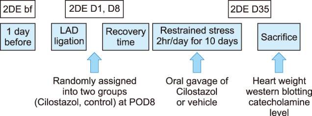
To produce chronic, intermittent stress, rats were restrained in rodent restraining devices (universal holder DJ-430, Dae Jong instrument, Seoul, Korea). The duration of the stress was usually 2 hours in the daytime and another 2 hours in the nighttime. Restraint stress was applied to experimental animals beginning 1 week after surgery and maintained for 10 days. Total restraint duration in each animal was 40 hours for 10 days (Fig. 1).
FIG. 1. Brief diagram of study design representing time for 2D echocardiography, restrained stress and administration of cilostazol. 2DE: 2 dimensional echocardiography, bf: before LAD ligation, POD: postoperative day.

4. Drug administration
Experimental rats received either a standard rat chow for 5 weeks alone or combined with cilostazol (Otsuka Pharmaceutical, Tokyo, Japan) 5 mg/kg for 5 weeks starting after recovery from surgery.
5. Assessment of myocardial remodeling
Two-dimensional echocardiography to measure left ventricular ejection fraction (LVEF) was performed at baseline, 1 days, 8 days, and 5 weeks after induction of MI. Echocardiography was performed with the VIVID 7 system (General Electric, USA), equipped with a 12-MHz electronic transducer. M-mode and 2-dimensional (2D) echocardiography images were obtained in the parasternal long- and short-axis views. Left ventricular (LV) dimensions were determined at the tips of the papillary muscle. LVEF was determined using Teichholz formula. Left ventricular mass index representing the extent of myocardial remodeling was estimated by total heart weight/total body weight ratio.
6. Histopathologic examination
After echocardiography at 5 weeks after MI, experimental animals were scarified under anesthesia with intraperitoneal injection of chloral hydrate, cardiac tissue was excised, and was fixed using 10% formaldehyde. Sections with a thickness of 4 µm were cut and stained with hematoxylin and eosin to examine the overall morphology.
7. BNP expression in cardiac tissue
Myocardial tissue was cleansed by phosphate buffered saline and homogenization using lysis buffer (25 mM Tris-HCl, 1 mM EGTA, 1 mM DTT (dithiothreitol), 0.1% Triton X-100, pH 7.4, phosphatase inhibitor cocktail, protease inhibitor cocktail). Thereafter, cytosol was extracted by centrifugation at 12,000 rpm and 30 minutes. Electrophoresis was performed with minigel electrophoresis device (mini-protean tetra cell, Bio-Rad Laboratories, Inc., Hercules, USA). After reaction with primary antibody for BNP (Abcam, Cambridge, UK) (1:1,000), a secondary antibody-goat rabbit Ig G conjugated Horse-Raddish Peroxidase (Santacruz Biotechnology Inc., Santa Cruz, USA) reaction was carried out for 1 hour. The degree of BNP expression was observed after reaction with an immobilon western chemiluminescent HRP substrate (Millipore, co., Billerica, MA, USA) and exposed to film.
8. Plasma catecholamine concentrations
To determine plasma norepinephrine and epinephrine levels, one milliliter of blood was collected through the catheter. Norepinephrine and epinephrine in the plasma were extracted by the method of Hallman et al. (1978)16 and assayed electrochemically by high performance liquid chromatography.
9. Statistical analysis
All results were expressed as mean±standard deviation. Statistical analysis was performed with SPSS (v 11.0 SPSS Inc., Chicago, USA.). Normality for distribution was tested with the Shapiro-Wilk test. Statistically significant effect of drug treatment was analyzed by ANOVA, followed by Bonferroni test when ANOVA was significant. A p value less than 0.05 was considered as significant.
RESULTS
1. LV systolic function
LV systolic function was assessed by LVEF with 2D echocardiography. Mean LVEF was not different between the two groups at baseline (78.76±4.78% in control, 79.42±5.85% in cilostazol, p=0.811) and immediate postoperative stage (48.33±10.93 % in control, 45.67±9.33% in cilostazol, p=0.835). At 5 week follow up, LVEF, however, was significant higher in the cilostazol group as compared with the control (66.93±14.33% vs. 47.00±17.18%, p=0.029) (Fig. 2A). There was no significant difference in the LV dimension at baseline and immediate postoperative stage. LV end diastolic dimension (9.2±0.9 mm in control vs. 8.0±1.5 mm in cilostzaol) and LV end systolic dimension (7.3±1.4 mm in control vs. 5.4±1.8 mm in cilostzaol) was smaller in the cilostazol group than the control at follow up exam. but there was no statistical significance (p>0.05).
FIG. 2. Effect of cilostazol treatment on left ventricular ejection fraction (LVEF) before (bf), and 1 (D1), 8 (D8) and 35 days (D35) after coronary artery ligation (A). Effect of cilostazol treatment on left ventricular mass index [heart weight/body weight] (B). Values are mean±SD. *p<0.05 vs. control group.
2. LV mass index
The LV mass index, expressed by total heart weight (g)/total body weight (kg) ratio, represents the degree of positive LV remodeling. Mean LV mass index was significantly lower in the cilostazol than in the control group (0.35±0.02 g/kg vs. 0.38±0.03 g/kg, p<0.05) (Fig. 2B).
3. Histopathologic findings
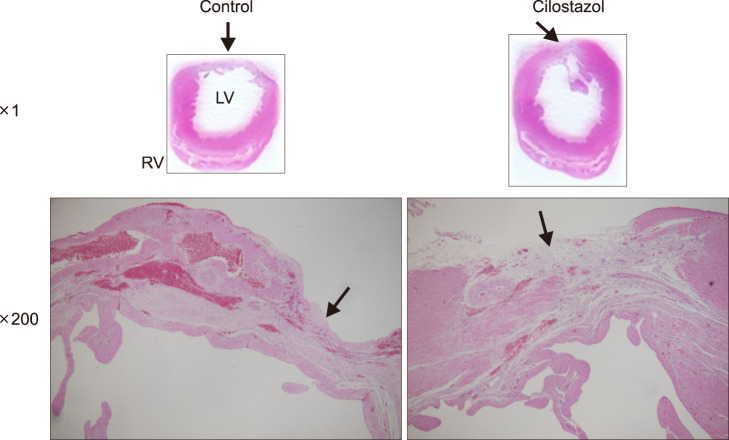
The cross sectional myocardial tissue image stained with hematoxylin and eosin showed macrophage infiltration, granulation tissue and fibrosis, representing ischemic necrosis of the myocardium (Fig. 3).
FIG. 3. Representative macroscopic (above) and microscopic (below) features of heart ventricle from control and cilostazol groups at 5 weeks after induction of myocardial infarct of the anterolateral left ventricular wall. LV: left ventricle, RV: right ventricle. Arrows indicate the infarcted area (×1). Note the macrophage infiltration, the formation of granulation tissue and the fibrotic change (×200), which were reduced by cilostazol treatment.
4. BNP expression in cardiac tissue
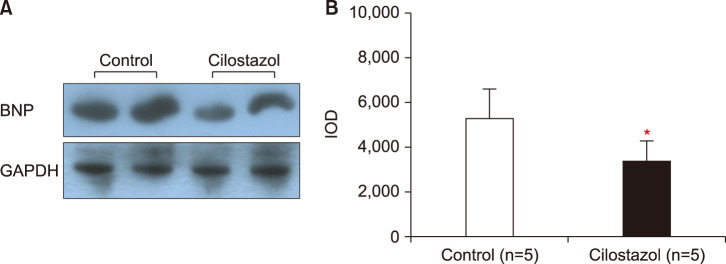
After scarification of experimental animals, both ventricles were excised and the level of BNP expression in ventricular tissue was assessed using the Western blot technique. Significantly higher expression was observed in the control group as compared with the cilostazol group (Fig. 4).
FIG. 4. Effects of cilostazol on the level of brain natriuretic peptide (BNP) expression in heart ventricles. The left panel shows representative immunoblots at 5 weeks after induction of myocardial infarct of the anterolateral left ventricular wall (A), and the right panel shows densitometric data (B). IOD: integrated optical density, GAPDH: Glyceraldehyde-3-Phosphate Dehydrogenase. Values are mean±SD. *p<0.05 vs. control group. Expression of BNP was significantly reduced by cilostazol treatment.
5. Plasma catecholamine levels
Plasma epinephrine (11.71±0.94 ng/ml vs. 12.89±1.20 ng/ml) and norepinephrine (6.18±1.97 ng/ml vs. 7.65±2.80 ng/ml) levels were significantly lower in the cliostazol than in the control group (p<0.05 each) (Fig. 5).
FIG. 5. Effect of cilostazol treatment on plasma concentrations of epinephrine (A) and norepinephrine (B). After 5 weeks of treatment with cilostazol, plasma epinephrine and norepinephrine concentrations were decreased compared with control group. Values are mean±SD. *p<0.05 vs. control group.
DISCUSSION
Heart failure is characterized by activation of the sympathetic nervous system leading to high circulating catecholamine concentrations which correlate with cardiovascular morbidity. Chronic stress is related to over-activation of the sympathetic nervous system which adds insult in subjects with heart failure. A previous related study demonstrated that chronic restraint stress was associated with adverse ventricular remodeling expressed by low LVEF and increased tissue BNP expression in MI animal models.17 Several agents have been proposed as candidates for management of stress associated cardiac insult. However, there is no established treatment strategy. As mentioned, cilostazol has a potential effect on inhibition of catecholamine secretion.13,14,15 Therefore, the present study was aimed to determine whether cilostazol has a protective effect against stress associated adverse LV remodeling and the degree of LV remodeling was assessed by imaging modality, measurement of heart weight, histopathologic examination, and tissue BNP expression.
LV systolic function is usually impaired after serious MI, but its evolution after MI varies among individuals. Well known contributing factors affecting change of myocardial function include: recovery of coronary circulation, extent of initial ischemic injury, and degree of adverse post infarction remodeling.18,19 As an imaging modality, two-dimensional echocardiography has been widely used for estimation of LV systolic function, which is expressed by LVEF. In the present study, 5 weeks of cilostazol treatment was associated with significant improvement of LVEF. Observed differences in the present study might be associated with various effects of cilostazol but considering the same extent of myocardial injury represented by no difference of immediate post-op LV function, it is possible that cilostazol attenuated stress-associated, adverse ventricular remodeling in the present study.
Total LV mass is increased as heart failure advances and reflects a degree of adverse LV remodeling. Total LV mass is correlated with total heart weight and usually shows a linear correlation with body weight. In the present study, therefore, the total heart weight to total body weight ratio was adopted to exclude confounding effects. As a result, the total heart weight to total body weight ratio was lower in the cilostazol group as compared with the controls, suggesting that cilostazol suppresses the myocardial remodeling.
Among biochemical markers reflecting the degree of LV dysfunction, BNP has been proposed as the most sensitive one. It is secreted in myocardial tissue and its level correlates with LV filling pressure. BNP is not only useful for diagnosis of heart failure, but also one of the important prognostic biomarkers. In the present study, the level of BNP expression was reduced by cilostazol suggesting the beneficial effects of cilostazol on hemodynamics after MI.
Although cilostazol treatments attenuated degree of adverse LV remodeling mediated by stress in the present study, its underlying mechanism remains to be determined. Increased catecholamine secretion was observed universally in patients with heart failure.20,21 This hormone has been proposed as a key factor in the downward spiral of heart failure deterioration. Moreover, several studies have demonstrated that increased plasma catecholamine is associated with poor clinical outcomes.17,22 In the present study, cilostazol suppressed catecholamine secretion induced by restraint stress in post-infarct rats. In addition, cilostazol was shown to inhibit catecholamine secretion in adrenal chromaffin cells in vitro.23 It is likely that cilostazol attenuated adverse LV remodeling induced by ST at least in part through the inhibition of catecholamine release in post-infarct rats.
There were some limitations in this study. One was the lack of evaluation on the other pleotrophic effects of cilostazol such as vasodilatation, decreased platelet aggregation, and antioxidant effects. Further study is be needed to identify relevant mechanisms for its protective effects. The other important limitation was the absence of a control group without stress. Restrained stress was presumed to be a factor accounting for adverse LV remodeling, but there was no assigned group for administration of cilostazol without stress in present study. Additional experiment could be helpful for resolving this issue.
The present study showed that cilostazol preserves left ventricular systolic function and attenuates restraint stress induced remodeling in postinfarct rats. The beneficial effect was associated with the reduced catecholamine release.
ACKNOWLEDGEMENTS
This study was supported by grant of the Korean Society of Cardiology (2007-2-170).
Footnotes
CONFLICT OF INTEREST STATEMENT: None declared.
References
- 1.Witte DR, Bots ML, Hoes AW, Grobbee DE. Cardiovascular mortality in Dutch men during 1996 European football championship: longitudinal population study. BMJ. 2000;321:1552–1554. doi: 10.1136/bmj.321.7276.1552. [DOI] [PMC free article] [PubMed] [Google Scholar]
- 2.Gelernt MD, Hochman JS. Acute myocardial infarction triggered by emotional stress. Am J Cardiol. 1992;69:1512–1513. doi: 10.1016/0002-9149(92)90918-o. [DOI] [PubMed] [Google Scholar]
- 3.Chi JS, Kloner RA. Stress and myocardial infarction. Heart. 2003;89:475–476. doi: 10.1136/heart.89.5.475. [DOI] [PMC free article] [PubMed] [Google Scholar]
- 4.Leor J, Poole WK, Kloner RA. Sudden cardiac death triggered by an earthquake. N Engl J Med. 1996;334:413–419. doi: 10.1056/NEJM199602153340701. [DOI] [PubMed] [Google Scholar]
- 5.Gullette EC, Blumenthal JA, Babyak M, Jiang W, Waugh RA, Frid DJ, et al. Effects of mental stress on myocardial ischemia during daily life. JAMA. 1997;277:1521–1526. [PubMed] [Google Scholar]
- 6.Krittayaphong R, Light KC, Biles PL, Ballenger MN, Sheps DS. Increased heart rate response to laboratory-induced mental stress predicts frequency and duration of daily life ambulatory myocardial ischemia in patients with coronary artery disease. Am J Cardiol. 1995;76:657–660. doi: 10.1016/s0002-9149(99)80192-1. [DOI] [PubMed] [Google Scholar]
- 7.Quirce CM, Odio M, Solano JM. The effects of predictable and unpredictable schedules of physical restraint upon rats. Life Sci. 1981;28:1897–1902. doi: 10.1016/0024-3205(81)90296-4. [DOI] [PubMed] [Google Scholar]
- 8.Armario A, Lopez-Calderon A, Jolin T, Balasch J. Response of anterior pituitary hormones to chronic stress. The specificity of adaptation. Neurosci Biobehav Rev. 1986;10:245–250. doi: 10.1016/0149-7634(86)90011-4. [DOI] [PubMed] [Google Scholar]
- 9.Mansi JA, Drolet G. Chronic stress induces sensitization in sympathoadrenal responses to stress in borderline hypertensive rats. Am J Physiol. 1997;272(3 Pt 2):R813–R820. doi: 10.1152/ajpregu.1997.272.3.R813. [DOI] [PubMed] [Google Scholar]
- 10.Kambayashi J, Liu Y, Sun B, Shakur Y, Yoshitake M, Czerwiec F. Cilostazol as a unique antithrombotic agent. Curr Pharm Des. 2003;9:2289–2302. doi: 10.2174/1381612033453910. [DOI] [PubMed] [Google Scholar]
- 11.Hashimoto A, Miyakoda G, Hirose Y, Mori T. Activation of endothelial nitric oxide synthase by cilostazol via a cAMP/protein kinase A- and phosphatidylinositol 3-kinase/Akt-dependent mechanism. Atherosclerosis. 2006;189:350–357. doi: 10.1016/j.atherosclerosis.2006.01.022. [DOI] [PubMed] [Google Scholar]
- 12.Shinoda-Tagawa T, Yamasaki Y, Yoshida S, Kajimoto Y, Tsujino T, Hakui N, et al. A phosphodiesterase inhibitor, cilostazol, prevents the onset of silent brain infarction in Japanese subjects with Type II diabetes. Diabetologia. 2002;45:188–194. doi: 10.1007/s00125-001-0740-2. [DOI] [PubMed] [Google Scholar]
- 13.Choi JM, Shin HK, Kim KY, Lee JH, Hong KW. Neuroprotective effect of cilostazol against focal cerebral ischemia via antiapoptotic action in rats. J Pharmacol Exp Ther. 2002;300:787–793. doi: 10.1124/jpet.300.3.787. [DOI] [PubMed] [Google Scholar]
- 14.Shin HK, Kim YK, Kim KY, Lee JH, Hong KW. Remnant lipoprotein particles induce apoptosis in endothelial cells by NAD(P)H oxidase-mediated production of superoxide and cytokines via lectin-like oxidized low-density lipoprotein receptor-1 activation: prevention by cilostazol. Circulation. 2004;109:1022–1028. doi: 10.1161/01.CIR.0000117403.64398.53. [DOI] [PubMed] [Google Scholar]
- 15.Nishio Y, Kashiwagi A, Takahara N, Hidaka H, Kikkawa R. Cilostazol, a cAMP phosphodiesterase inhibitor, attenuates the production of monocyte chemoattractant protein-1 in response to tumor necrosis factor-alpha in vascular endothelial cells. Horm Metab Res. 1997;29:491–495. doi: 10.1055/s-2007-979086. [DOI] [PubMed] [Google Scholar]
- 16.Hallman H, Farnebo LO, Hamberger B, Johnsson G. A sensitive method for the determination of plasma catecholamines using liquid chromatography with electrochemical detection. Life Sci. 1978;23:1049–1052. doi: 10.1016/0024-3205(78)90665-3. [DOI] [PubMed] [Google Scholar]
- 17.Bonnefont-Rousselot D, Mahmoudi A, Mougenot N, Varoquaux O, Le Nahour G, Fouret P, et al. Catecholamine effects on cardiac remodelling, oxidative stress and fibrosis in experimental heart failure. Redox Rep. 2002;7:145–151. doi: 10.1179/135100002125000389. [DOI] [PubMed] [Google Scholar]
- 18.Masci PG, Ganame J, Francone M, Desmet W, Lorenzoni V, Iacucci I, et al. Relationship between location and size of myocardial infarction and their reciprocal influences on post-infarction left ventricular remodelling. Eur Heart J. 2011;32:1640–1648. doi: 10.1093/eurheartj/ehr064. [DOI] [PubMed] [Google Scholar]
- 19.Wu E, Ortiz JT, Tejedor P, Lee DC, Bucciarelli-Ducci C, Kansal P, et al. Infarct size by contrast enhanced cardiac magnetic resonance is a stronger predictor of outcomes than left ventricular ejection fraction or end-systolic volume index: prospective cohort study. Heart. 2008;94:730–736. doi: 10.1136/hrt.2007.122622. [DOI] [PubMed] [Google Scholar]
- 20.Venugopalan P, Agarwal AK. Plasma catecholamine levels parallel severity of heart failure and have prognostic value in children with dilated cardiomyopathy. Eur J Heart Fail. 2003;5:655–658. doi: 10.1016/s1388-9842(03)00109-0. [DOI] [PubMed] [Google Scholar]
- 21.Agarwal AK, Venugopalan P, Woodhouse C, de Bono D. Catecholamine levels in heart failure due to dilated cardiomyopathy and their relationship to the severity of heart failure. Eur J Heart Fail. 2000;2:261–263. doi: 10.1016/s1388-9842(00)00066-0. [DOI] [PubMed] [Google Scholar]
- 22.Viquerat CE, Daly P, Swedberg K, Evers C, Curran D, Parmley WW, et al. Endogenous catecholamine levels in chronic heart failure. Relation to the severity of hemodynamic abnormalities. Am J Med. 1985;78:455–460. doi: 10.1016/0002-9343(85)90338-9. [DOI] [PubMed] [Google Scholar]
- 23.Azuma M, Houchi H, Mizuta M, Kinoshita M, Teraoka K, Minakuchi K. Inhibitory action of cilostazol, a phosphodiesterase III inhibitor, on catecholamine secretion from cultured bovine adrenal chromaffin cells. J Cardiovasc Pharmacol. 2003;41 Suppl 1:S29–S32. [PubMed] [Google Scholar]


