ABSTRACT
Aims and Objectives:
The purpose of this study is to compare digital and conventional impression methods by preclinical students in terms of time and ease and to evaluate their preferences and future expectations.
Materials and Methods:
Twenty volunteered, 2nd year preclinical students (11 females and 9 males) participated in this study. Students took digital and conventional impressions of the left lower first molar which was made full ceramic crown preparation and opposite full arch from a typodont model (Frasaco, Frasaco GmbH, Tettnang, Germany). They used intraoral scanner (CEREC Omnicam, Sirona Dental GmbH, Bensheim, Germany) for digital impression and also used additional type (Express XT Penta H, 3M ESPE, Seefeld, Germany) and condensation type (Zetaplus, Zhermack SpA, Badia Polesine, Italy) silicones for conventional impression. Their taking impression time was measured. Before taking impression and after taking impression, two kinds of questionnaires were conducted to students about their preference, ease of impression methods, and their future expectations. Statistical analysis was performed by IBM SPSS 23 and Excel 2010 version. Differences between conventional and digital impression in terms of time were analyzed by student’s-t paired test and effect of gender was analyzed by students’s-t independent test.
Results:
There were statistically significant differences between digital and conventional impression methods in terms of taking impression and total impression time (P < 0.001). But there wasn’t any statistically significant difference between two methods in terms of preparation time. About 85% of students preferred the digital impression method and also 85% of students found that the digital impression method was easy. 95% of students expected to find intraoral scanner in the clinic where working first time.
Conclusions:
As a result of this study, it has been seen that the students preferred the digital impression method to the conventional impression method and found that the digital impression method was easier.
KEYWORDS: Computer-aided design/computer-aided manufacturing, conventional impression, digital impression, experience of dental students, impression methods
INTRODUCTION
According to Glossary of Prosthodontic Terms, the impression is “a negative likeness or copy in reverse of the surface of an object; an imprint of the teeth and adjacent structures for use in dentistry.[1] Taking impression is a very important step in the production of fixed prosthetic restorations such as crowns, bridges, inlays, onlays, and implants as well as removable dentures for a long time in dentistry.[2,3] In dentistry, impression was taken with conventional methods for many years and nowadays elastomeric impression materials especially polyvinyl siloxane and polyether are used very reliably in terms of impression accuracy.[3,4,5]
At the beginning of the 1980s, digital impression systems occurred as Werner Mörmann began to think about what could be done to develop one session treatment. He shared this idea with his electronic engineer friend, Marco Brandestini. In this way, it has been started to develop digital impression instruments with optical reading systems.[6,7,8]
Digital and conventional impression methods have some advantages and disadvantages compared to each other.[9,10,11] In conventional impression method, having a greater number of steps increases the possibility of making extra mistakes.[12,13,14] Standardization of the milling stage in the digital impression method and less step numbers reduce the possibility of mistakes and improves adaptability.[10,15,16,17] Digital methods are more preferable in terms of time and preference of clinicians.[18,19,20]
In the digital impression method, the possibility of a problem because of inadequency of impression details is less than conventional method. Even if there are fewer scanned places in the digital impression, only the missing areas can be scanned without making re-impression. Intraoral camera has less effect on the gag reflex than the impression tray. It is easier to store digital impression.[21]
The difficulty of scanning the distal part in the digital impression and requirement of titanium oxide powder spray for contrast (such as CEREC Bluecam systems) are some disadvantages of the digital system.[21,22]
In addition, the other disadvantages of the digital impression method are cost and requirement of extra education for using.
Dental students learn the conventional impression method in the dentistry education. It is also necessary to be informed the students about the technological innovations such as digital impression systems and how to apply them in their professional life.
The aim of this study is to compare the digital and conventional impression methods by preclinical students who have not taken digital impressions before and to investigate future expectations regarding the digital impression method. The null hypothesis of this search is that there are not any differences between taking digital and conventional impression time of students and also there are not any differences between preferences of students about conventional and digital impression methods.
MATERIALS AND METHODS
This study was reviewed and approved by the Istanbul Medipol University Non-Interventional Clinical Research Ethics Board (108-40098-604.01.01-E.20043).
20 volunteered 2nd year preclinical students (11 females and 9 males) which have any experiences about digital impression attended to this search. Informed consent forms were given to the students, and they were asked to sign. An instructor mentioned to students about the digital impression method; scanning, designing, and milling.
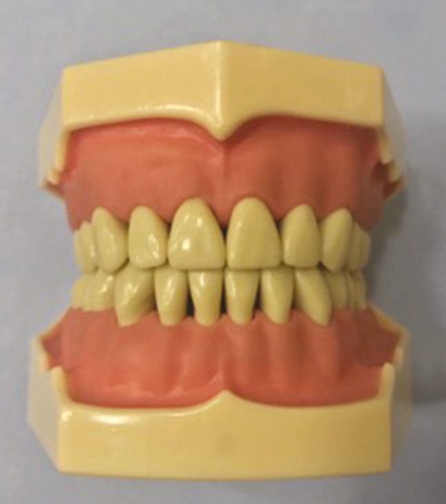
The lower typodont model (Frasaco, Frasaco GmbH, Tettnang, Germany) which had left lower first molar full-crown preparation was mounted on the simulated patient in the preclinical laboratory [Figure 1]. They made impression both digital and conventional methods on this simulated patient.
Figure 1.

Typodont model (Frasaco, Frasaco GmbH, Tettnang, Germany)
CONVENTIONAL IMPRESSION
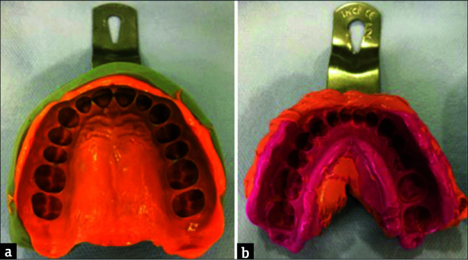
For the conventional impression method, the ideal impression tray was selected, and the lower jaw impression was taken using polyvinyl siloxane impression material (Express XT Penta H, 3M ESPE, Seefeld, Germany) with two-step impression technique in accordance with the manufacturer’s recommendation. The opposite arch impression was taken using condensation type silicone material (Zetaplus, Zhermack SpA, Badia Polesine, Italy) with one-step impression technique [Figure 2].
Figure 2.

(A) Conventional impression of upper jaw using condensation type silicone (left side), (B) conventional impression of lower jaw using additional type silicone (right side)
In the conventional impression method, preparation time included selecting impression tray, mixing of the impression material. Taking impression time started from the placement of the impression tray to mouth and ended when the impression item set. At the end, the tray is removed from the mouth. The total taking impression time was the total duration of these.
DIGITAL IMPRESSION
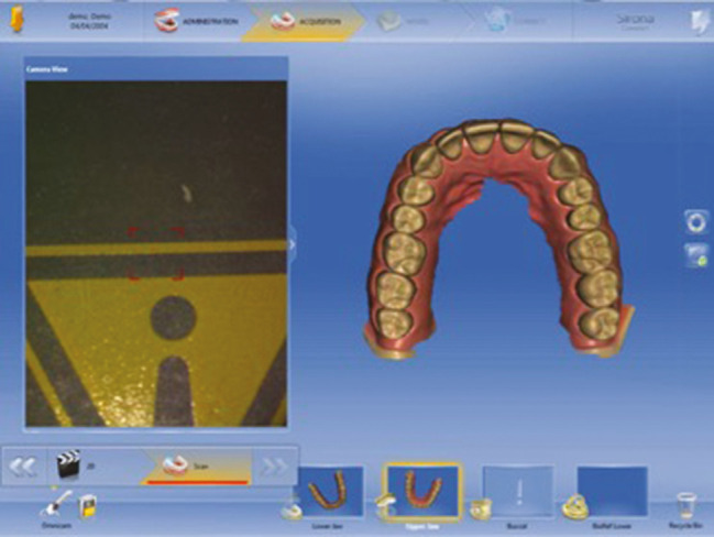
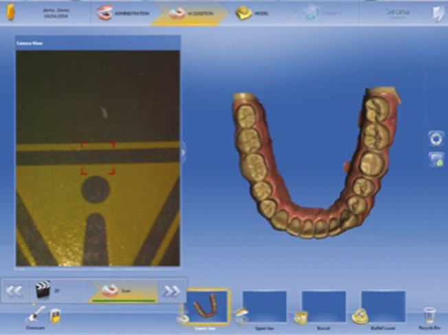
Intraoral scanner (CEREC Omnicam, Sirona Dental GmbH, Bensheim, Germany) was used for digital impression. An instructor showed to the student how they should hold intraoral scanner during the scanning. Then the upper and lower jaws were scanned individually [Figures 3 and 4]. Instructor assessed whether there was any deficiency in the impression.
Figure 3.

Digital impression of upper jaw using intraoral scanner
Figure 4.

Digital impression of lower jaw using intraoral scanner
In the digital impression method, preparation time included entering the patient’s information into system, the stages of describing how the intraoral scanner should be held during the scanning. The digital impression time included the time from the beginning of the intraoral scanning to the end of the scanning. Total taking impression time was the total duration of these.
QUESTIONNAIRE
Before taking impression, a questionnaire which asking about their expectations about conventional and digital impression methods was filled by students.
After taking impression, a questionnaire which was about the students’ experiences of taking impressions with digital and conventional methods and their expectations about the future method of taking digital impression was filled.
STATISTICAL ANALYSIS
Statistical analysis was performed by IBM SPSS 23 and Excel 2010 version (IBM Corporation, New York, NY, USA). Shapiro-Wilk test was used for checking the normality distribution of data. Differences between conventional and digital impression in terms of time and differences between upper and lower jaw in terms of time were analyzed by student’s-t paired test and differences between gender were analyzed by students’s-t independent test. The statistical significance level was set at P ≤ 0.001. The mean and standart deviation values of the conventional and digital impression preparation times were given. The power analysis was done by G*Power software (Ver. 3.0.10, Franz Faul, Universitat Kiel, Kiel, Germany) at a significance level of α = 0.05.
RESULTS
PREPARATION TIME
The mean preparation time of conventional impression for upper jaw were 1.73 (±0.678) minutes and the mean preparation time of digital impression for upper jaw were 1.54 (±0.265) minutes. The mean preparation time of conventional impression for lower jaw were 1.57 (±0.613) minutes and the mean preparation time of digital impression for lower jaw were 1.53 (±0.265) minutes. There were no statistically significant differences between conventional and digital impression methods in terms of the preparation time for upper jaw (P = 0.200) and lower jaw (P = 0.803). Table 1 includes the preparation time of conventional and digital impression methods for upper and lower jaw.
Table 1.
The preperation time of conventional and digital impression for upper and lower jaw
| Conventional impression (mean ± SD) | Digital impression (mean ± SD) | P | |
|---|---|---|---|
| Preperation time (for upper jaw/minutes) | 1.73 ± 0.678 | 1.54 ± 0.265 | 0.200 |
| Preperation time (for lower jaw/minutes) | 1.57 ± 0.613 | 1.53 ± 0.265 | 0.803 |
SD = standart deviation, statistical significance P ≤ 0.001
TAKING IMPRESSION TIME
The mean taking impression time of conventional impression for upper jaw were 11.33 (±3.54) minutes and the mean taking impression time of digital impression for upper jaw were 3.11 (±1.89) minutes. The mean taking impression time of conventional impression for lower jaw were 10.65 (±2.65) minutes and the mean taking impression time of digital impression for lower jaw were 4.54 (±2.03) minutes. There were statistically significant differences between conventional and digital impression methods in terms of the total impression time for upper jaw (P < 0.001) and lower jaw (P < 0.001). Table 2 includes the taking impression time of conventional and digital impression methods for upper and lower jaw.
Table 2.
The taking impression time of conventional and digital impression for upper and lower jaw
| Conventional impression (mean ± SD) | Digital impression (mean ± SD) | P | |
|---|---|---|---|
| Taking impression time (for upper jaw/minutes) | 11.33 ± 3.54 | 3.11 ± 1.89 | <0.001 |
| Taking impression time (for lower jaw/minutes) | 10.65 ± 2.65 | 4.54 ± 2.03 | <0.001 |
SD =: standart deviation, statistical significance P ≤ 0.001
TOTAL IMPRESSION TIME
The mean total impression time of conventional impression for upper jaw were 13.06 (±3.57) minutes and the mean total impression time of digital impression for upper jaw were 4.65 (±1.83) minutes. The mean total impression time of conventional impression for lower jaw were 12.21 (±2.54) minutes and the mean total impression time of digital impression for lower jaw were 6.07 (±2.06) minutes. There were statistically significant differences between conventional and digital impression methods in terms of the taking impression time for upper jaw (P < 0.001) and lower jaw (P < 0.001). Table 3 includes the total impression time of conventional and digital impression methods for upper and lower jaw.
Table 3.
The total impression time of conventional and digital impression for upper and lower jaw
| Conventional impression (mean ± SD) | Digital impression (mean ± SD) | P | |
|---|---|---|---|
| Total impression time (for upper jaw/minutes) | 13.06 ± 3.57 | 4.65 ± 1.83 | <0.001 |
| Total impression time (for lower jaw/minutes) | 12.21 ± 2.54 | 6.07 ± 2.06 | <0.001 |
SD = standart deviation, statistical significance P ≤ 0.001
EFFECT OF JAW POSITION ON THE TOTAL IMPRESSION TIME
There was no statistically significant difference between the upper jaw and lower jaw in terms of total impression time in conventional impression methods (P = 0.296) and digital impression methods (P = 0.009). Table 4 includes the total impression time of conventional and digital impression methods for upper and lower jaw.
Table 4.
The effect of jaw position on the total impression time of the conventional and digital impression methods
| Upper jaw (mean ± SD) | Lower jaw (mean ± SD) | P | |
|---|---|---|---|
| Total impression time (conventional/minutes) | 13.06 ± 3.57 | 12.21 ± 2.54 | 0.296 |
| Total impression time (digital/minutes) | 4.65 ± 1.83 | 6.07 ± 2.06 | 0.009 |
SD = standart deviation, statistical significance P ≤ 0.001
EFFECT OF GENDER ON THE TOTAL IMPRESSION TIME
There was no statistically significant difference between female and male in terms of total impression time taken with both impression methods from the upper and lower jaw. Table 5 includes the total impression time of conventional and digital impression methods for upper and lower jaw divided into groups of female and male.
Table 5.
The effect of gender on the total impression time of the conventional and digital impression methods
| Gender | N | Mean ± SD | P | |
|---|---|---|---|---|
| Conventional – total impression time (for upper jaw/minutes) | Male | 9 | 14.61 ± 3.168 | 0.079 |
| Female | 11 | 11.80 ± 3.51 | ||
| Conventional – total impression time (for lower jaw/minutes) | Male | 9 | 13.47 ± 2.656 | 0.040 |
| Female | 11 | 11.17 ± 2.00 | ||
| Digital – total impression time (for upper jaw/minutes) | Male | 9 | 4.37 ± 0.727 | 0.551 |
| Female | 11 | 4.88 ± 2.41 | ||
| Digital – total impression time (for lower jaw/minutes) | Male | 9 | 6.87 ± 2.117 | 0.121 |
| Female | 11 | 5.42 ± 1.86 |
SD = standart deviation, statistical significance P ≤ 0.001
EXPECTATIONS OF THE STUDENTS BEFORE TAKING IMPRESSION
About 55% of the participants were females. According to the results of the study, digital impression method was known <45% and conventional impression method was well known at 55%. Before taking impression, it is expected that the digital impression method would be easy at 75% and the conventional impression method would be difficult at 60% [Table 6].
Table 6.
Pretaking impression questionnaire
| n (%) | |
|---|---|
| Do you know digital impression method? | |
| I do not know | 3 (15) |
| I heard only its name | 8 (40) |
| I know a little bit | 9 (45) |
| I know moderately | 0 (0) |
| I know very well | 0 (0) |
| Do you know conventional impression method? | |
| I do not know | 0 (0) |
| I heard only its name | 0 (0) |
| I know a little bit | 0 (0) |
| I know moderately | 9 (45) |
| I know very well | 11 (55) |
| What kind of expectation do you have about the ease/difficulty of the digital impression method? | |
| Very easy | 2 (10) |
| Easy | 15 (75) |
| Very difficult | 0 (0) |
| Difficult | 0 (0) |
| No idea | 3 (15) |
| What kind of expectation do you have about the ease/difficulty of the conventional impression method? | |
| Very easy | 0 (0) |
| Easy | 8 (40) |
| Very difficult | 0 (0) |
| Difficult | 12 (60) |
| No idea | 0 (0) |
OPINIONS AFTER TAKING IMPRESSION
After taking impression, 45% of students found that the digital measurement method was easy. Conventional impression method was difficult at 55% [Table 7].
Table 7.
Posttaking impression questionnaire
| n (%) | |
|---|---|
| How was the ease/difficulty of the digital impression method? | |
| Very easy | 9 (45) |
| Easy | 8 (40) |
| Very difficult | 0 (0) |
| Difficult | 3 (15) |
| No idea | 0 (0) |
| How was the ease/difficulty of the conventional impression method? | |
| Very easy | 0 (0) |
| Easy | 9 (45) |
| Very difficult | 0 (0) |
| Difficult | 11 (55) |
| No idea | 0 (0) |
FUTURE EXPECTATIONS OF STUDENTS ABOUT DIGITAL IMPRESSION
About 95% of participants wanted to have a system that they could work with digital impression in the clinic where working the first time. 85% of participants thought that digital impression method would be the first choice impression method during their professional life [Table 8].
Table 8.
Future expectations of studentst
| n (%) | |
|---|---|
| Do you want to have a system that you can work with digital impression in the clinic where working the first time? | |
| Yes | 19 (95) |
| No | 1 (5) |
| Do you think that the digital impression method will be the first impression method for you in your professional life? | |
| Yes | 17 (85) |
| No | 3 (15) |
DISCUSSION
In the conventional impression method, preparation time included selecting impression tray, mixing of the impression material. Taking impression time started from the placement of the impression tray to mouth and ended when the impression item set. At the end, the tray is removed from the mouth. The total taking impression time was the total duration of these. In the digital impression method, preparation time included entering the patient’s information into system, the stages of describing how the intraoral scanner should be held during the scanning. The digital impression time included the time from the beginning of the intraoral scanning to the end of the scanning. Total taking impression time was the total duration of these. In this study, it was observed that the students took digital impression in a shorter time compared to the conventional method. It has proved that digital method was better in term of effectiveness. Thus, the first null hypothesis of the study was rejected. In our surveys, carried out before and after taking conventional and digital impressions, our goal was to investigate the change of ideas and preferences of the students about the impression method. Students preferred digital impression to conventional impression method. The second null hypothesis is also rejected.
In this study, typodont jaws were placed in the mouth of the model patients in the preclinic as they were in the studies of Lee and Gallucci,[19] Zitzmann et al.[23] and Marti et al.[24] Although the impression of implant was taken on the typodont jaw in the study of Lee and Gallucci.[19] and Zitzmann et al.,[23] impression of prepared left lower first molar was taken by conventional and digital methods in this study consistent with study of Marti et al.[24]
Only Omnicam system (CEREC Omnicam, Sirona Dental GmbH, Bensheim, Germany) was used as intraoral scanner in this study. Omnicam is a continuous video process. Lee and Gallucci[19] used iTero (Align Technology Inc., San Jose, CA, USA). iTero has taking still-frame mechanism. Marti et al.[24] used LAVA C. O. S. (3M ESPE, St. Paul, MN, USA) which has also video capturing mechanism. Zitzmann et al.[23] used TRIOS Pod System (3 Shape, Copenhagen, Denmark). This has single-shot photo-mode the as capturing technique. In our clinic, we already have Omnicam as a digital scanner. For this reason, Omnicam has been preferred.
Marti et al.,[24] Lee and Gallucci[19] and Zitzmann et al.[23] used alginate for conventional impression. For taking lower jaw impression, polyvinil siloxan (Express XT Penta H, 3M ESPE, Seefeld, Germany) was used and for taking upper jaw impression, condensation silicon (Zetaplus, Zhermach SpA, Badia Polesive, Italy) was used in our study. Our aim was to give details of prepared left lower first molar in the conventional impression. Opposite jaw impression did notneed details. The accuracy of condensation type impression is better than alginate, and we take impression from real patients in our clinic like this procedure. For these reason, polyvinil siloxan and condensation type silicon were used for upper jaw inconsistent with the other studies.
In contrast to the results of Marti et al.[24] and consistent with Lee and Gallucci.,[19] Zitzmann et al.,[23] the time of digital impression method was shorter than the conventional impression method. Marti et al.[24] mentioned that the training given before taking digital impression increased the duration of the digital impression method. It was thought that one of the reasons for the difference was the video lecture in addition to the training given by the trainer. Although studies of Lee and Gallucci.[19] and Zitzmann et al.[23] are the implant-based study, the results of this study are similar with them. The total digital impression time is shorter than conventional impression method.
Students were more familiar with the conventional method before taking the impression. This situation is thought to be due to the fact that the students took polyvinyl siloxane and condensation type silicone in the prosthetic courses at the preclinical laboratory while they did not take digital impression. They knew digital impression only as a theoretical course.
Students found the digital method easier than conventional method in this study, consistent with Lee and Gallucci.[19] and Zitzmann et al.[23] In the study of Marti et al.,[24] the students found the digital method more easily before they started to take impression. After completing the impression process, the students found the conventional method easier. In literature, the preference frequency of the digital measurement method is very high.[19,20,21]
In this study, the students reported that they wanted to work with digital impression method as 95% in the clinic where working first time and 85% of students would be the first choice of digital impression method in their professional life. Accompanied by this evidence, dentistry faculties should review the education systems and take technological innovations into their education by considering student’s preferences.
The limitations of this study are about using of a single intraoral scanner, working with any assistant, absence of steps such as die preparation, painting die spacer, and also patient’s satisfaction and attitude cannot be evaluated. In addition, pouring and mounting the cast did not measure in the conventional method. Our main limitation was that students learned conventional impression method at preclinical practical training. However, they had no idea about taking impression with digital impression methods. In future studies, it will be able to give theoretical and practical information about digital impression method to students during 6–8 months. After the information process, search should be started. In this study, only one intraoral scanner was used. In the future research, different intraoral scanner should be used, and these researches should identify differences between systems. Also in the future research, restorations should be manufactured from different type impression methods and compared with each other in terms of adaptation.
CONCLUSIONS
As a result of this study, it has been seen that the students preferred the digital impression method to the conventional impression method and found the digital impression method easier. The majority of students want to the digital impression system at first-time clinical experience in their professional lives. This choice is related that they want to see the reflection of the extremely developing digital age on their profession. Lecturers and dentistry faculties also should pay attention to add developing technologies to their educational contents and make their students well equipped about technological innovations.
FINANCIAL SUPPORT AND SPONSORSHIP
This study has not received any financial support or sponsorship.
CONFLICTS OF INTEREST
No potential conflict of interest was reported by the authors.
AUTHORS CONTRIBUTIONS
Halenur Bilir has to provide contributions planning stages of the study and Ceren Aygüzen has to provide contributions processing of the data. Both of them have to provide contributions reviewing the literature and writing the article.
ETHICAL POLICY AND INSTITUTIONAL REVIEW BOARD STATEMENT
This study was reviewed and approved by the Istanbul Medipol University Non-Interventional Clinical Research Ethics Board (108-40098-604.01.01-E.20043)
PATIENT DECLARATION OF CONSENT
Not applicable.
DATA AVAILABILITY STATEMENT
The authors confirm that the data supporting the findings of this study are available within the article
ACKNOWLEDGEMENT
No contribution to this study from any scientific research fund.
REFERENCES
- 1.The glossary of prosthodontic terms. J Prosthet Dent. (Ninth edition) 2017;117:e1–105. doi: 10.1016/j.prosdent.2016.12.001. [DOI] [PubMed] [Google Scholar]
- 2.Sailer I, Mühlemann S, Fehmer V, Hämmerle CH, Benic GI. Randomized controlled clinical trial of digital and conventional workflows for the fabrication of zirconia-ceramic fixed partial dentures. Part I: Time efficiency of complete-arch digital scans versus conventional impressions. J Prosthet Dent. 2018:S0022–3913(18) 30363–9. doi: 10.1016/j.prosdent.2018.04.021. [DOI] [PubMed] [Google Scholar]
- 3.Rau CT, Olafsson VG, Delgado AJ, Ritter AV, Donovan TE. The quality of fixed prosthodontic impressions: An assessment of crown and bridge impressions received at commercial laboratories. J Am Dent Assoc. 2017;148:654–60. doi: 10.1016/j.adaj.2017.04.038. [DOI] [PubMed] [Google Scholar]
- 4.Seelbach P, Brueckel C, Wöstmann B. Accuracy of digital and conventional impression techniques and workflow. Clin Oral Investig. 2013;17:1759–64. doi: 10.1007/s00784-012-0864-4. [DOI] [PubMed] [Google Scholar]
- 5.Mehl A, Ender A, Mörmann W, Attin T. Accuracy testing of a new intraoral 3D camera. Int J Comput Dent. 2009;12:11–28. [PubMed] [Google Scholar]
- 6.Mörmann WH. The origin of the cerec method: A personal review of the first 5 years. Int J Comput Dent. 2004;7:11–24. [PubMed] [Google Scholar]
- 7.Mörmann WH. The evolution of the CEREC system. J Am Dent Assoc. 2006;137:7S–13S. doi: 10.14219/jada.archive.2006.0398. [DOI] [PubMed] [Google Scholar]
- 8.Rekow ED. Dental CAD/CAM systems: A 20-year success story. J Am Dent Assoc. 2006;137 Suppl:5S–6S. doi: 10.14219/jada.archive.2006.0396. [DOI] [PubMed] [Google Scholar]
- 9.Su TS, Sun J. Comparison of marginal and internal fit of 3-unit ceramic fixed dental prostheses made with either a conventional or digital impression. J Prosthet Dent. 2016;116:362–7. doi: 10.1016/j.prosdent.2016.01.018. [DOI] [PubMed] [Google Scholar]
- 10.Abdel-Azim T, Rogers K, Elathamna E, Zandinejad A, Metz M, Morton D. Comparison of the marginal fit of lithium disilicate crowns fabricated with CAD/CAM technology by using conventional impressions and two intraoral digital scanners. J Prosthet Dent. 2015;114:554–9. doi: 10.1016/j.prosdent.2015.04.001. [DOI] [PubMed] [Google Scholar]
- 11.Amin S, Weber HP, Finkelman M, El Rafie K, Kudara Y, Papaspyridakos P. Digital vs. Conventional full-arch implant impressions: A comparative study. Clin Oral Implants Res. 2017;28:1360–7. doi: 10.1111/clr.12994. [DOI] [PubMed] [Google Scholar]
- 12.Wöstmann B, Rehmann P, Trost D, Balkenhol M. Effect of different retraction and impression techniques on the marginal fit of crowns. J Dent. 2008;36:508–12. doi: 10.1016/j.jdent.2008.03.013. [DOI] [PubMed] [Google Scholar]
- 13.Ender A, Mehl A. In-vitro evaluation of the accuracy of conventional and digital methods of obtaining full-arch dental impressions. Quintessence Int. 2015;46:9–17. doi: 10.3290/j.qi.a32244. [DOI] [PubMed] [Google Scholar]
- 14.Chochlidakis KM, Papaspyridakos P, Geminiani A, Chen CJ, Feng IJ, Ercoli C. Digital versus conventional impressions for fixed prosthodontics: A systematic review and meta-analysis. J Prosthet Dent. 2016;116:184–190.e12. doi: 10.1016/j.prosdent.2015.12.017. [DOI] [PubMed] [Google Scholar]
- 15.Lin WS, Harris BT, Elathamna EN, Abdel-Azim T, Morton D. Effect of implant divergence on the accuracy of definitive casts created from traditional and digital implant-level impressions: An in vitro comparative study. Int J Oral Maxillofac Implants. 2015;30:102–9. doi: 10.11607/jomi.3592. [DOI] [PubMed] [Google Scholar]
- 16.Gimenez-Gonzalez B, Hassan B, Özcan M, Pradíes G. An in vitro study of factors influencing the performance of digital intraoral impressions operating on active wavefront sampling technology with multiple implants in the edentulous maxilla. J Prosthodont. 2017;26:650–5. doi: 10.1111/jopr.12457. [DOI] [PubMed] [Google Scholar]
- 17.Papaspyridakos P, Gallucci GO, Chen CJ, Hanssen S, Naert I, Vandenberghe B. Digital versus conventional implant impressions for edentulous patients: Accuracy outcomes. Clin Oral Implants Res. 2016;27:465–72. doi: 10.1111/clr.12567. [DOI] [PubMed] [Google Scholar]
- 18.Yuzbasioglu E, Kurt H, Turunc R, Bilir H. Comparison of digital and conventional impression techniques: Evaluation of patients’ perception, treatment comfort, effectiveness and clinical outcomes. BMC Oral Health. 2014;14:10. doi: 10.1186/1472-6831-14-10. [DOI] [PMC free article] [PubMed] [Google Scholar]
- 19.Lee SJ, Gallucci GO. Digital vs. Conventional implant impressions: Efficiency outcomes. Clin Oral Implants Res. 2013;24:111–5. doi: 10.1111/j.1600-0501.2012.02430.x. [DOI] [PubMed] [Google Scholar]
- 20.Schepke U, Meijer HJ, Kerdijk W, Cune MS. Digital versus analog complete-arch impressions for single-unit premolar implant crowns: Operating time and patient preference. J Prosthet Dent. 2015;114:403–6.e1. doi: 10.1016/j.prosdent.2015.04.003. [DOI] [PubMed] [Google Scholar]
- 21.Ahlholm P, Sipilä K, Vallittu P, Jakonen M, Kotiranta U. Digital versus conventional impressions in fixed prosthodontics: A review. J Prosthodont. 2018;27:35–41. doi: 10.1111/jopr.12527. [DOI] [PubMed] [Google Scholar]
- 22.Mühlemann S, Benic GI, Fehmer V, Hämmerle CH, Sailer I. Randomized controlled clinical trial of digital and conventional workflows for the fabrication of zirconia-ceramic posterior fixed partial dentures. Part II: Time efficiency of CAD-CAM versus conventional laboratory procedures. J Prosthet Dent. 2018:S0022–3913(18) 30362–7. doi: 10.1016/j.prosdent.2018.04.020. [DOI] [PubMed] [Google Scholar]
- 23.Zitzmann NU, Kovaltschuk I, Lenherr P, Dedem P, Joda T. Dental students’ perceptions of digital and conventional impression techniques: A randomized controlled trial. J Dent Educ. 2017;81:1227–32. doi: 10.21815/JDE.017.081. [DOI] [PubMed] [Google Scholar]
- 24.Marti AM, Harris BT, Metz MJ, Morton D, Scarfe WC, Metz CJ, et al. Comparison of digital scanning and polyvinyl siloxane impression techniques by dental students: Instructional efficiency and attitudes towards technology. Eur J Dent Educ. 2017;21:200–5. doi: 10.1111/eje.12201. [DOI] [PubMed] [Google Scholar]
Associated Data
This section collects any data citations, data availability statements, or supplementary materials included in this article.
Data Availability Statement
The authors confirm that the data supporting the findings of this study are available within the article


