Abstract
We report a case of a 53-year-old lady who was incidentally diagnosed to have giant anterior mediastinal mass while undergoing preoperative evaluation for another surgery. She came for surgery after 2 years when she became symptomatic. A large 6.7-lb (2800 g) tumor occupying both hemithoraces and engulfing heart was excised in its entirety through a clamshell thoracotomy under cardiopulmonary bypass standby. Histopathology revealed the final diagnosis as well as differentiated liposarcoma. She is now able to walk 2 km without any symptoms at the end of a 24-month follow-up.
Keywords: Mediastinal tumors, Liposarcoma, Clamshell thoracotomy
Introduction
Mediastinal liposarcoma is a rare entity and only accounts for 0.1 to.75% of all mediastinal tumors [1]. Liposarcomas are classified histopathologically into 4 subtypes: well-differentiated, dedifferentiated, myxoid, and pleomorphic [1] as per the World Health Organization. Among reported cases, 85% present with symptoms and 15% are found incidentally on imaging [2]. Symptoms typically arise from compression of intrathoracic structures and include dyspnea, tachypnea, chest pain, wheezing, hoarseness, superior vena cava compression, arrhythmias, heart failure, and syncope. We report a case of incidentally detected liposarcoma in a lady which grew into a giant tumor and became symptomatic after 2 years of diagnosis. She successfully underwent complete excision of this tumor by clamshell thoracotomy approach. Histopathology revealed it to be a well-differentiated variety of liposarcoma.
Case report
A 53-year-old lady with known case of hypertension, hyper-reactive airway disease, and status post hysterectomy 10 years back developed incisional hernia at previous operative site. While undergoing preoperative evaluation for incisional hernioplasty, she was detected to have large superior and anterior mediastinal mass on chest roentgenography. As she had no symptoms related to this lesion, she refused further investigation and surgical treatment. Two years later she presented with history of nonproductive cough and dyspnea on exertion New York Heart Association class II which rapidly progressed within a week to class IV. Chest computed tomography (CT) revealed a 20 × 21 × 21 cm anterior mediastinal mass abutting pericardium and aortic arch, involving both hemithoraces with variable degree of segmental and subsegmental atelectasis of the lungs (Fig. 1). Histopathology report of CT-guided biopsy done at the same time was suggestive of thymolipoma or liposarcoma.
Fig. 1.
Preoperative radiological images. a Preoperative chest X-ray. b Computed tomography scan in lateral decubitus showing a huge mediastinal mass engulfing heart and great vessels extending bilaterally. c CT images of serial axial sections depicting the tumor.
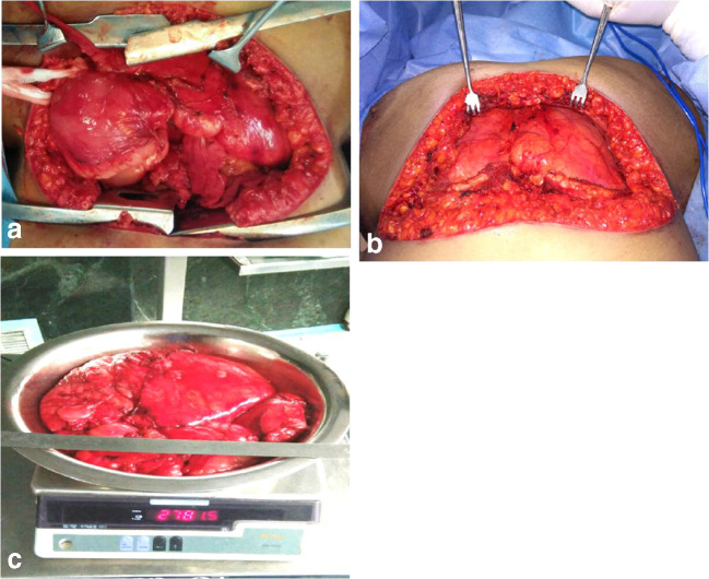
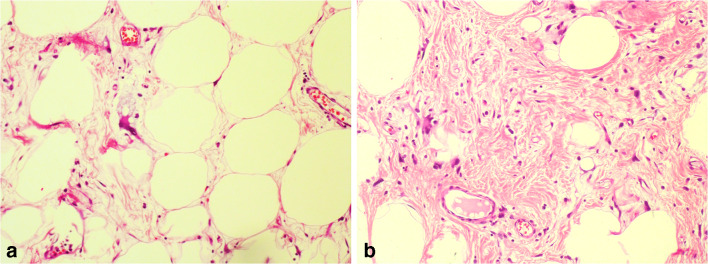
Surgical excision of the mass was planned with clamshell thoracotomy approach under cardiopulmonary bypass standby in view of extension of the lesion into both hemithoraces. She became unstable on induction of anesthesia, developed bradycardia, and unrecordable blood pressure in supine position. She was quickly shifted to semi-recumbent position and bilateral anterior thoracotomy was done expeditiously to decompress the chest. Femoral sites were simultaneously exposed for instituting cardiopulmonary bypass although it was not required as she quickly stabilized hemodynamically. Bilateral thoracotomy was converted to clamshell incision electively and proceeded. A huge mass weighing 6.7 lb (2.8 kg) was excised in toto (Fig. 2). She was extubated on first postoperative day and subsequent recovery was uneventful. She was able to walk out of the hospital on the eighth postoperative day. Surgical histopathology demonstrated a 6.17-lb (2800 g), well-differentiated liposarcoma (Fig. 3). At follow-up at the end of 24 months, her preoperative symptoms were completely resolved and are now able to carry out her routine physical activities and walk 2 km comfortably.
Fig. 2.
Operative images. a Operative image showing the giant tumor approached through clamshell thoracotomy. b Lungs expanding bilaterally after complete excision of tumor. c Surgical specimen of the tumor
Fig. 3:
Histopathological images. a Histopathology slide showing atypical adipocytes amidst normal adipocytes. b Histopathology slide showing atypical adipocytes in fibrous septa
Discussion
Mediastinal liposarcomas account for 0.1 to 0.75% of all mediastinal tumors [1]. They usually present in the fifth decade and the male to female distribution is equal [3]. Among four described varieties by World Health Organization, myxoid and pleomorphic tumors are associated with increased mortality [4], while differentiated liposarcomas are known to have better prognosis. However, well-differentiated liposarcomas are associated with high risk of local recurrence with 5-year local recurrence rates of approximately 50% [5, 6]. Hence, complete resection of the tumor is necessary. Mediastinal liposarcomas grow slowly and are unlikely to invade surrounding organs. Many patients remain asymptomatic until their tumor is very large becoming a “giant” liposarcoma encroaching on to the great vessels or the heart or the lungs. Small liposarcomas of the mediastinum can be resected by videothoracoscopy or robotic-assisted thoracoscopy. Larger tumors often require an open approach such as sternotomy, thoracotomy, clamshell, or hemiclamshell incisions. Clamshell or bilateral thoracotomy will be required if the tumor is extensive involving both the hemithoraces as in our case.
Conclusion
Giant mediastinal liposarcomas are rare tumors. Epidemiologically, they are most common in the fifth or sixth decade of life with equal sex distribution. By the time the patient becomes symptomatic, tumors invariably become “giant” encroaching vital structures in the chest and creating a challenge for surgical excision and may often require cardiopulmonary bypass support. Surgical approaches for optimal resection are debatable and varied and literature on it is scarce. We present a 53-year-old lady with well-differentiated liposarcoma who was incapacitated by symptoms and successfully underwent excision via a clamshell thoracotomy. She had an uneventful postoperative course and now at the end of the 24-month follow-up, she is able to walk 2 km comfortably. Three important lessons can be learned from this case. First, we should have cardiopulmonary bypass on standby and used expeditiously if required. Second, clamshell thoracotomy is the desired and unparalleled approach for tumors with this type of anatomical location. Third, complete excision of the tumor is of paradigm importance to avoid recurrence proved by a 2-year follow-up in this case.
Funding information
We declare that no funding is involved in repoting of this article.
Compliance with Ethical Standards
Conflict of Interest
We declare that there is no conflict of interest.
Human and Animal Rights and Informed Consent
We declared that there is no violation of Human /Animal rights during this case reporting.
Informed consent could not be obtained from the patient. Detailed explanation and all required documents along with ethical statement had been submiited already.
Footnotes
Publisher’s note
Springer Nature remains neutral with regard to jurisdictional claims in published maps and institutional affiliations.
References
- 1.World Health Organization In . In: World Health Organization classification of tumours, pathology and genetics of tumours of soft tissue and bone. Fletcher CD, Unni KK, Mertend F, editors. Lyon: IARC Press; 2002. [Google Scholar]
- 2.Schweitzer DL, Aguam AS. Primary liposarcoma of the mediastinum. Report of a case and review of the literature. J Thorac Cardiovasc Surg. 1977;74:83–97. doi: 10.1016/S0022-5223(19)41418-9. [DOI] [PubMed] [Google Scholar]
- 3.Chen M, Yang J, Zhu L, Zhou C, Zhao H. Primary intrathoracic liposarcoma: a clinicopathologic study and prognostic analysis of 23 cases. J Cardiothorac Surg. 2014;9:119. doi: 10.1186/1749-8090-9-119. [DOI] [PMC free article] [PubMed] [Google Scholar]
- 4.Boland Jennifer M., Colby Thomas V., Folpe Andrew L. Liposarcomas of the Mediastinum and Thorax. The American Journal of Surgical Pathology. 2012;36(9):1395–1403. doi: 10.1097/PAS.0b013e3182562bc1. [DOI] [PubMed] [Google Scholar]
- 5.Dalal Kimberly Moore, Antonescu Cristina R., Singer Samuel. Diagnosis and management of lipomatous tumors. Journal of Surgical Oncology. 2008;97(4):298–313. doi: 10.1002/jso.20975. [DOI] [PubMed] [Google Scholar]
- 6.Park James O., Qin Li-Xuan, Prete Francesco P., Antonescu Cristina, Brennan Murray F., Singer Samuel. Predicting Outcome by Growth Rate of Locally Recurrent Retroperitoneal Liposarcoma. Annals of Surgery. 2009;250(6):977–982. doi: 10.1097/SLA.0b013e3181b2468b. [DOI] [PMC free article] [PubMed] [Google Scholar]


