Abstract
Lichen sclerosus (LS) was first described by Hallopeau in 1887. It is a chronic inflammatory condition most commonly involving the anogenital region with a relapsing course and a potential for destruction, functional impairment, atrophy, and malignant changes. LS affects both sexes with a female preponderance of 5:1. The exact prevalence of the disease is difficult to predict as the lesions are asymptomatic in the initial phase and later when the complications arise patients might visit the surgeon, pediatrician, gynecologist, or urologist. The etiology of LS has a complex interplay of genetic factors, autoimmunity, infections, and trauma. Physical examination to assess the extent of the disease and decide the line of management is the most crucial step in the management. Corticosteroids, calcineurin inhibitor, retinoids, phototherapy, and surgery can be helpful. Self-examination and long-term follow-up are necessary.
Keywords: Balanitis xerotica, kraurosis vulvae, lichen sclerosus
INTRODUCTION
Lichen sclerosus (LS) was first described by Hallopeau in 1887.[1] It is a chronic inflammatory condition most commonly involving the anogenital region with a relapsing course and a potential for destruction, functional impairment, atrophy, and malignant changes.
Synonyms such as “lichen sclerosus et atrophicus,” “guttate scleroderma,” “white spot disease,” “kraurosis vulvae,” and “balanitis xerotica” have been abandoned and replaced by “Lichen sclerosus.”[2]
EPIDEMIOLOGY
LS affects both sexes with a female preponderance of 5:1. The exact prevalence of the disease is difficult to predict as the lesions are asymptomatic in the initial phase and later when the complications arise patients might visit the surgeon, pediatrician, gynecologist, or urologist. The estimated prevalence was 0.1%–0.3% of all the patients referred to dermatology department.[3] A prepubertal onset was observed in 9% cases, 40% in the reproductive age, and 50% in postmenopausal females.[4] The disease occurs in prepubertal children and postmenopausal women. It affects men in their fourth decade. A retrospective study by Kizer et al. estimated an incidence of 0.07% in men.[5] The highest incidence is between the age of 9–11 in boys.[6]
ETIOLOGY
The etiology of LS has a complex interplay of genetic factors, autoimmunity, infections, and trauma.
GENETIC FACTORS
Familial cases have been reported in LS. The disease is commonly seen in Caucasians, as compared to other ethnicities.[7]
Positive family history is noted in 12% of women with vulvar LS.[8] The prevalence of vulvar carcinoma was reported to be high in LS patients with a positive family history. Familial cases of LS are at an increased risk of developing other autoimmune disorders.
Human leukocyte antigen associated with disease were HLA DQ7 (in both men and women), HLA DQ8, HLA DQ9 (more in women), HLA 11 and HLA 12 (more in males).[9,10]
AUTOIMMUNITY
Various autoimmune diseases have been reported, especially in women with vulvar LS, hinting toward autoimmunity as an etiological factor in LS.[11] In a large study, 21.5% of patients with vulvar LS suffered from autoimmune diseases with thyroiditis being the most common (12%) followed by alopecia areata (9%), vitiligo (6%), and pernicious anemia (2%).[12] Another study conducted on females with biopsy-proven LS showed 30% incidence of autoimmune thyroid disease.[13] A family history of other autoimmune diseases has been found in 21% LS patients.[14]
Autoimmunity is not very important in male LS. Lipscombe et al. reported that 19% of their patients had a family history of other autoimmune diseases and only 6% of patients had an associated autoimmune disease.[15]
Anti-BP180 and anti-BP 230 antibodies have been identified in the sera of 30% of the affected patients.[16,17]
Immunoglobulin G autoantibodies against extracellular matrix protein 1 were found to be higher in males with LS compared to females.[9] The level was high in patients with the disease duration of >1 year suggesting its major role in progression and not in the development of the disease.
TRAUMA
The relative sparing of the uncovered areas in LS and rare occurrence of LS in men circumcised at birth[10] suggests the role of occlusion in causing the disease. Accumulation of smegma under the foreskin also contributes in the development of LS in uncircumcised males.
Genital instrumentation, trauma, genital jewelry, and surgery may lead to the disease, especially in circumcised males.[18]
Similarly, in females with early disease, the lesions remain sharply confined to the opposing mucosal surface.[19] Tissue damage during delivery may be one of the factors for predisposition, as the occurrence is found to be less in the nulliparous female.
Chronic irritation from underclothing and irritation caused by the urine have been implicated in the causation of LS. This is due to the micro-incontinence generally from a dysfunctional valve.[20] The pooled urine triggers chronic inflammation which later leads to sclerosis.
Trauma due to sexual abuse may be a trigger for the disease, especially in young girls.
INFECTIONS
Various infections have been implicated in causing LS, but their role is controversial.
There have been conflicting reports regarding the role of Borrelia burgdorferi in LS. Some authors from Europe have found evidence of Borrelia infection in the lesions of LS, but US-based studies have refuted this association.[21,22]
Human papillomavirus has been isolated from lesions of penile LS in the pediatric age group.[23]
Epstein-Barr Virus DNA has been found in around 25%–30% of patients with vulvar LS.[24]
Hepatitis C infection has been reported in some cases of LS.[25]
HORMONES
The occurrence of LS in prepubertal girls and postmenopausal women points toward the role of low estrogens.[3]
Friedrich observed low levels of dihydrotestosterone and suggested that a decreased activity of 5α-reductase was responsible for LS.[26] Clifton et al. reported low levels of androgen receptors on immunohistochemical tests performed on the lesions of both genital and extragenital LS.[27] This suggested a relationship between decreased androgen receptors and the progression of the disease.
Oral contraceptive pills with anti-androgenic properties are linked to the early onset of the disease in women.[28]
DRUGS
Rare reports of LS after treatment with carbamazepine for leprosy in a male patient have been noted and Imatinib for treatment in leukemia has been noted.[29]
CLINICAL FEATURES
Anogenital lichen sclerosus in women
The patient usually presents with itching which is worse at night and severe enough to disturb sleep. Chronic itching may lead to superimposed lichen simplex. Pain, dysuria, dyspareunia, and soreness are common complaints. A large number of patients develop sexual problems due to scarring from long-standing disease. The severity of the disease is not always related to the severity of the symptoms.[30,31] Many a times, the disease may be asymptomatic and detected as an incidental finding during examination.
The lesion starts as a well-defined erythematous plaque around the periclitoral fold along with edema. The affected skin becomes fragile and develops fissures, erosions, purpura, and ecchymoses. Fissuring is particularly noted between the urethra and clitoris, the interlabial space and the base of the posterior fourchette. Porcelain white papules and plaques with follicular delling and hyperkeratosis are pathognomonic. If left untreated the lesions become dry, sclerotic, hypopigmented, and atrophic giving an appearance of cellophane paper [Figure 1]. Excessive scarring results in the destruction of the labia along with the burying of the clitoral head.
Figure 1.
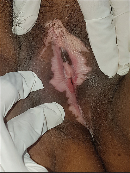
Atrophic porcelain white, cellophane paper skin with erosions; female genital lichen sclerosus
Periclitoral hood is the most common site (70%) followed by perineum (68%) and perianal fold (32%).[32] Other sites that may get affected are interlabial sulci, labia minora, and labia majora. The involvement of labial, perineal and anal areas gives the characteristic “figure of eight” shape also termed as “hourglass” or “keyhole.”[33] Extremely descriptive terms like “butterfly wings” and “lotus flower” are used by dermatologists with poetic propensities.[34] Lesions may extend till the genitocrural folds. Lesions can arise in an episiotomy scar. Mucosal involvement does not occur in LS and thus cervix and vagina are always spared unlike in lichen planus.[35]
Chronic adhesions can result in smegmatic pseudocyst.[36] Introital narrowing [Figure 2] due to fibrosis causes dyspareunia. Vulvar melanosis and genital lentiginosis with variegated pigmentation and irregular borders may be seen in long-standing cases which may mimic malignant melanoma clinically and histologically.[37]
Figure 2.
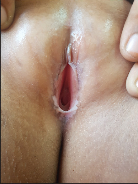
Late changes showing atrophy and clitoral resorption
Anogenital lichen sclerosus in girls
LS in girls can easily be misdiagnosed for sexual abuse.[36] However, it is believed that sexual abuse may trigger the disease in young girls. Furthermore, a diagnosis of LS does not rule out sexual abuse.
The mean age for the development of the disease in young girls is 5 years.[7] Itching and soreness are the most common presenting complaints. Erosions may result in dysuria, painful defecation, constipation, and bleeding. The sexually transmitted disease should be ruled out in cases with a poor response to treatment in older prepubertal girls[35] Figure 3.
Figure 3.
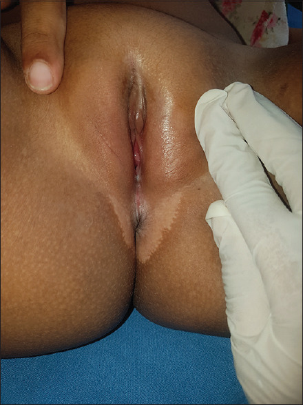
Lichen sclerosus in a young girl with hypopigmented plaque and fissuring
Common site affected is the perianal region causing infantile perineal protrusion (IPP).[38] It appears as a soft-tissue protrusion in the midline anterior to the anus. LS symptomatically improves with time but may not resolve by puberty. Only 25% of cases have shown resolution by menarche.[39]
The affected skin shows purpura and ecchymoses. Edema may or may not be present.
Genital lichen sclerosus in men
Men usually present with difficulty in retracting the preputial skin, dysuria, and sexual complaints. In the early course of the disease, there is grayish to bluish-white discoloration [Figure 4] of the inner surface of the prepuce and/or the glans. Many patients develop telangiectasia and the skin becomes thinned out and atrophic and results in nonretracting, tightened preputial skin leading to phimosis. Aynaud et al. have documented that 40% of the secondary phimosis in older males is due to LS. Atrophic skin is more prone to sexual trauma.[40]
Figure 4.
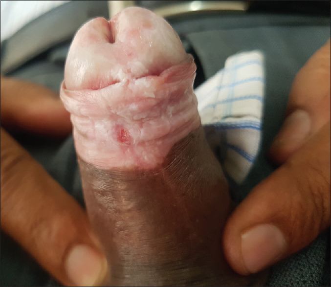
Tightened preputial skin with erythema and fissure
Preputial skin is affected in 70% cases, glans in 60%, both prepuce and glans in 40% and external meatus and urethra in 17%.[5] Meatus is first to get involved in the case of urethral disease [Figure 5] and may lead to inflammatory scarring, stenosis, and obstruction. Surgical intervention is required in such cases. Perianal involvement is a rare finding in males.[41]
Figure 5.
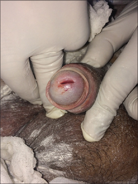
Lichen sclerosus in male showing inflammatory urethral involvement
Erosions, ulceration, purpura, and bulla may even be present. Long-standing disease may cause priapism and erectile dysfunction in 55% of cases, whereas urological symptoms such as dysuria and poor urinary flow may occur in 18%.[42]
Genital lichen sclerosus in boys
Phimosis is the presenting symptom in 40% of the cases. Glans, foreskin, and urethra are usually involved.[6] Perianal involvement is extremely rare. The evolution of the lesion is similar to adult males [Figure 6].
Figure 6.
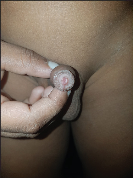
Early lichen sclerosus in a 6-year-old boy presenting with phimosis
Differential diagnoses
The two most common differential diagnoses are vitiligo and mucosal lichen planus [Tables 1 and 2],[43] Other differentials are plasma cell balanitis/vulvitis, genital graft vs. host reaction, Bowen's disease, candidial intertrigo, nevus depigmentosus, and idiopathic guttate hypomelanosis.
Table 1.
Difference between vitiligo and lichen sclerosus
| Vitiligo | LS |
|---|---|
| Lesions are asymptomatic | Lesions are associated with intense pruritus |
| Lesions are not indurated | Lesions are indurated |
| There is no atrophy in untreated lesions | Atrophy is characteristic |
| There are no destructive consequences | Narrowing of the introitus in females and phimosis in males is seen |
| Telangiectasia, ecchymoses and bullae are absent | Telangiectasia, ecchymoses and bullae may be present |
LS=Lichen sclerosus
Table 2.
Difference between mucosal lichen planus and lichen sclerosus
| Mucosal Lichen planus | LS |
|---|---|
| Violaceus papules are seen | Papules are atrophic and ivory white |
| Vagina and cervix are involved (vulvo-vaginal syndrome) | Vagina and cervix are not involved |
| Lesions are present in oral cavity | Oral cavity is infrequently involved |
LS=Lichen sclerosus
Genital lichen sclerosus and risk of malignancy
In girls with LS, the risk of squamous cell carcinoma (SCC) is present only if the disease persists past puberty. The estimated risk is around 4%–7%.[7,44] An immune dysregulation present in most cases of genital LS may precipitate the development of SCC.[45]
In postpubertal uncircumcised males, a pseudohyperplastic variant of SCC is commonly seen. Lesions are well-differentiated and nonverruciform, occurring over preexisting LS of the penile foreskin. The incidence may range from 2% to 50%. Malignant transformation takes around 10 years to develop. A high incidence of HPV 16 is associated with SCC occurring in cases of penile LS.[46]
Impact of social/sexual life
Quality of life is highly impaired in men and women of all ages. Sexual distress was revealed significantly in individuals. Due to persisting chronic pain, pruritus and inflammation, patient's private as well as work life is affected.[29]
Follow up and prognosis
LS runs a chronic course with progression or cyclical remission. Pruritus can be suppressed by almost 80% with therapy in female patients. Scarring in the late phase is irreversible.[4]
Males have an advantage over the females, as early interventions such as circumcision and application of potent topical steroids can cause cure rate of 50%–100%.[42,47,48]
The disease runs an unpredictable course in children. If it persists past puberty, close monitoring is required, as there is a greater chance of malignant transformation.
Regular follow-up is crucial in both males and females to detect early malignant transformation of the preexisting lesion.
Indications for biopsy:
To confirm the diagnosis
To differentiate early edematous stage from the late stage of scarring to decide the line of management
For early detection of carcinoma in situ or SCC.[35]
Histopathology
In the early stages, the histological features are uncharacteristic and subtle. Acanthosis, irregular hypergranulosis, and hyperkeratosis may be present with subepithelial edema, dilated blood vessels, and homogenized collagen. Infiltrate constitutes of lymphocytes with epidermal lymphocytic exocytosis. Lymphohistiocytic or lymphocytic vasculitis may be present.
In the advanced stage, there is a “trilayered” or “striped” appearance. Epidermis shows hyperkeratosis, follicular delling and hydropic basal cell degeneration. Upper dermis shows pallor with homogenization of collagen and mid-dermis shows band-like infiltrate composed of lymphoid cells admixed with plasma cells and histiocytes. Acanthosis is seen in long-standing vulvar LS.
Few complicated cases present with eosinophilic spongiosis and marked exocytosis; such cases are more resistant to treatment. Pathology of the disease is not conclusive for reaching a diagnosis and a clinicopathological co-relation is a must.
Management
Physical examination to assess the extent of the disease and decide the line of management is the most crucial step. A photographic evaluation of the lesion is a must for documentation.
In the maintenance phase of the disease, the patient is taught the technique of genital self-examination and instructed to report back to the physician in case of any perceived activity.
Aims of the treatment are:
To achieve symptomatic relief
To improve the appearance of the skin
To slow down or halt the progression of the disease and decrease the risk of complications.[25]
Corticosteroids
Highly potent topical corticosteroids (TCS) are the treatment of choice for genital LS in both adults as well as children due to their anti-inflammatory effects.[49]
British Association of Dermatologists recommends application of clobetasol propionate 0.05% ointment once daily at night for the first 4 weeks followed by alternate night application for another 4 weeks and further tapered to twice a week for 4 weeks.[50] In the tapering phase, the frequency of application may be increased if symptoms recur. Symptomatic improvement in 70% of the women has been noted with the therapy.[51] evidence level: 1–2; recommendation Grade: A.
Ecchymosis, erosions, and fissuring resolve; whereas pallor, atrophy, and scarring persist.
In men, topical steroids minimize the need for circumcision.[19]
Mid potent steroids such as mometasone furoate (0.1%) are effective in children in the early phase of the disease as well as for maintenance.[52,53,54]
Steroid-induced atrophy has not been reported in LS even with long-term use.
Intralesional steroids are useful in TCS resistant cases.[55] Evidence level: 1+; recommendation Grade: B.[29]
Topical calcineurin inhibitors
Topical calcineurin inhibitors (TCIs) are generally used for their anti-inflammatory and immunomodulatory effects.
Both pimecrolimus (1%) and tacrolimus (0.03%, 0.1%) are safe and effective in children and adults with severe genital LS. Twice daily application is recommended.[56,57]
However, clobetasol propionate has been found to be more effective than pimecrolimus.[58]
Transient burning and tingling sensation at the site of contact are common during the initial therapy and disappear gradually. The theoretical oncogenic potential of TCI does not make this modality an attractive first-line option. However, in one study, no lymphomas or other malignancies were reported.[59] Evidence level: 3; recommendation Grade: D (tacrolimus).
Retinoids
Tretinoin (0.025%) gel applied once daily is effective in genital LS, but it can cause irritation.[60]
Oral etretinate (1 mg/kg/day)[61] and acitretin (20–30 mg/day)[62] have shown improvement in clinical signs and symptoms, but the results are not very promising.
Although effective, topical, and oral retinoids have not been widely accepted due to their side effects and the need for long-term treatment. They are highly teratogenic and should be considered as maintenance therapy or in case of treatment failure with steroids. Evidence level: 3; recommendation Grade: D.
Hormones
Topical sex hormone creams such as estrogen (0.1%), testosterone (2%), and progesterone (2%) are not effective in genital LS.[63] Evidence level: 1+; recommendation Grade: A
Vitamins
Topical Vitamin E cream administered as an adjuvant has no added advantage over symptom control.[64]
Combined oral therapy with Vitamin A and E has shown improvement in an anecdotal case report of genital LS.[65] Evidence level: 2+; recommendation Grade: D.
Moisturizers
Moisturizers give symptomatic relief when used as an adjunct with other topical agents.[66]
Methotrexate
Methotrexate 10–15 mg/week in combination with systemic steroids over a period of 6 months has shown improvement in resistant generalized LS.[67] Evidence level: 3; recommendation Grade: D.
Cyclosporine
Cyclosporine (3–4 mg/kg/day) has shown improvement in refractory genital LS which may be sustained even after cessation of the drug.[68] The adverse effects of the drug were generally minor. Evidence level: 3; recommendation Grade: D.
Biologics
A single intralesional injection of adalimumab, a TNF-α binding biologic, was effective in a male with refractory LS.[69] Evidence level: 3; recommendation Grade: D.
Hydroxyurea
A serendipitous discovery by Tomson et al. in a woman suffering from vulvar LS with polycythemia rubra vera given hydroxyurea showed improvement in vulvar soreness and pruritus.[70] Evidence level: 3; recommendation Grade: D.
PHOTOTHERAPY
Psoralens with ultraviolet light
Topical psoralens with ultraviolet light therapy using 8-methoxypsoralen are effective in genital LS.[71,72]
The well-documented carcinogenic potential of phototherapy precludes its use as a first-line therapy.[29] Evidence level: 1+; recommendation Grade: B.
Photodynamic therapy
Various studies have shown the use of 5-aminolevulinic acid and argon dye lasers as a reasonably effective therapy in resistant genital LS in women.[73] However, pain, burning, and stinging sensation may occur in some patients. Evidence level: 3; recommendation Grade: D.
Surgery
Surgery should only be performed when the disease is inactive. It is curative in most cases of male genital LS.[74] Complete circumcision is the treatment of choice in uncomplicated LS of the glans and prepuce. Certain other surgeries include meatal dilatation, meatotomy, and meatoplasty. Urethral surgeries are performed in cases of hypospadias.[75]
In persistent and relapsing cases, procedures such as skin grafting or ablative laser resurfacing of the glans have been successful.[76]
In women, surgical interventions are mutilating and hence reserved for end-stage disease, particularly for cases of vulvar malignancies.[77] Surgeries such as vulvectomy, de-adhesiolysis/Z-plasty, CO2 hydro-dissection, dermabrasion, and cryosurgeries have been tried. Evidence level: 3; recommendation Grade: D.
CONCLUSION
Genital lesions are no man's land. Many specialists including urologists, gynaecologists, surgeons, and dermatologists handle genital problems. LS is an under-reported problem in both males and females. However, it can have important structural and functional connotations. Hence, early diagnosis and management are of utmost importance, especially in view of the small, but significant risk of malignancy.
Declaration of patient consent
The authors certify that they have obtained all appropriate patient consent forms. In the form the patient(s) has/have given his/her/their consent for his/her/their images and other clinical information to be reported in the journal. The patients understand that their names and initials will not be published and due efforts will be made to conceal their identity, but anonymity cannot be guaranteed.
Financial support and sponsorship
Nil.
Conflicts of interest
There are no conflicts of interest.
REFERENCES
- 1.Ridley C M. Genital lichen sclerosus (lichen sclerosus et atrophicus) in childhood and adolescence. Journal of the Royal Society of Medicine. 1993;86:69–75. [PMC free article] [PubMed] [Google Scholar]
- 2.Friedrich EG. New nomenclature for vulvar disease. Obstet Gynecol. 1976;124:147–54. [PubMed] [Google Scholar]
- 3.Wallace HJ. Lichen sclerosus et atrophicus. Trans St Johns Hosp Dermatol Soc. 1971;57:9–30. [PubMed] [Google Scholar]
- 4.Cooper SM, Gao XH, Powell JJ, Wojnarowska F. Does treatment of vulvar lichen sclerosus influence its prognosis? Arch Dermatol. 2004;140:702–6. doi: 10.1001/archderm.140.6.702. [DOI] [PubMed] [Google Scholar]
- 5.Nelson DM, Peterson AC. Lichen sclerosus: Epidemiological distribution in an equal access health care system. J Urol. 2011;185:522–5. doi: 10.1016/j.juro.2010.09.107. [DOI] [PubMed] [Google Scholar]
- 6.Kiss A, Király L, Kutasy B, Merksz M. High incidence of balanitis xerotica obliterans in boys with phimosis: Prospective 10-year study. Pediatr Dermatol. 2005;22:305–8. doi: 10.1111/j.1525-1470.2005.22404.x. [DOI] [PubMed] [Google Scholar]
- 7.Powell JJ, Wojnarowska F. Lichen sclerosus. Lancet. 1999;353:1777–83. doi: 10.1016/s0140-6736(98)08228-2. [DOI] [PubMed] [Google Scholar]
- 8.Powell J, Wojnarowska F. Childhood vulvar lichen sclerosus: An increasingly common problem. J Am Acad Dermatol. 2001;44:803–6. doi: 10.1067/mjd.2001.113474. [DOI] [PubMed] [Google Scholar]
- 9.Edmonds EV, Oyama N, Chan I, Francis N, McGrath JA, Bunker CB. Extracellular matrix protein 1 autoantibodies in male genital lichen sclerosus. Br J Dermatol. 2011;165:218–9. doi: 10.1111/j.1365-2133.2011.10326.x. [DOI] [PubMed] [Google Scholar]
- 10.Bunker CB, Edmonds E, Hawkins D, Francis N, Dinneen M. Re: Lichen sclerosus: Review of the literature and current recommendations for management: J M Pugliese, A F Morey and A C Peterson. J Urol. 2007;178:2268–2276. doi: 10.1016/j.juro.2007.08.024. J Urol 2009;181:1502-3. [DOI] [PubMed] [Google Scholar]
- 11.Powell J, Wojnrowska F, Winsey S, Marren P, Welsh K. Lichen sclerosus pre-menarche: Auto immunity and immunogenetics. Br J Dermatol. 2000;141:481–4. doi: 10.1046/j.1365-2133.2000.03360.x. [DOI] [PubMed] [Google Scholar]
- 12.Murphy R. Lichen sclerosus. Dermatol Clin. 2010;28:707–15. doi: 10.1016/j.det.2010.07.006. [DOI] [PubMed] [Google Scholar]
- 13.Leese GP, Flynn RV, Jung RT, Macdonald TM, Murphy MJ, Morris AD. Increasing prevalence and incidence of thyroid disease in Tayside, Scotland: The Thyroid Epidemiology Audit and Research Study (TEARS) Clin Endocrinol (Oxf) 2008;68:311–6. doi: 10.1111/j.1365-2265.2007.03051.x. [DOI] [PubMed] [Google Scholar]
- 14.Meyrick Thomas RH, Ridley CM, McGibbon DH, Black MM. Lichen sclerosus et atrophicus and autoimmunity – A study of 350 women. Br J Dermatol. 1988;118:41–6. doi: 10.1111/j.1365-2133.1988.tb01748.x. [DOI] [PubMed] [Google Scholar]
- 15.Lipscombe TK, Wayte J, Wojnarowska F, Marren P, Luzzi G. A study of clinical and aetiological factors and possible associations of lichen sclerosus in males. Australas J Dermatol. 1997;38:132–6. doi: 10.1111/j.1440-0960.1997.tb01129.x. [DOI] [PubMed] [Google Scholar]
- 16.Oyama N, Chan I, Neill SM, Hamada T, South AP, Wessagowit V, et al. Autoantibodies to extracellular matrix protein 1 in lichen sclerosus. Lancet. 2003;362:118–23. doi: 10.1016/S0140-6736(03)13863-9. [DOI] [PubMed] [Google Scholar]
- 17.Howard A, Dean D, Cooper S, Kirtshig G, Wojnarowska F. Circulating basement membrane zone antibodies are found in lichen sclerosus of the vulva. Australas J Dermatol. 2004;45:12–5. doi: 10.1111/j.1440-0960.2004.00026.x. [DOI] [PubMed] [Google Scholar]
- 18.Bunker CB. Male genital lichen sclerosus and tacrolimus. Br J Dermatol. 2007;157:1079–80. doi: 10.1111/j.1365-2133.2007.08179.x. [DOI] [PubMed] [Google Scholar]
- 19.Riddell L, Edwards A, Sherrard J. Clinical features of lichen sclerosus in men attending a department of genitourinary medicine. Sex Transm Infect. 2000;76:311–3. doi: 10.1136/sti.76.4.311. [DOI] [PMC free article] [PubMed] [Google Scholar]
- 20.Bunker CB, Sanjay Kulkarni, Guido Barbagli, Deepak Kirpekar, et al. Lichen sclerosus of the male genitalia and urethra: Surgical options and results in a multicenter international experience with 215 patients. Eur Urol. 2009;55:945–56. doi: 10.1016/j.eururo.2008.07.046. Eur Urol 2010;58:e55-6. [DOI] [PubMed] [Google Scholar]
- 21.DeVito JR, Merogi AJ, Vo T, Boh EE, Fung HK, Freeman SM, et al. Role of Borrelia burgdorferi in pathogenesis of morphoea and scleroderma and lichen sclerosus et atrophicus: A PCR study of thirty-five cases.Br. J Dermatol. 2000;142:481–4. [Google Scholar]
- 22.Fujiwara H, Fujiwara K, Hashimoto K, Mehregan AH, Schaumburg-Lever G, Lange R, et al. Detection of Borrelia burgdorferi DNA (B garinii or B afzelii) in morphea and lichen sclerosus et atrophicus tissues of German and Japanese but not of US patients. Arch Dermatol. 1997;133:41–4. [PubMed] [Google Scholar]
- 23.Drut RM, Gómez MA, Drut R, Lojo MM. Human papillomavirus is present in some cases of childhood penile lichen sclerosus: An in situ hybridization and SP-PCR study. Pediatr Dermatol. 1998;15:85–90. doi: 10.1046/j.1525-1470.1998.1998015085.x. [DOI] [PubMed] [Google Scholar]
- 24.Aidé S, Lattario FR, Almeida G, do Val IC, da Costa Carvalho M. Epstein-Barr virus and human papillomavirus infection in vulvar lichen sclerosus. J Low Genit Tract Dis. 2010;14:319–22. doi: 10.1097/LGT.0b013e3181d734f1. [DOI] [PubMed] [Google Scholar]
- 25.Val I, Almeida G. An overview of lichen sclerosus. Clin Obstet Gynecol. 2005;48:808–17. doi: 10.1097/01.grf.0000179635.64663.3d. [DOI] [PubMed] [Google Scholar]
- 26.Friedrich EG, Jr, Kalra PS. Serum levels of sex hormones in vulvar lichen sclerosus, and the effect of topical testosterone. N Engl J Med. 1984;310:488–91. doi: 10.1056/NEJM198402233100803. [DOI] [PubMed] [Google Scholar]
- 27.Clifton MM, Garner IB, Kohler S, Smoller BR. Immunohistochemical evaluation of androgen receptors in genital and extragenital lichen sclerosus: Evidence for loss of androgen receptors in lesional epidermis. J Am Acad Dermatol. 1999;41:43–6. doi: 10.1016/s0190-9622(99)70404-4. [DOI] [PubMed] [Google Scholar]
- 28.Günthert AR, Faber M, Knappe G, Hellriegel S, Emons G. Early onset vulvar lichen sclerosus in premenopausal women and oral contraceptives. Eur J Obstet Gynecol Reprod Biol. 2008;137:56–60. doi: 10.1016/j.ejogrb.2007.10.005. [DOI] [PubMed] [Google Scholar]
- 29.Kirtschig G, Cooper S, Aberer W, Gunthert A, Becker K, Jasaitiene D, et al. Evidence-based (S3) Guideline on (anogenital) Lichen sclerosus. JEADV. 2015;29:1–43. doi: 10.1111/jdv.13136. [DOI] [PubMed] [Google Scholar]
- 30.Breier F, Khanakah G, Stanek G, Kunz G, Aberer E, Schmidt B, et al. Isolation and polymerase chain reaction typing of Borrelia afzelii from a skin lesion in a seronegative patient with generalized ulcerating bullous lichen sclerosus et atrophicus. Br J Dermatol. 2001;144:387–92. doi: 10.1046/j.1365-2133.2001.04034.x. [DOI] [PubMed] [Google Scholar]
- 31.Dalziel KL. Effect of lichen sclerosus on sexual function and parturition. J Reprod Med. 1995;40:351–4. [PubMed] [Google Scholar]
- 32.Lorenz B, Kaufman RH, Kutzner SK. Lichen sclerosus. Therapy with clobetasol propionate. J Reprod Med. 1998;43:790–4. [PubMed] [Google Scholar]
- 33.Taussig FJ. Chronic leukoplakic vulvitis followed by cancer. Surg Clin North Am. 1922;2:1559–70. [Google Scholar]
- 34.Friedrich EG. Vulvar Disease. Philadelphia: WB Saunders; 1983. pp. 71–4. 129-42. [Google Scholar]
- 35.Neill SM, Lewis FM, Tatnall FM, Cox NH British Association of Dermatologists. British Association of dermatologists' guidelines for the management of lichen sclerosus 2010. Br J Dermatol. 2010;163:672–82. doi: 10.1111/j.1365-2133.2010.09997.x. [DOI] [PubMed] [Google Scholar]
- 36.Funaro D. Lichen sclerosus: A review and practical approach. Dermatol Ther. 2004;17:28–37. doi: 10.1111/j.1396-0296.2004.04004.x. [DOI] [PubMed] [Google Scholar]
- 37.Schaffer JV, Orlow SJ. Melanocytic proliferations in the setting of vulvar lichen sclerosus: Diagnostic considerations. Pediatr Dermatol. 2005;22:276–8. doi: 10.1111/j.1525-1470.2005.22325.x. [DOI] [PubMed] [Google Scholar]
- 38.Khachemoune A, Guldbakke KK, Ehrsam E. Infantile perineal protrusion. J Am Acad Dermatol. 2006;54:1046–9. doi: 10.1016/j.jaad.2006.02.029. [DOI] [PubMed] [Google Scholar]
- 39.Smith SD, Fischer G. Childhood onset vulvar lichen sclerosus does not resolve at puberty: A prospective case series. Pediatr Dermatol. 2009;26:725–9. doi: 10.1111/j.1525-1470.2009.01022.x. [DOI] [PubMed] [Google Scholar]
- 40.Aynaud O, Piron D, Casanova JM. Incidence of preputial lichen sclerosus in adults: Histologic study of circumcision specimens. J Am Acad Dermatol. 1999;41:923–6. doi: 10.1016/s0190-9622(99)70247-1. [DOI] [PubMed] [Google Scholar]
- 41.Fistarol SK, Itin PH. Diagnosis and treatment of lichen sclerosus: An update. Am J Clin Dermatol. 2013;14:27–47. doi: 10.1007/s40257-012-0006-4. [DOI] [PMC free article] [PubMed] [Google Scholar]
- 42.Edmonds EV, Hunt S, Hawkins D, Dinneen M, Francis N, Bunker CB. Clinical parameters in male genital lichen sclerosus: A case series of 329 patients. J Eur Acad Dermatol Venereol. 2012;26:730–7. doi: 10.1111/j.1468-3083.2011.04155.x. [DOI] [PubMed] [Google Scholar]
- 43.Kirtschig G, Becker K, Günthert A, Jasaitiene D, Cooper S, Chi CC, et al. Evidence-based (S3) guideline on (anogenital) lichen sclerosus. J Eur Acad Dermatol Venereol. 2015;29:e1–43. doi: 10.1111/jdv.13136. [DOI] [PubMed] [Google Scholar]
- 44.Powell J, Robson A, Cranston D, Wojnarowska F, Turner R. High incidence of lichen sclerosus in patients with squamous cell carcinoma of the penis. Br J Dermatol. 2001;145:85–9. doi: 10.1046/j.1365-2133.2001.04287.x. [DOI] [PubMed] [Google Scholar]
- 45.Cubilla AL, Velazquez EF, Young RH. Pseudohyperplastic squamous cell carcinoma of the penis associated with lichen sclerosus. An extremely well-differentiated, nonverruciform neoplasm that preferentially affects the foreskin and is frequently misdiagnosed: A report of 10 cases of a distinctive clinicopathologic entity. Am J Surg Pathol. 2004;28:895–900. doi: 10.1097/00000478-200407000-00008. [DOI] [PubMed] [Google Scholar]
- 46.Hagedorn M, Golüke T, Mall G. Lichen sclerosus and squamous cell carcinoma of the vulva. J Dtsch Dermatol Ges. 2003;1:864–8. doi: 10.1046/j.1439-0353.2003.03035.x. [DOI] [PubMed] [Google Scholar]
- 47.Becker K. Lichen sclerosus in boys. Dtsch Arztebl Int. 2011;108:53–8. doi: 10.3238/arztebl.2011.053. [DOI] [PMC free article] [PubMed] [Google Scholar]
- 48.Dalziel KL, Wojnarowska F. Long-term control of vulval lichen sclerosus after treatment with a potent topical steroid cream. J Reprod Med. 1993;38:25–7. [PubMed] [Google Scholar]
- 49.Dalziel KL, Millard PR, Wojnarowska F. The treatment of vulval lichen sclerosus with a very potent topical steroid (clobetasol propionate 0.05%) cream. Br J Dermatol. 1991;124:461–4. doi: 10.1111/j.1365-2133.1991.tb00626.x. [DOI] [PubMed] [Google Scholar]
- 50.Lagos BR, Maibach HI. Frequency of application of topical corticosteroids: An overview. Br J Dermatol. 1998;139:763–6. doi: 10.1046/j.1365-2133.1998.02498.x. [DOI] [PubMed] [Google Scholar]
- 51.Garzon MC, Paller AS. Ultrapotent topical corticosteroid treatment of childhood genital lichen sclerosus. Arch Dermatol. 1999;135:525–8. doi: 10.1001/archderm.135.5.525. [DOI] [PubMed] [Google Scholar]
- 52.Virgili A, Minghetti S, Borghi A, Corazza M. Proactive maintenance therapy with a topical corticosteroid for vulvar lichen sclerosus: Preliminary results of a randomized study. Br J Dermatol. 2013;168:1316–24. doi: 10.1111/bjd.12273. [DOI] [PubMed] [Google Scholar]
- 53.Kiss A, Csontai A, Pirót L, Nyirády P, Merksz M, Király L. The response of balanitis xerotica obliterans to local steroid application compared with placebo in children. J Urol. 2001;165:219–20. doi: 10.1097/00005392-200101000-00062. [DOI] [PubMed] [Google Scholar]
- 54.Patrizi A, Gurioli C, Medri M, Neri I. Childhood lichen sclerosus: A long-term follow-up. Pediatr Dermatol. 2010;27:101–3. doi: 10.1111/j.1525-1470.2009.01050.x. [DOI] [PubMed] [Google Scholar]
- 55.Baggish MD, Ventolini G. Lichen sclerosus: Subepidermal steroid injection therapy. A large, long term follow-up study. J Gynecol Surg. 2006;22:137–41. [Google Scholar]
- 56.Nissi R, Eriksen H, Risteli J, Niemimaa M. Pimecrolimus cream 1% in the treatment of lichen sclerosus. Gynecol Obstet Invest. 2007;63:151–4. doi: 10.1159/000096736. [DOI] [PubMed] [Google Scholar]
- 57.Li Y, Xiao Y, Wang H, Li H, Luo X. Low-concentration topical tacrolimus for the treatment of anogenital lichen sclerosus in childhood: Maintenance treatment to reduce recurrence. J Pediatr Adolesc Gynecol. 2013;26:239–42. doi: 10.1016/j.jpag.2012.11.010. [DOI] [PubMed] [Google Scholar]
- 58.Goldstein AT, Creasey A, Pfau R, Phillips D, Burrows LJ. A double-blind, randomized controlled trial of clobetasol versus pimecrolimus in patients with vulvar lichen sclerosus. J Am Acad Dermatol. 2011;64:e99–104. doi: 10.1016/j.jaad.2010.06.011. [DOI] [PubMed] [Google Scholar]
- 59.Böhm M, Frieling U, Luger TA, Bonsmann G. Successful treatment of anogenital lichen sclerosus with topical tacrolimus. Arch Dermatol. 2003;139:922–4. doi: 10.1001/archderm.139.7.922. [DOI] [PubMed] [Google Scholar]
- 60.Virgili A, Corazza M, Bianchi A, Mollica G, Califano A. Open study of topical 0025% tretinoin in the treatment of vulvar lichen sclerosus One year of therapy. J Reprod Med. 1995;40:614–8. [PubMed] [Google Scholar]
- 61.Romppanen U, Tuimala R, Ellmén J, Lauslahti K. Oral treatment of vulvar dystrophy with an aromatic retinoid, etretinate. Geburtshilfe Frauenheilkd. 1986;46:242–7. doi: 10.1055/s-2008-1035907. [DOI] [PubMed] [Google Scholar]
- 62.Bousema MT, Romppanen U, Geiger JM, Baudin M, Vähä-Eskeli K, Vartiainen J, et al. Acitretin in the treatment of severe lichen sclerosus et atrophicus of the vulva: A double-blind, placebo-controlled study. J Am Acad Dermatol. 1994;30:225–31. doi: 10.1016/s0190-9622(94)70021-4. [DOI] [PubMed] [Google Scholar]
- 63.Bracco GL, Carli P, Sonni L, Maestrini G, De Marco A, Taddei GL, et al. Clinical and histologic effects of topical treatments of vulval lichen sclerosus. A critical evaluation. J Reprod Med. 1993;38:37–40. [PubMed] [Google Scholar]
- 64.Virgili A, Minghetti S, Borghi A, Corazza M. Long-term maintenance therapy for vulvar lichen sclerosus: The results of a randomized study comparing topical Vitamin E with an emollient. Eur J Dermatol. 2013;23:189–94. doi: 10.1684/ejd.2013.1987. [DOI] [PubMed] [Google Scholar]
- 65.Calista D, Cappelli MC, Fonglietta F, Gambi A. Vitamin A and E in the treatment of atrophic lichen sclerosus of the vulva. [Italian] Specializzati Oggi Dermatol. 1994;3:12–4. [Google Scholar]
- 66.Simonart T, Lahaye M, Simonart JM. Vulvar lichen sclerosus: Effect of maintenance treatment with a moisturizer on the course of the disease. Menopause. 2008;15:74–7. doi: 10.1097/gme.0b013e3180616689. [DOI] [PubMed] [Google Scholar]
- 67.Nayeemuddin F, Yates VM. Lichen sclerosus et atrophicus responding to methotrexate. Clin Exp Dermatol. 2008;33:651–2. doi: 10.1111/j.1365-2230.2008.02721.x. [DOI] [PubMed] [Google Scholar]
- 68.Bulbul Baskan E, Turan H, Tunali S, Toker SC, Saricaoglu H. Open-label trial of cyclosporine for vulvar lichen sclerosus. J Am Acad Dermatol. 2007;57:276–8. doi: 10.1016/j.jaad.2007.03.006. [DOI] [PubMed] [Google Scholar]
- 69.Lowenstein EB, Zeichner JA. Intralesional adalimumab for the treatment of refractory balanitis xerotica obliterans. JAMA Dermatol. 2013;149:23–4. doi: 10.1001/2013.jamadermatol.39. [DOI] [PubMed] [Google Scholar]
- 70.Tomson N, Sterling JC. Hydroxycarbamide: A treatment for lichen sclerosus? Br J Dermatol. 2007;157:622. doi: 10.1111/j.1365-2133.2007.07991.x. [DOI] [PubMed] [Google Scholar]
- 71.Reichrath J, Reinhold U, Tilgen W. Treatment of genito-anal lesions in inflammatory skin diseases with PUVA cream photochemotherapy: An open pilot study in 12 patients. Dermatology. 2002;205:245–8. doi: 10.1159/000065858. [DOI] [PubMed] [Google Scholar]
- 72.Beattie PE, Dawe RS, Ferguson J, Ibbotson SH. UVA1 phototherapy for genital lichen sclerosus. Clin Exp Dermatol. 2006;31:343–7. doi: 10.1111/j.1365-2230.2006.02082.x. [DOI] [PubMed] [Google Scholar]
- 73.Hillemanns P, Untch M, Pröve F, Baumgartner R, Hillemanns M, Korell M. Photodynamic therapy of vulvar lichen sclerosus with 5-aminolevulinic acid. Obstet Gynecol. 1999;93:71–4. doi: 10.1016/s0029-7844(98)00321-4. [DOI] [PubMed] [Google Scholar]
- 74.Kulkarni S, Barbagli G, Kirpekar D, Mirri F, Lazzeri M. Lichen sclerosus of the male genitalia and urethra: Surgical options and results in a multicenter international experience with 215 patients. Eur Urol. 2009;55:945–54. doi: 10.1016/j.eururo.2008.07.046. [DOI] [PubMed] [Google Scholar]
- 75.Gargollo PC, Kozakewich HP, Bauer SB, Borer JG, Peters CA, Retik AB, et al. Balanitis xerotica obliterans in boys. J Urol. 2005;174:1409–12. doi: 10.1097/01.ju.0000173126.63094.b3. [DOI] [PubMed] [Google Scholar]
- 76.Garaffa G, Shabbir M, Christopher N, Minhas S, Ralph DJ. The surgical management of lichen sclerosus of the glans penis: Our experience and review of the literature. J Sex Med. 2011;8:1246–53. doi: 10.1111/j.1743-6109.2010.02165.x. [DOI] [PubMed] [Google Scholar]
- 77.Regauer S. Residual anogenital lichen sclerosus after cancer surgery has a high risk for recurrence: A clinicopathological study of 75 women. Gynecol Oncol. 2011;123:289–94. doi: 10.1016/j.ygyno.2011.07.010. [DOI] [PubMed] [Google Scholar]


